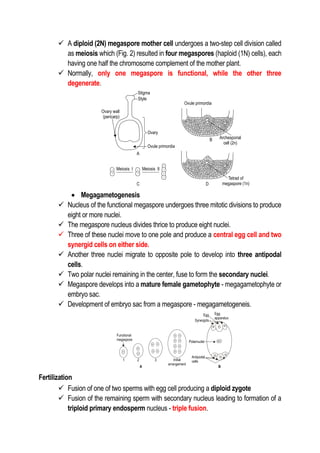
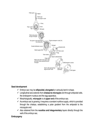
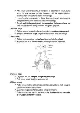
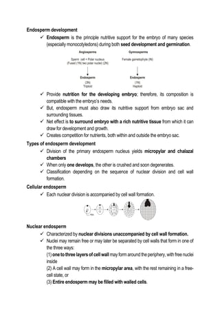
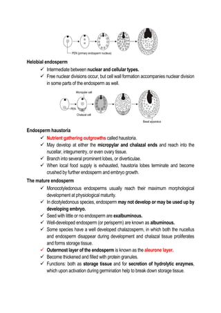
ICAR AIEEA JRF & SRF for PG admissions
ICAR NET, ARS & STO (T-6)
IBPS – AFO
PLANT SCIENCE: Seed Science and Technology
TITBITS 2: Sporogenesis, Gametogenesis, Fertilization & Seed
Development
Prepared by
Dr. K. Vanangamudi
Formerly Dean (Agriculture),
Dean Adhiparasakthi Agricultural College,
Professor & Head,
Seed Science & Technology, TNAU, Coimbatore
 Reproduction - process by which living organisms give rise to offspring of
similar kind (species).
 In crop plants, mode of reproduction is of two types, viz.
1) Sexual reproduction
2) Asexual reproduction
Sexual reproduction
 Multiplication of plants through embryos which have developed by fusion of male
and female gametes
Ovule development
 Development of ovule occurs within the ovary, which provides a location for
nurture and development of the female gametophyte
 Ovule growth begins as a small outgrowth within the nucellus.
 Secondary outgrowths,orcollars(integuments),soonappear aroundtheperiphery
of the nucellar outgrowths and envelop it.
 Consist of the inner and outer integuments and ultimately become the testa
(seed coat) of the mature ovule.
Funicle, hilum, chalaza and raphe
 Developing ovule is commonly attached to placenta by the funiculus.
 Scar on the ovule made where the funiculus detaches at maturity - hilum.
 Point where the integuments meets at the nucellar apex is the micropyle, and
the region of integumentary origin and attachment, usually opposite the
micropyle, is the chalaza.
 Between chalaza and hilum of many species is an area known as the raphe.
Nucellus
 Nucellus provides tissue for the origin and nature of the female gametophyte,
from archesporial cell to the mature megagametophyte.
 Originates from ovary tissues and provides site of archesporial cell origin.
Integuments
 Nature and thickness of the integuments vary considerably among species,
depending on their role in contributing to embryo sac and ovule development.
Micropyle
 Integumentary pore or opening in the ovule through which the pollen tube
grows to fertilize the egg cell of the female gametophyte.
Epistase
 Development of well-defined nucellar or integumentary tissue in the micropylar
region of the seed of certain species.
 Cells adjacent to the micropyle may show a marked elongation.
SEXUAL REPRODUCTION
Sporogenesis
 Production of microspores and megaspores
 In anthers, microspores are formed through microsporogensis
 In ovules, the megaspores are formed through megasporogenesis.
Gametogenesis
 Production of male and female gametes in the microspores and megaspores.
 Microsporogenesis
 Sporophytic cells in the pollen sacs of anther which undergo meiotic division to
form haploid i.e., microspores are called microspore (MMC) or pollen mother
cell (PMC)
 Each PMC produce four microspores and each microspore after thickening of
the wall transforms into pollen grain.
 Microgametogenesis
 Production of male gametes or sperm.
 On maturation of the pollen, microspore nucleus divides mitotically to produce a
generative and a vegetative or tube nucleus.
 Pollen is generally released in this binucleate stage.
 Reach of pollen over the stigma - pollination.
 Generative nucleus undergoes another mitotic division to produce two male
gametes or sperm nuclei.
 Pollen along with pollen tube possessing a pair of sperm nuclei -
microgametophyte.
 Pollen tube enters the embryo sac through micropyle and discharges the two
sperm nuclei.
 Megasporogenesis
 In angiosperms, seeds originate from meristematic tissue of the ovary wall
called ovule primordial.
 From nucellus, one cell develops special characteristics that distinguish it from
adjacent cells.
 As this cell increases in size, its nucleus becomes larger and its cytoplasm grows
denser in preparation for cell division (archesporial cell).
 First division results in a megaspore mother cell and a parietal cell. Usually the
parietal cell remains undivided and soon deteriorates; however, in some
species, it undergoes further division and contributes to seed formation.
 A diploid (2N) megaspore mother cell undergoes a two-step cell division called
as meiosis which (Fig. 2) resulted in four megaspores (haploid (1N) cells), each
having one half the chromosome complement of the mother plant.
 Normally, only one megaspore is functional, while the other three
degenerate.
 Megagametogenesis
 Nucleus of the functional megaspore undergoes three mitotic divisions to produce
eight or more nuclei.
 The megaspore nucleus divides thrice to produce eight nuclei.
 Three of these nuclei move to one pole and produce a central egg cell and two
synergid cells on either side.
 Another three nuclei migrate to opposite pole to develop into three antipodal
cells.
 Two polar nuclei remaining in the center, fuse to form the secondary nuclei.
 Megaspore develops into a mature female gametophyte - megagametophyte or
embryo sac.
 Development of embryo sac from a megaspore - megagametogeneis.
Fertilization
 Fusion of one of two sperms with egg cell producing a diploid zygote
 Fusion of the remaining sperm with secondary nucleus leading to formation of a
triploid primary endosperm nucleus - triple fusion.
Seed development
 Embryo sac may be ellipsoidal, elongated or variously bent in shape.
 Longitudinal axis extends from chalaza to micropyle and through antipodal cells,
the endosperm nucleus and the egg apparatus.
 Morphologically, micropyle is at upper end of the embryo sac.
 As embryo sac is growing, it requires a constant nutritive supply, which is provided
through the chalaza, establishing a polar gradient from the antipodal to the
micropylar end.
 Also obtained from the nucellus and integumentary layers directly through the
wall of the embryo sac.
Embryogeny
 After sexual fusion or syngamy, a brief period of reorganization occurs, during
which the large vacuole gradually disappears, with the zygote cytoplasm
becoming more homogeneous and the nucleus large.
 Lines of polarity in preparation for future division and growth already exist in
embryo sac having been established in the unfertilized egg.
 The still undivided zygote typically elongates along the horizontal axis, and
small vacuoles become evenly distributed through the cytoplasm.
1. Globular stage
 Globular stage of embryo development precedes the cotyledon development.
 Embryo is spherical in shape. Suspensor also develops along with embryo.
2. Heart stage
 Globular embryo develops into two lobed form and looks like a heart.
 Suspensor acts as an “umbilical cord” providing nutrients to the embryo.
3. Torpedo stage
 Cotyledons and axis elongate, enlarge and grow longer.
 Embryo may remain straight or became curved.
4. Mature embryo
 As the embryo mature, cotyledons curve and provide nutrition to plant, enough to
get plant started with photosynthesis.
 Suspensor disappears when cotyledons enlarge and mature.
 Endosperm has been used for nutrients for the development and maturation
of cotyledons and embryonic axis.
Endosperm development
 Endosperm is the principle nutritive support for the embryo of many species
(especially monocotyledons) during both seed development and germination.
 Provide nutrition for the developing embryo; therefore, its composition is
compatible with the embryo’s needs.
 But, endosperm must also draw its nutritive support from embryo sac and
surrounding tissues.
 Net effect is to surround embryo with a rich nutritive tissue from which it can
draw for development and growth.
 Creates competition for nutrients, both within and outside the embryo sac.
Types of endosperm development
 Division of the primary endosperm nucleus yields micropylar and chalazal
chambers
 When only one develops, the other is crushed and soon degenerates.
 Classification depending on the sequence of nuclear division and cell wall
formation.
Cellular endosperm
 Each nuclear division is accompanied by cell wall formation.
Nuclear endosperm
 Characterized by nuclear divisions unaccompanied by cell wall formation.
 Nuclei may remain free or may later be separated by cell walls that form in one of
the three ways:
(1) one to three layers of cell wall may form around the periphery, with free nuclei
inside
(2) A cell wall may form in the micropylar area, with the rest remaining in a free-
cell state, or
(3) Entire endosperm may be filled with walled cells.
Helobial endosperm
 Intermediate between nuclear and cellular types.
 Free nuclear divisions occur, but cell wall formation accompanies nuclear division
in some parts of the endosperm as well.
Endosperm haustoria
 Nutrient gathering outgrowths called haustoria.
 May develop at either the micropylar and chalazal ends and reach into the
nucellar, integumentry, or even ovary tissue.
 Branch into several prominent lobes, or diverticulae.
 When local food supply is exhausted, haustoria lobes terminate and become
crushed by further endosperm and embryo growth.
The mature endosperm
 Monocotyledonous endosperms usually reach their maximum morphological
development at physiological maturity.
 In dicotyledonous species, endosperm may not develop or may be used up by
developing embryo.
 Seed with little or no endosperm are exalbuminous.
 Well-developed endosperm (or perisperm) are known as albuminous.
 Some species have a well developed chalazosperm, in which both the nucellus
and endosperm disappear during development and chalazal tissue proliferates
and forms storage tissue.
 Outermost layer of the endosperm is known as the aleurone layer.
 Become thickened and filled with protein granules.
 Functions: both as storage tissue and for secretion of hydrolytic enzymes,
which upon activation during germination help to break down storage tissue.
Life cycle of plant

Sporogenesis, Gametogenesis, Fertilization & Seed Development.pdf

  • 1.
    ICAR AIEEA JRF& SRF for PG admissions ICAR NET, ARS & STO (T-6) IBPS – AFO PLANT SCIENCE: Seed Science and Technology TITBITS 2: Sporogenesis, Gametogenesis, Fertilization & Seed Development Prepared by Dr. K. Vanangamudi Formerly Dean (Agriculture), Dean Adhiparasakthi Agricultural College, Professor & Head, Seed Science & Technology, TNAU, Coimbatore
  • 2.
     Reproduction -process by which living organisms give rise to offspring of similar kind (species).  In crop plants, mode of reproduction is of two types, viz. 1) Sexual reproduction 2) Asexual reproduction Sexual reproduction  Multiplication of plants through embryos which have developed by fusion of male and female gametes Ovule development  Development of ovule occurs within the ovary, which provides a location for nurture and development of the female gametophyte  Ovule growth begins as a small outgrowth within the nucellus.  Secondary outgrowths,orcollars(integuments),soonappear aroundtheperiphery of the nucellar outgrowths and envelop it.  Consist of the inner and outer integuments and ultimately become the testa (seed coat) of the mature ovule. Funicle, hilum, chalaza and raphe  Developing ovule is commonly attached to placenta by the funiculus.  Scar on the ovule made where the funiculus detaches at maturity - hilum.  Point where the integuments meets at the nucellar apex is the micropyle, and the region of integumentary origin and attachment, usually opposite the micropyle, is the chalaza.  Between chalaza and hilum of many species is an area known as the raphe.
  • 3.
    Nucellus  Nucellus providestissue for the origin and nature of the female gametophyte, from archesporial cell to the mature megagametophyte.  Originates from ovary tissues and provides site of archesporial cell origin. Integuments  Nature and thickness of the integuments vary considerably among species, depending on their role in contributing to embryo sac and ovule development. Micropyle  Integumentary pore or opening in the ovule through which the pollen tube grows to fertilize the egg cell of the female gametophyte. Epistase  Development of well-defined nucellar or integumentary tissue in the micropylar region of the seed of certain species.  Cells adjacent to the micropyle may show a marked elongation. SEXUAL REPRODUCTION Sporogenesis  Production of microspores and megaspores  In anthers, microspores are formed through microsporogensis  In ovules, the megaspores are formed through megasporogenesis. Gametogenesis  Production of male and female gametes in the microspores and megaspores.  Microsporogenesis  Sporophytic cells in the pollen sacs of anther which undergo meiotic division to form haploid i.e., microspores are called microspore (MMC) or pollen mother cell (PMC)  Each PMC produce four microspores and each microspore after thickening of the wall transforms into pollen grain.
  • 4.
     Microgametogenesis  Productionof male gametes or sperm.  On maturation of the pollen, microspore nucleus divides mitotically to produce a generative and a vegetative or tube nucleus.  Pollen is generally released in this binucleate stage.  Reach of pollen over the stigma - pollination.  Generative nucleus undergoes another mitotic division to produce two male gametes or sperm nuclei.  Pollen along with pollen tube possessing a pair of sperm nuclei - microgametophyte.  Pollen tube enters the embryo sac through micropyle and discharges the two sperm nuclei.  Megasporogenesis  In angiosperms, seeds originate from meristematic tissue of the ovary wall called ovule primordial.  From nucellus, one cell develops special characteristics that distinguish it from adjacent cells.  As this cell increases in size, its nucleus becomes larger and its cytoplasm grows denser in preparation for cell division (archesporial cell).  First division results in a megaspore mother cell and a parietal cell. Usually the parietal cell remains undivided and soon deteriorates; however, in some species, it undergoes further division and contributes to seed formation.
  • 5.
     A diploid(2N) megaspore mother cell undergoes a two-step cell division called as meiosis which (Fig. 2) resulted in four megaspores (haploid (1N) cells), each having one half the chromosome complement of the mother plant.  Normally, only one megaspore is functional, while the other three degenerate.  Megagametogenesis  Nucleus of the functional megaspore undergoes three mitotic divisions to produce eight or more nuclei.  The megaspore nucleus divides thrice to produce eight nuclei.  Three of these nuclei move to one pole and produce a central egg cell and two synergid cells on either side.  Another three nuclei migrate to opposite pole to develop into three antipodal cells.  Two polar nuclei remaining in the center, fuse to form the secondary nuclei.  Megaspore develops into a mature female gametophyte - megagametophyte or embryo sac.  Development of embryo sac from a megaspore - megagametogeneis. Fertilization  Fusion of one of two sperms with egg cell producing a diploid zygote  Fusion of the remaining sperm with secondary nucleus leading to formation of a triploid primary endosperm nucleus - triple fusion.
  • 6.
    Seed development  Embryosac may be ellipsoidal, elongated or variously bent in shape.  Longitudinal axis extends from chalaza to micropyle and through antipodal cells, the endosperm nucleus and the egg apparatus.  Morphologically, micropyle is at upper end of the embryo sac.  As embryo sac is growing, it requires a constant nutritive supply, which is provided through the chalaza, establishing a polar gradient from the antipodal to the micropylar end.  Also obtained from the nucellus and integumentary layers directly through the wall of the embryo sac. Embryogeny
  • 7.
     After sexualfusion or syngamy, a brief period of reorganization occurs, during which the large vacuole gradually disappears, with the zygote cytoplasm becoming more homogeneous and the nucleus large.  Lines of polarity in preparation for future division and growth already exist in embryo sac having been established in the unfertilized egg.  The still undivided zygote typically elongates along the horizontal axis, and small vacuoles become evenly distributed through the cytoplasm. 1. Globular stage  Globular stage of embryo development precedes the cotyledon development.  Embryo is spherical in shape. Suspensor also develops along with embryo. 2. Heart stage  Globular embryo develops into two lobed form and looks like a heart.  Suspensor acts as an “umbilical cord” providing nutrients to the embryo. 3. Torpedo stage  Cotyledons and axis elongate, enlarge and grow longer.  Embryo may remain straight or became curved. 4. Mature embryo  As the embryo mature, cotyledons curve and provide nutrition to plant, enough to get plant started with photosynthesis.  Suspensor disappears when cotyledons enlarge and mature.  Endosperm has been used for nutrients for the development and maturation of cotyledons and embryonic axis.
  • 8.
    Endosperm development  Endospermis the principle nutritive support for the embryo of many species (especially monocotyledons) during both seed development and germination.  Provide nutrition for the developing embryo; therefore, its composition is compatible with the embryo’s needs.  But, endosperm must also draw its nutritive support from embryo sac and surrounding tissues.  Net effect is to surround embryo with a rich nutritive tissue from which it can draw for development and growth.  Creates competition for nutrients, both within and outside the embryo sac. Types of endosperm development  Division of the primary endosperm nucleus yields micropylar and chalazal chambers  When only one develops, the other is crushed and soon degenerates.  Classification depending on the sequence of nuclear division and cell wall formation. Cellular endosperm  Each nuclear division is accompanied by cell wall formation. Nuclear endosperm  Characterized by nuclear divisions unaccompanied by cell wall formation.  Nuclei may remain free or may later be separated by cell walls that form in one of the three ways: (1) one to three layers of cell wall may form around the periphery, with free nuclei inside (2) A cell wall may form in the micropylar area, with the rest remaining in a free- cell state, or (3) Entire endosperm may be filled with walled cells.
  • 9.
    Helobial endosperm  Intermediatebetween nuclear and cellular types.  Free nuclear divisions occur, but cell wall formation accompanies nuclear division in some parts of the endosperm as well. Endosperm haustoria  Nutrient gathering outgrowths called haustoria.  May develop at either the micropylar and chalazal ends and reach into the nucellar, integumentry, or even ovary tissue.  Branch into several prominent lobes, or diverticulae.  When local food supply is exhausted, haustoria lobes terminate and become crushed by further endosperm and embryo growth. The mature endosperm  Monocotyledonous endosperms usually reach their maximum morphological development at physiological maturity.  In dicotyledonous species, endosperm may not develop or may be used up by developing embryo.  Seed with little or no endosperm are exalbuminous.  Well-developed endosperm (or perisperm) are known as albuminous.  Some species have a well developed chalazosperm, in which both the nucellus and endosperm disappear during development and chalazal tissue proliferates and forms storage tissue.  Outermost layer of the endosperm is known as the aleurone layer.  Become thickened and filled with protein granules.  Functions: both as storage tissue and for secretion of hydrolytic enzymes, which upon activation during germination help to break down storage tissue.
  • 10.