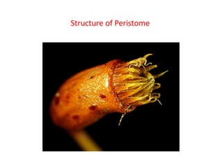
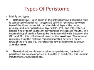
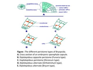
PERISTOME TEETH IN MOSS
Deboshree Das
Roll No.- 11
3rd sem BOTANY
Introduction
• Peristome derived from greek word – ‘peri’ meaning
‘around’ or ‘about’ and ‘stoma’, meaning ‘mouth’.
• The peristome is a teeth-like projection that surrounds
the mouth of capsule in most of the members of
Bryidae (Mosses) of Bryophytes. So, it is an important
characteristic of the Bryopsida.
• The peristome teeth spring from the rim or diaphragm.
The peristome teeth are triangular two-ply structures
which operate like trap-doors.
• The peristome is different in morphology across
species and is used as a classification tools.
Cont..
• In mosses, the peristome is a specialized
structure in the sporangium that allows for
gradual spore discharge.
• The teeth may be solid cellular tissue or
composed only of thickened portion of the cell
walls of adjacent cells.
On the basis of absence and presence of
peristome teeth ,mosses are classified into two
types
• Stegocarpous – The bryidae in which apical
portion of the capsule differentiates into
operculum and peristome.
• Cleistocarpous – In few genera of the true
mosses,both operculum and peristome teeths
are absent.
Structure of Peristome
Types Of Peristome
• Mainly two types :
 Orthodontous - Each tooth of the arthrodontous peristome type
is composed of periclinal (tangential) cell wall remnants between
two of the three concentric peristomial cell layers: the outer,
primary, and inner peristomial layers (OPL, PPL, and IPL). Often, a
double ring of teeth is present surrounding the capsule mouth . The
external ring of teeth is formed by the tangential walls between the
OPL and PPL; it is collectively known as the exostome. The internal
row consists of the cell wall remnants located between the cell
rings of the PPL and IPL, therefore the row of segments is known
as endostome.
 Nematodontous - In nematodontous peristome, the teeth of
peristome are solid composed of bundles of dead cells. It is found in
Polytrichum, Pogonatum etc.
Structures
• Depending on the number of cell columns articulated to form the
length of a tooth, two primary types of arthrodontous peristomes
are known: diplolepidous and haplolepidous.
• In haplolepidous peristomes the exostome is missing, but the
endostome is well developed and strongly thickened. The outer
surface of an endostome segment is made up of just one column of
cells from the PPL (Figure 2C). The haplolepidous peristome is
characteristic of the Grimmiales, Seligeriales, Archidiales, Pottiales,
and Dicranales.
• In diplolepidous peristomes two columns of adjacent OPL cells
participate in the formation of an exostome tooth (Figure 2B, D, E).
A medial vertical line marking the two cells can be observed along
the outer surface of each tooth. This type of peristome can be
observed in taxa of the Funariales, Bryales and Hypnales, among
others.
• Two basic peristome "types" have long been recognized in the
diplolepidous mosses: diplolepidous-opposite, and diplolepidous-
alternate. In diplolepidous peristomes usually both the exostome
and the endostome are present, but in several families the
endostome is lacking or reduced. When the two concentric rows
are present, each external tooth can be directly opposite or
alternated to the internal segments of the endostome.
• In the diplolepidous-opposite or "Funaria type", the anticlinal walls
of the IPL and PPL are almost perfectly aligned (co-radial), so the
endostome segments and the exostome teeth are opposite each
other. This peristome organization in found in the Funariales.
• In the diplolepidous-alternate or "Bryum type", anticlinal cell
divisions in the IPL are asymmetric, producing narrow cells adjacent
to large cells. Later in development, the IPL ring shifts displacing the
position of radial IPL cell walls relative to those in the PPL cells. This
results in the placement of the endostome segments alternating
with the exostome teeth. The diplolepidous alternate peristome is
common in the Bryales, Rhizogoniales, Hookeriales, and Hypnales.
Figure : The different peristome types of Bryopsida.
A. Cross section of an embryonic sporophyte capsule.
B. Diplolepidous opposite peristome (Funaria type).
C. Haplolepidous peristome (Dicranum type).
D. Diplolepidous alternate (Orthotrichum type).
E. Diplolepidous alternate (Bryum type).
Functions
• Peristome teeth serve as a valve that allows the spores
to exit the capsule. The peristome teeth open and
splay outward during times of low humidity, allowing
the spores to float away and disperse. One layer readily
absorbs and releases water, while the other layer does
not react very much to varying humidity levels. This
causes the teeth to bend outward during times of low
humidity.
• Some peristome teeth work in other ways; example,
Funaria mosses have slits between the teeth that
expand during dry periods. In other mosses, such as
Polystrichum mosses, the peristome teeth do not
respond to moisture.
References
Thank you

Peristome teeth in moss

  • 1.
    PERISTOME TEETH INMOSS Deboshree Das Roll No.- 11 3rd sem BOTANY
  • 2.
    Introduction • Peristome derivedfrom greek word – ‘peri’ meaning ‘around’ or ‘about’ and ‘stoma’, meaning ‘mouth’. • The peristome is a teeth-like projection that surrounds the mouth of capsule in most of the members of Bryidae (Mosses) of Bryophytes. So, it is an important characteristic of the Bryopsida. • The peristome teeth spring from the rim or diaphragm. The peristome teeth are triangular two-ply structures which operate like trap-doors. • The peristome is different in morphology across species and is used as a classification tools.
  • 3.
    Cont.. • In mosses,the peristome is a specialized structure in the sporangium that allows for gradual spore discharge. • The teeth may be solid cellular tissue or composed only of thickened portion of the cell walls of adjacent cells.
  • 4.
    On the basisof absence and presence of peristome teeth ,mosses are classified into two types • Stegocarpous – The bryidae in which apical portion of the capsule differentiates into operculum and peristome. • Cleistocarpous – In few genera of the true mosses,both operculum and peristome teeths are absent.
  • 5.
  • 7.
    Types Of Peristome •Mainly two types :  Orthodontous - Each tooth of the arthrodontous peristome type is composed of periclinal (tangential) cell wall remnants between two of the three concentric peristomial cell layers: the outer, primary, and inner peristomial layers (OPL, PPL, and IPL). Often, a double ring of teeth is present surrounding the capsule mouth . The external ring of teeth is formed by the tangential walls between the OPL and PPL; it is collectively known as the exostome. The internal row consists of the cell wall remnants located between the cell rings of the PPL and IPL, therefore the row of segments is known as endostome.  Nematodontous - In nematodontous peristome, the teeth of peristome are solid composed of bundles of dead cells. It is found in Polytrichum, Pogonatum etc.
  • 8.
  • 9.
    • Depending onthe number of cell columns articulated to form the length of a tooth, two primary types of arthrodontous peristomes are known: diplolepidous and haplolepidous. • In haplolepidous peristomes the exostome is missing, but the endostome is well developed and strongly thickened. The outer surface of an endostome segment is made up of just one column of cells from the PPL (Figure 2C). The haplolepidous peristome is characteristic of the Grimmiales, Seligeriales, Archidiales, Pottiales, and Dicranales. • In diplolepidous peristomes two columns of adjacent OPL cells participate in the formation of an exostome tooth (Figure 2B, D, E). A medial vertical line marking the two cells can be observed along the outer surface of each tooth. This type of peristome can be observed in taxa of the Funariales, Bryales and Hypnales, among others.
  • 10.
    • Two basicperistome "types" have long been recognized in the diplolepidous mosses: diplolepidous-opposite, and diplolepidous- alternate. In diplolepidous peristomes usually both the exostome and the endostome are present, but in several families the endostome is lacking or reduced. When the two concentric rows are present, each external tooth can be directly opposite or alternated to the internal segments of the endostome. • In the diplolepidous-opposite or "Funaria type", the anticlinal walls of the IPL and PPL are almost perfectly aligned (co-radial), so the endostome segments and the exostome teeth are opposite each other. This peristome organization in found in the Funariales. • In the diplolepidous-alternate or "Bryum type", anticlinal cell divisions in the IPL are asymmetric, producing narrow cells adjacent to large cells. Later in development, the IPL ring shifts displacing the position of radial IPL cell walls relative to those in the PPL cells. This results in the placement of the endostome segments alternating with the exostome teeth. The diplolepidous alternate peristome is common in the Bryales, Rhizogoniales, Hookeriales, and Hypnales.
  • 11.
    Figure : Thedifferent peristome types of Bryopsida. A. Cross section of an embryonic sporophyte capsule. B. Diplolepidous opposite peristome (Funaria type). C. Haplolepidous peristome (Dicranum type). D. Diplolepidous alternate (Orthotrichum type). E. Diplolepidous alternate (Bryum type).
  • 12.
    Functions • Peristome teethserve as a valve that allows the spores to exit the capsule. The peristome teeth open and splay outward during times of low humidity, allowing the spores to float away and disperse. One layer readily absorbs and releases water, while the other layer does not react very much to varying humidity levels. This causes the teeth to bend outward during times of low humidity. • Some peristome teeth work in other ways; example, Funaria mosses have slits between the teeth that expand during dry periods. In other mosses, such as Polystrichum mosses, the peristome teeth do not respond to moisture.
  • 13.
  • 14.