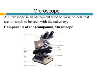
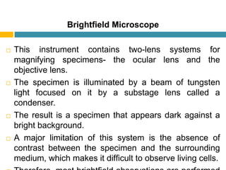
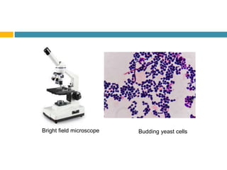
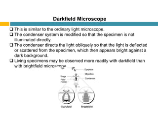
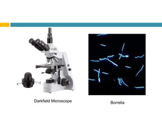
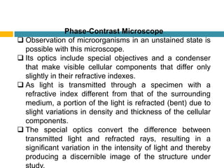
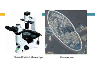
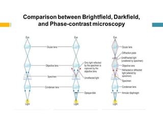
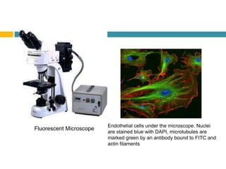
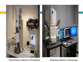
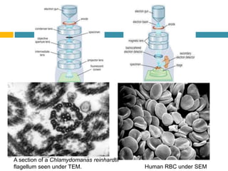
MICROBIOLOGY QUICK LEARN
MICROSCOPE
COMPONENTS
PRINCIPLES
TYPES
APPLICATIONS
Ms Saajida Sultaana Mahusook
Microscope
A microscope is an instrument used to view objects that
are too small to be seen with the naked eye.
Components of the (compound)Microscope
Theoretical Principles of Microscopy
Magnification
• It is the ability of a microscope to enlarge an object.
• Magnification is the function of a two-lens system; the ocular lens
and the objective lens.
• Magnification power is calculated by multiplying the powers of the
ocular lens and the objective lens.
Resolving power
• It is how far apart two adjacent objects must be before a given
lens shows them as discrete entities.
• The resolving power of a lens is dependent on the wavelength of
light used and the numerical aperture.
• Resolving power =
wavelength of light
2 ∗ numerical aperture
Numerical aperture
• Numerical aperture is defined as a function of the
diameter of the objective lens in relation to its focal
length.
Refractive index
• This is the bending power of light passing through air
from the glass slide to the objective lens.
Illumination
Effective illumination is required for efficient
magnification and resolving power.
Types of microscopes
Microscopes are designated as either light microscopes or
electron microscopes.
 Light microscopes use visible light or ultraviolet rays to
illuminate specimens.
 They include
 brightfield,
 darkfield,
 phase-contrast, and
 fluorescent instruments.
 Electron microscopes use electron beams (instead of light
rays) and magnets (instead of lenses) to observe
submicroscopic particles.
Brightfield Microscope
 This instrument contains two-lens systems for
magnifying specimens- the ocular lens and the
objective lens.
 The specimen is illuminated by a beam of tungsten
light focused on it by a substage lens called a
condenser.
 The result is a specimen that appears dark against a
bright background.
 A major limitation of this system is the absence of
contrast between the specimen and the surrounding
medium, which makes it difficult to observe living cells.
Bright field microscope Budding yeast cells
Darkfield Microscope
 This is similar to the ordinary light microscope.
 The condenser system is modified so that the specimen is not
illuminated directly.
 The condenser directs the light obliquely so that the light is deflected
or scattered from the specimen, which then appears bright against a
dark background.
 Living specimens may be observed more readily with darkfield than
with brightfield microscopy.
BorreliaDarkfield Microscope
Phase-Contrast Microscope
 Observation of microorganisms in an unstained state is
possible with this microscope.
 Its optics include special objectives and a condenser
that make visible cellular components that differ only
slightly in their refractive indexes.
 As light is transmitted through a specimen with a
refractive index different from that of the surrounding
medium, a portion of the light is refracted (bent) due to
slight variations in density and thickness of the cellular
components.
 The special optics convert the difference between
transmitted light and refracted rays, resulting in a
significant variation in the intensity of light and thereby
producing a discernible image of the structure under
Phase-Contrast Microscope Paramecium
Fluorescent Microscope
 This microscope is used most frequently to visualize specimens
that are chemically tagged with a fluorescent dye.
 The source of illumination is an ultraviolet (UV) light obtained from
a high-pressure mercury lamp or hydrogen quartz lamp.
 The ocular lens is fitted with a filter that permits the longer
ultraviolet wavelengths to pass, while the shorter wavelengths are
blocked or eliminated.
 Ultraviolet radiations are absorbed by the fluorescent label, and
the energy is re-emitted in the form of a different wavelength in the
visible light range.
 The fluorescent dyes absorb at wavelengths between 230 and
350 nanometers (nm) and emit orange, yellow, or greenish light.
 This microscope is used primarily for the detection of antigen-
antibody reactions. Antibodies are conjugated with a fluorescent
dye that becomes excited in the presence of ultraviolet light, and
the fluorescent portion of the dye becomes visible against a black
background.
Endothelial cells under the microscope. Nuclei
are stained blue with DAPI, microtubules are
marked green by an antibody bound to FITC and
actin filaments
Fluorescent Microscope
Electron Microscope
 This instrument provides a revolutionary method of microscopy, with
magnifications up to 1 million X.
 This permits visualization of submicroscopic cellular particles as well
as viral agents. In the electron microscope, the specimen is
illuminated by a beam of electrons rather than light, and the focusing
is carried out by electromagnets instead of a set of optics.
 These components are sealed in a tube in which a complete vacuum
is established.
 Transmission electron microscopes require specimens that are
prepared as thin filaments, fixed and dehydrated for the electron
beam to pass freely through them.
 As the electrons pass through the specimen, images are formed by
directing the electrons onto photographic film, thus making internal
cellular structures visible.
 Scanning electron microscopes are used for visualizing surface
characteristics rather than intracellular structures.
 A narrow beam of electrons scans back and forth, producing a
three-dimensional image as the electrons are reflected off the
specimen’s surface.
Transmission electron microscope Scanning electron microscope
Human RBC under SEM
A section of a Chlamydomanas reinhardtii
flagellum seen under TEM.
Thank You

Microbiology Quick Learn microscope

  • 1.
  • 2.
    Microscope A microscope isan instrument used to view objects that are too small to be seen with the naked eye. Components of the (compound)Microscope
  • 3.
    Theoretical Principles ofMicroscopy Magnification • It is the ability of a microscope to enlarge an object. • Magnification is the function of a two-lens system; the ocular lens and the objective lens. • Magnification power is calculated by multiplying the powers of the ocular lens and the objective lens. Resolving power • It is how far apart two adjacent objects must be before a given lens shows them as discrete entities. • The resolving power of a lens is dependent on the wavelength of light used and the numerical aperture. • Resolving power = wavelength of light 2 ∗ numerical aperture
  • 4.
    Numerical aperture • Numericalaperture is defined as a function of the diameter of the objective lens in relation to its focal length. Refractive index • This is the bending power of light passing through air from the glass slide to the objective lens. Illumination Effective illumination is required for efficient magnification and resolving power.
  • 5.
    Types of microscopes Microscopesare designated as either light microscopes or electron microscopes.  Light microscopes use visible light or ultraviolet rays to illuminate specimens.  They include  brightfield,  darkfield,  phase-contrast, and  fluorescent instruments.  Electron microscopes use electron beams (instead of light rays) and magnets (instead of lenses) to observe submicroscopic particles.
  • 6.
    Brightfield Microscope  Thisinstrument contains two-lens systems for magnifying specimens- the ocular lens and the objective lens.  The specimen is illuminated by a beam of tungsten light focused on it by a substage lens called a condenser.  The result is a specimen that appears dark against a bright background.  A major limitation of this system is the absence of contrast between the specimen and the surrounding medium, which makes it difficult to observe living cells.
  • 7.
    Bright field microscopeBudding yeast cells
  • 8.
    Darkfield Microscope  Thisis similar to the ordinary light microscope.  The condenser system is modified so that the specimen is not illuminated directly.  The condenser directs the light obliquely so that the light is deflected or scattered from the specimen, which then appears bright against a dark background.  Living specimens may be observed more readily with darkfield than with brightfield microscopy.
  • 9.
  • 10.
    Phase-Contrast Microscope  Observationof microorganisms in an unstained state is possible with this microscope.  Its optics include special objectives and a condenser that make visible cellular components that differ only slightly in their refractive indexes.  As light is transmitted through a specimen with a refractive index different from that of the surrounding medium, a portion of the light is refracted (bent) due to slight variations in density and thickness of the cellular components.  The special optics convert the difference between transmitted light and refracted rays, resulting in a significant variation in the intensity of light and thereby producing a discernible image of the structure under
  • 11.
  • 13.
    Fluorescent Microscope  Thismicroscope is used most frequently to visualize specimens that are chemically tagged with a fluorescent dye.  The source of illumination is an ultraviolet (UV) light obtained from a high-pressure mercury lamp or hydrogen quartz lamp.  The ocular lens is fitted with a filter that permits the longer ultraviolet wavelengths to pass, while the shorter wavelengths are blocked or eliminated.  Ultraviolet radiations are absorbed by the fluorescent label, and the energy is re-emitted in the form of a different wavelength in the visible light range.  The fluorescent dyes absorb at wavelengths between 230 and 350 nanometers (nm) and emit orange, yellow, or greenish light.  This microscope is used primarily for the detection of antigen- antibody reactions. Antibodies are conjugated with a fluorescent dye that becomes excited in the presence of ultraviolet light, and the fluorescent portion of the dye becomes visible against a black background.
  • 14.
    Endothelial cells underthe microscope. Nuclei are stained blue with DAPI, microtubules are marked green by an antibody bound to FITC and actin filaments Fluorescent Microscope
  • 15.
    Electron Microscope  Thisinstrument provides a revolutionary method of microscopy, with magnifications up to 1 million X.  This permits visualization of submicroscopic cellular particles as well as viral agents. In the electron microscope, the specimen is illuminated by a beam of electrons rather than light, and the focusing is carried out by electromagnets instead of a set of optics.  These components are sealed in a tube in which a complete vacuum is established.  Transmission electron microscopes require specimens that are prepared as thin filaments, fixed and dehydrated for the electron beam to pass freely through them.  As the electrons pass through the specimen, images are formed by directing the electrons onto photographic film, thus making internal cellular structures visible.  Scanning electron microscopes are used for visualizing surface characteristics rather than intracellular structures.  A narrow beam of electrons scans back and forth, producing a three-dimensional image as the electrons are reflected off the specimen’s surface.
  • 16.
    Transmission electron microscopeScanning electron microscope
  • 17.
    Human RBC underSEM A section of a Chlamydomanas reinhardtii flagellum seen under TEM.
  • 18.