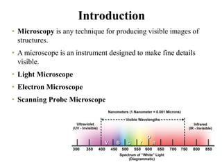
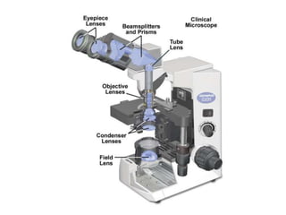
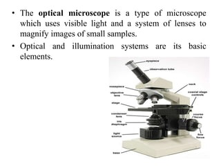
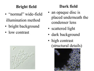
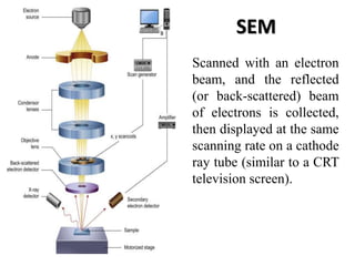
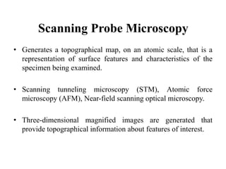
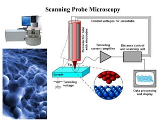
Microscopy
Types of microscopes
Light microscopes –
UV, Dark field, Phase contrast, Fluorescent
and
Electron microscope.
Dr. S. PARTHASARATHY, M.Sc. (Agri)., Ph.D.
Assistant Professor (Plant Pathology)
Introduction
• Microscopy is any technique for producing visible images of
structures.
• A microscope is an instrument designed to make fine details
visible.
• Light Microscope
• Electron Microscope
• Scanning Probe Microscope
• The optical microscope is a type of microscope
which uses visible light and a system of lenses to
magnify images of small samples.
• Optical and illumination systems are its basic
elements.
Types of light microscope
– Bright-field microscope
– Dark-field microscope
– Phase-contrast microscope
– Fluorescence microscopes
Bright field
• “normal” wide-field
illumination method
• bright background
• low contrast
Dark field
• an opaque disc is
placed underneath the
condenser lens
• scattered light
• dark background
• high contrast
(structural details)
Polarizing microscope Brightfield/ Darkfield
Stereo Microscope
Phase contrast
• Principle: Different densities and refractive
indices of different parts of cells.
Fluorescent microscope
• Fluorescent compound which absorbs light
at one wavelength and then emits light at
longer wavelength.
Epi-Fluorescence microscope with 3 filters 4
Semi Apochromatic objective lenses
Inverted Fluorescence Compound
Microscope
Inverted Microscopes have the condenser
and light source above the stage and the
objective lenses below.
SEM
Scanned with an electron
beam, and the reflected
(or back-scattered) beam
of electrons is collected,
then displayed at the same
scanning rate on a cathode
ray tube (similar to a CRT
television screen).
TEM
Contrasts in the image are
produced by differences in
beam scattering or diffraction
produced between various
elements of the
microstructure.
Scanning electron microscope Transmission electron
microscope
Morphology character Internal structure
Electron beams scan over the
sample
Electron beams pass through
the sample
Magnification 1,00,00 0X Magnification 5,00,000 X
Three dimensional black and
white images
Produce two dimensional black
and white structure
Based on scattered electrons or
produce images by detecting
secondary electron which are
emitted from the surface of the
sample
Based on the transmitted
electron two produce image
Scanning Probe Microscopy
• Generates a topographical map, on an atomic scale, that is a
representation of surface features and characteristics of the
specimen being examined.
• Scanning tunneling microscopy (STM), Atomic force
microscopy (AFM), Near-field scanning optical microscopy.
• Three-dimensional magnified images are generated that
provide topographical information about features of interest.
Scanning Probe Microscopy
Microscopic techniques

Microscopic techniques

  • 1.
    Microscopy Types of microscopes Lightmicroscopes – UV, Dark field, Phase contrast, Fluorescent and Electron microscope. Dr. S. PARTHASARATHY, M.Sc. (Agri)., Ph.D. Assistant Professor (Plant Pathology)
  • 3.
    Introduction • Microscopy isany technique for producing visible images of structures. • A microscope is an instrument designed to make fine details visible. • Light Microscope • Electron Microscope • Scanning Probe Microscope
  • 8.
    • The opticalmicroscope is a type of microscope which uses visible light and a system of lenses to magnify images of small samples. • Optical and illumination systems are its basic elements.
  • 10.
    Types of lightmicroscope – Bright-field microscope – Dark-field microscope – Phase-contrast microscope – Fluorescence microscopes
  • 11.
    Bright field • “normal”wide-field illumination method • bright background • low contrast Dark field • an opaque disc is placed underneath the condenser lens • scattered light • dark background • high contrast (structural details)
  • 12.
    Polarizing microscope Brightfield/Darkfield Stereo Microscope
  • 14.
    Phase contrast • Principle:Different densities and refractive indices of different parts of cells.
  • 16.
    Fluorescent microscope • Fluorescentcompound which absorbs light at one wavelength and then emits light at longer wavelength.
  • 17.
    Epi-Fluorescence microscope with3 filters 4 Semi Apochromatic objective lenses Inverted Fluorescence Compound Microscope Inverted Microscopes have the condenser and light source above the stage and the objective lenses below.
  • 18.
    SEM Scanned with anelectron beam, and the reflected (or back-scattered) beam of electrons is collected, then displayed at the same scanning rate on a cathode ray tube (similar to a CRT television screen).
  • 19.
    TEM Contrasts in theimage are produced by differences in beam scattering or diffraction produced between various elements of the microstructure.
  • 20.
    Scanning electron microscopeTransmission electron microscope Morphology character Internal structure Electron beams scan over the sample Electron beams pass through the sample Magnification 1,00,00 0X Magnification 5,00,000 X Three dimensional black and white images Produce two dimensional black and white structure Based on scattered electrons or produce images by detecting secondary electron which are emitted from the surface of the sample Based on the transmitted electron two produce image
  • 22.
    Scanning Probe Microscopy •Generates a topographical map, on an atomic scale, that is a representation of surface features and characteristics of the specimen being examined. • Scanning tunneling microscopy (STM), Atomic force microscopy (AFM), Near-field scanning optical microscopy. • Three-dimensional magnified images are generated that provide topographical information about features of interest.
  • 23.