Submitted By:
Waseem Sajjad
0654-Mphil-Z-22
SESSION2022-2024
Submitted To:
Dr. Shafaat
Government college University Lahore
Electron Microscope Principle
Scanning, Transmission Electron
Microscope and Microphotography
2.
CONTENT
1. Microscopy
2. ElectronMicroscopy
3. Principle of Electron Microscopy
4. SEM
5. TEM
6. SEM VS TEM
7. Microphotography
8. Applications
3.
MICROSCOPY
Microorganisms are muchtoo small to be seen with the unaided eye; they must be
observed with a microscope.
“The word microscope is derived from the Latin word micro (small) and the
Greek word skopos (to look at)”.
Modern microbiologists use microscopes that produce, with great clarity,
magnifications that range from ten to thousands of times greater than those of
van Leeuwenhoek’s single lens.
4.
MICROSCOPY
History
• The simplemicroscope used by van Leeuwenhoek in the seventeenth century had
only one lens and was similar to a magnifying glass.
• However, van Leeuwenhoek was the best lens grinder in the world in his day. His
lenses were ground with such precision that a single lens could magnify a microbe
300 times.
• His simple microscopes enabled him to be the first person to see bacteria.
• Contemporaries of van Leeuwenhoek, such as Robert Hooke, built compound
microscopes, which have multiple lenses.
5.
ELECTRON MICROSCOPY
• Objectssmaller than about 0.2 μm, such as viruses or the internal structures of
cells, must be examined with an electron microscope.
• In electron microscopy, a beam of electrons is used instead of light.
• Like light, free electrons travel in waves.
• The resolving power of the electron microscope is far greater than that of the other
microscopes described here so far. The better resolution of electron microscopes is
due to the shorter wavelengths of electrons; the wavelengths of electrons are about
100,000 times smaller than the wavelengths of visible light.
6.
PRINCIPLE OF ELECTRONMICROSCOPY
• Thus, electron microscopes are used to examine structures too small to be resolved
with light microscopes. Images produced by electron microscopes are always black
and white.
• Instead of using glass lenses, an electron microscope uses electromagnetic lenses to
focus a beam of electrons onto a specimen.
• There are two types of electron microscopes:
1. Scanning electron microscope
2. Transmission electron microscope
7.
SCANNING ELECTRON MICROSCOPY
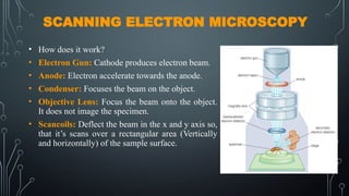
•How does it work?
• Electron Gun: Cathode produces electron beam.
• Anode: Electron accelerate towards the anode.
• Condenser: Focuses the beam on the object.
• Objective Lens: Focus the beam onto the object.
It does not image the specimen.
• Scancoils: Deflect the beam in the x and y axis so,
that it’s scans over a rectangular area (Vertically
and horizontally) of the sample surface.
8.
SCANNING ELECTRON MICROSCOPY
•Sample: Focused by electron beam.
• Backscattered Electron Detector: BSE are beam of
electrons that are reflected from the sample. Since
heavy elements (high atomic number) backscattered
electrons more strongly then light elements (Low
atomic number) and thus appears brighter in the image.
• Imaging: Converts the electron image in to sum form
perceptible to the human eye. Imaging is digitally
captured for display on a computer monitor.
• Note: All lenses are electromagnet. Snice the electron
beam is negatable chartered, and electro magnetic force
can be use as lens.
9.
ADVANTAGES AND DISADVANTAGES
ADVANTAGES
1.Power full magnification
2. High quality and resolution
3. Provide information on sample’s
surface and composition
DISADVANTAGES
1. Larger and very expensive
2. Operation and analysis required
special maintenance
3. Images are black and white. They can
not display living specimens in natural
colors
10.
TRANSMISSION ELECTRON MICROSCOPY
•How does it work?
• Electron Gun: Cathode produces electron
beam.
• Anode: Electron accelerate towards the
anode.
• Condenser: Focuses the beam on the object.
• Sample: Focused by electron beam.
• Objective Lens: Focuses and magnifies the
image
• Imaging: Converts the electron image in to
sum form perceptible to the human eye.
Imaging is digitally captured for display on a
computer monitor.
11.
HOW DOES THEIMAGE FORM?
• Contrast formation: The contrast between two adjacent areas in an image can be formed due
the difference in electron densities.
• During the interaction of electron beam with the sample, some of the electrons will be lost
due to the absorption.
• The darker areas of the image represent those areas of the sample where more electrons are
absorbed.
• While the lighter areas of the image represent those areas of the sample where less electrons
are absorbed.
• Sample and electron beams should be places in vacuum.
• Specimen should be very thin and dehydrated.
12.
ADVANTAGES AND DISADVANTAGES
ADVANTAGES
1.Power full magnification
2. High quality and resolution
3. Provide information on compound
structure
DISADVANTAGES
1. Larger and very expensive
2. Operation and analysis required special
maintenance
3. Required special training
4. Images are black and white. They can not
display living specimens in natural colors
13.
SEM VS TEM
SEM
1.Resolution 0.5nm
2. Information about the sample surface
3. Uses electrons that are reflected on
by the sample surface to create an
image
TEM
1. Resolution 50pm
2. Offers information about the inner
structure of sample
3. Uses Electrons that are passing
through the sample to create an
image
APPLICATIONS
1. Cell ultrastructure:Electron microscopes can reveal the ultrastructure of cells,
including the internal organelles such as mitochondria, ribosomes, and the
endoplasmic reticulum.
2. Tissue structure: Electron microscopes can be used to examine the structure of
tissues, revealing the arrangement of cells, extracellular matrix, and blood vessels.
3. Microbial structure: Electron microscopy can provide a detailed look at the
structure of microorganisms, such as bacteria, viruses, and fungi.
4. Pathology: Electron microscopy is used in the field of pathology to examine the
structure of tissues and cells in order to diagnose diseases and understand their
underlying mechanisms.