Downloaded 264 times


![ STOOL EXAMINATION: (Microscopic examination)
Normal saline preparation: Actively motile
trophozoites.
Iodine preparation: Cysts or dead trophozoites.
[Excretion of cysts in the stool is often intermittent,
therefore, at least three consecutive specimens should
be examined]
Charcot-Leyden crystals may appear in saline
preparation. These are diamond-shaped crystals, clear
and refractile.
Concentration method such as formal ether may be
used for concentration of amoebic cysts in the stool
when the number of amoebae are scanty.
STOOL ANTIGEN DETECTION:
ELISA is used to detect antigens of E.histolytica in
faeces.](https://image.slidesharecdn.com/diarrhoea-140815103826-phpapp02/75/Microbiological-Aspects-Of-Diarrhoea-22-2048.jpg)


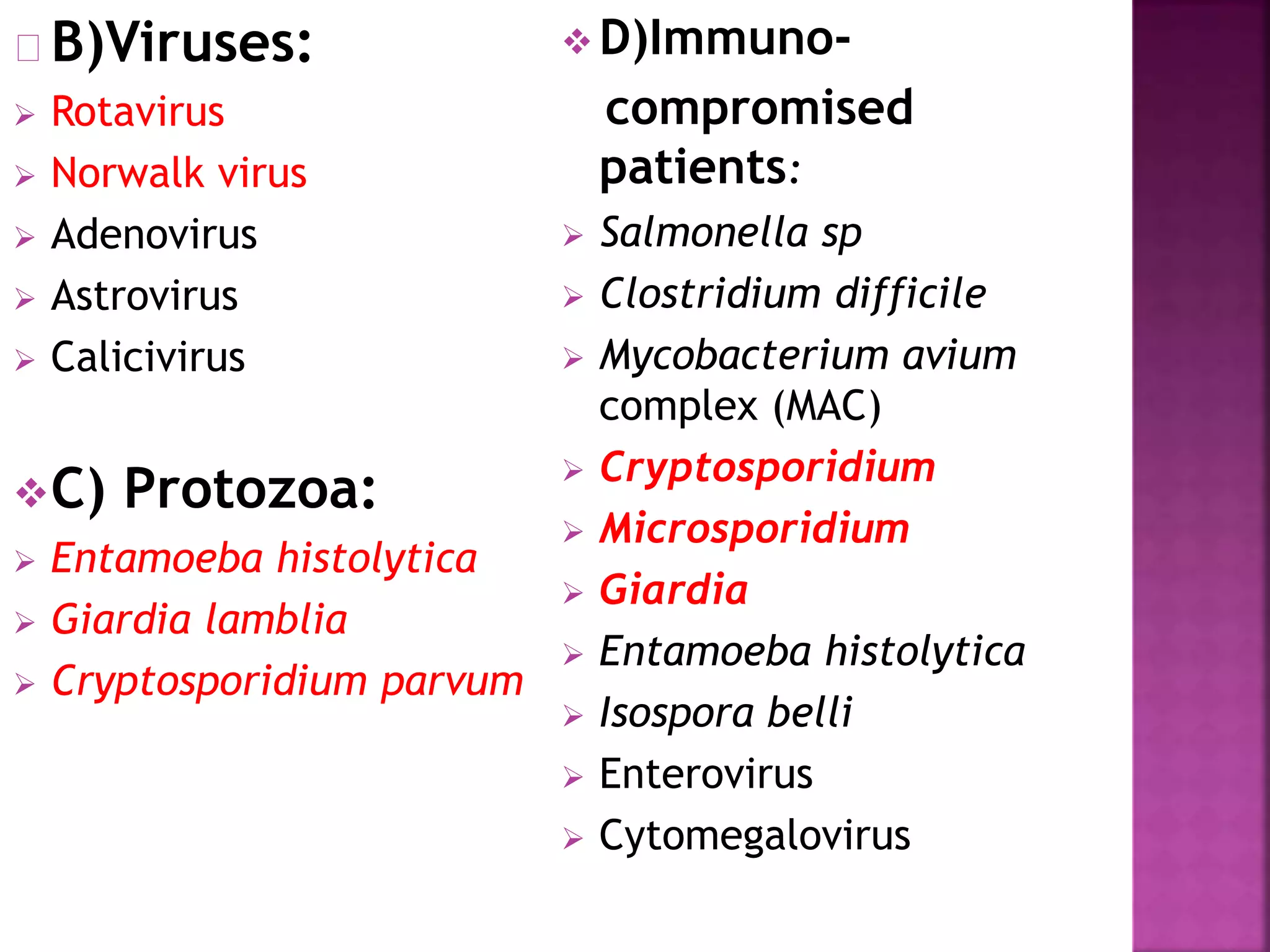
This document discusses the definitions, etiology, and laboratory diagnosis of diarrheal diseases. It defines diarrhea, gastroenteritis, and dysentery. It lists common bacterial, viral, protozoan, and opportunistic pathogens that can cause these diseases. It then provides details on specimen collection and testing methods for identifying various pathogens, including culture-based and antigen/toxin detection techniques. These laboratory methods are described for bacteria like Salmonella, Shigella, Campylobacter, C. difficile, as well as parasites like Giardia, Cryptosporidium, and Entamoeba histolytica.


![Laboratory_Diagnosis_of_Enteric_Fever_Detailed[1].pptx](https://cdn.slidesharecdn.com/ss_thumbnails/laboratorydiagnosisofentericfeverdetailed1-250920160313-94756c84-thumbnail.jpg?width=640&height=640&fit=bounds)


