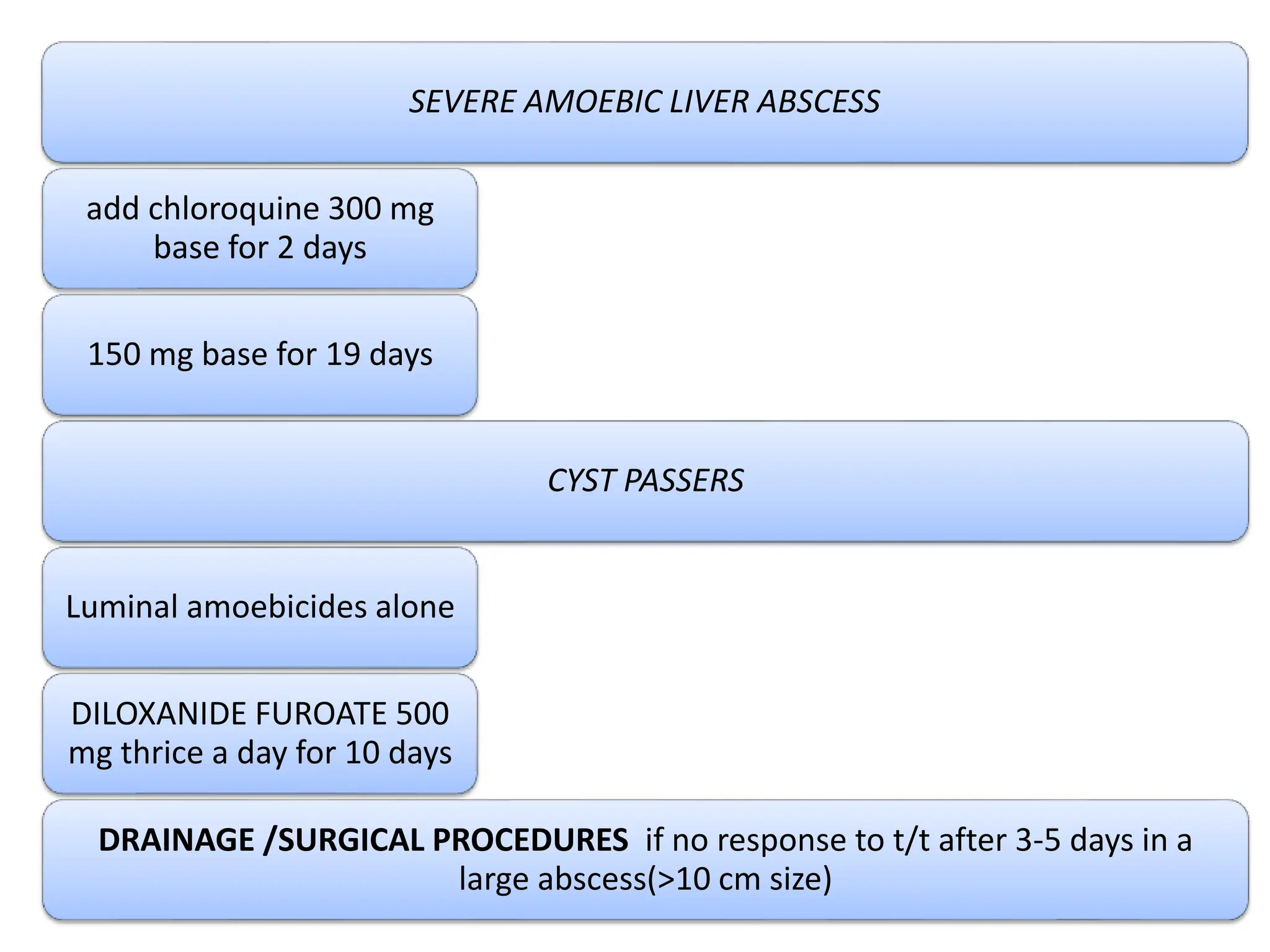
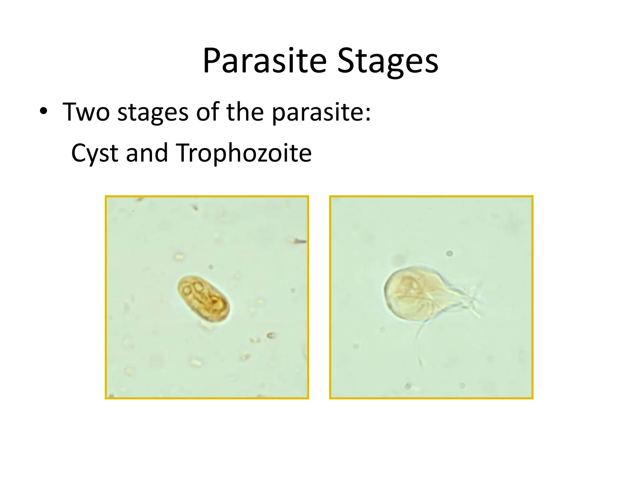
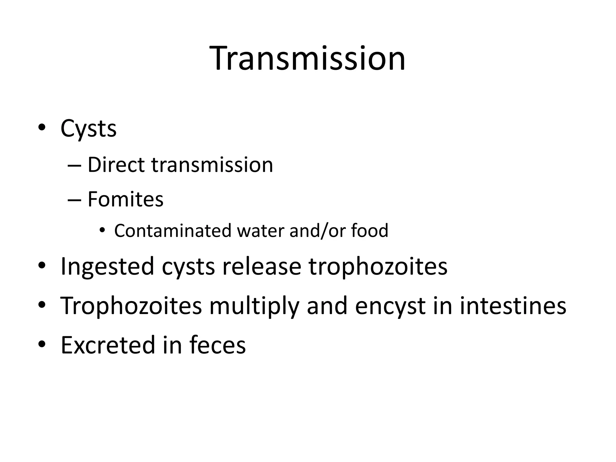
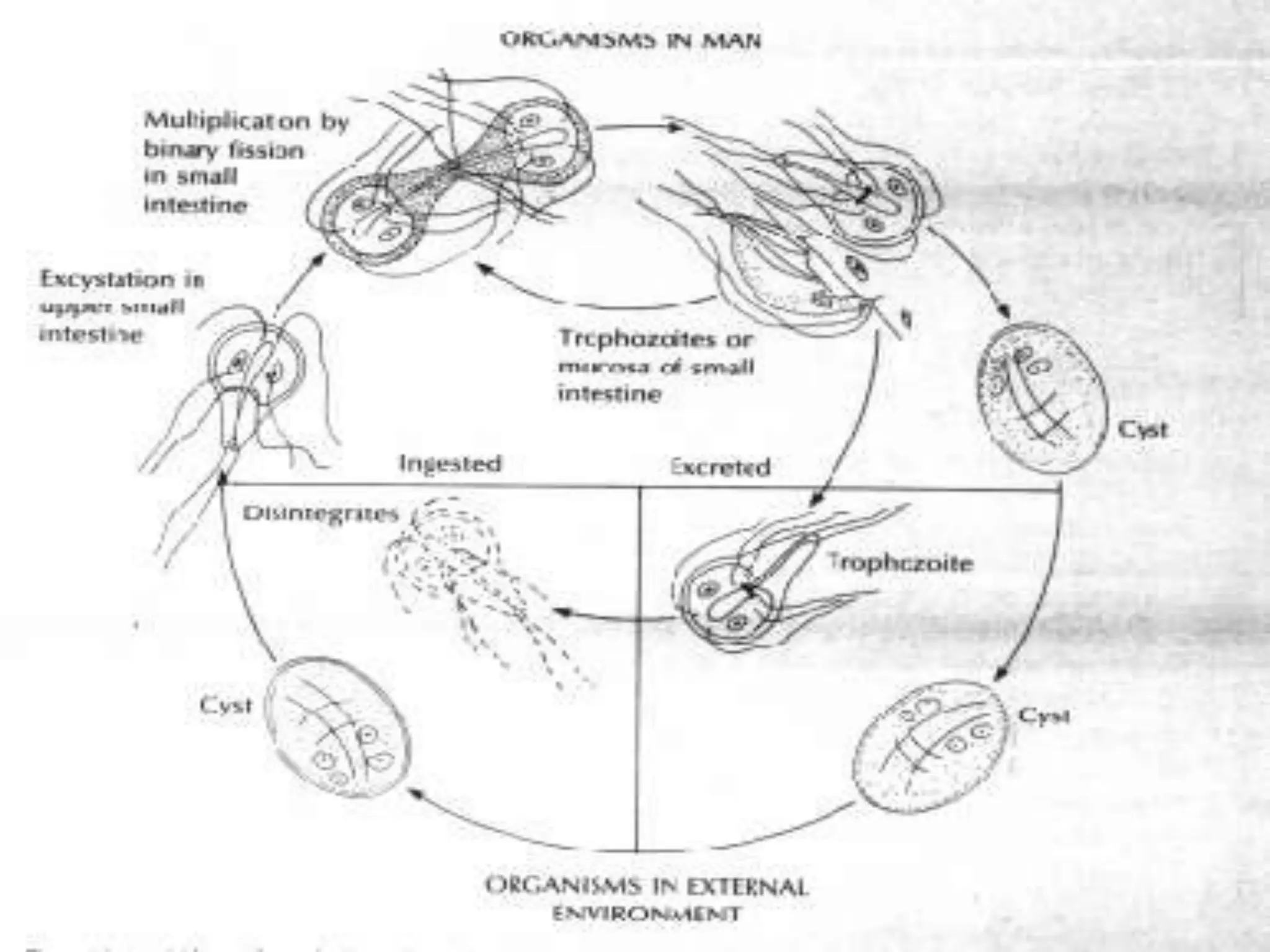
AMEBIASIS
DR. JYOTI VERMA
ASSISTANT PROFESSOR
DEPARTMENT OF MEDICINE
DEFINATION
• The term “Amebiasis” is used clinically to denote all
those conditions which are produced in the human
host by infection with E.Histolytica at different areas
of its invasion.
• Causal agent: Entamoeba histolytica is well
recognized as a pathogenic amoeba.
• People in developing countries that have poor
sanitary conditions
• Immigrants from developing countries
• Travellers to developing countries
• People who live in institutions that have poor
sanitary conditions
• HIV-positive patients
• homosexuals
RISK FACTORS
• 1-direct contact of person to person( fecal-oral)
• 2- Veneral transmission among homosexual
males(oral-anal)
• 3- Food or drink contaminated with feces containing
the E.his. cyst
• 4- Use of human feces (night soil) for soil fertilizer
• 5- contamination of foodstuffs by flies, and possibly
cockroaches
TRANSMISSION
LIFE CYCLE OF ENTAMEBOE HISTOLYTICA
PATHOGENESIS CAUSED BY E.HISTOLYTICA
Noninvasive species: Entamoeba dispar or Entamoeba
moshkovskii does not causes disease.
• E. histolytica infection can cause disease.
• Endemic areas parts of Mexico, India, and nations in the
tropical regions of Africa, South and Central America, and
Asia.
• 1-10% - industrialised countries.
• 50-80% - developing countries.
• It is called a10% disease- 10% of world population affected
of which 10% have active amebic disease from which 10%
die every year.
• In the intestine 10% of Entamoeba species is pathogenic
( E.histolytica),rest is E.dispar.
EPIDEMIOLOGY
• PRIMARY /INTESTINAL LESIONS – LARGE INTESTINE
• SECONDARY /METASTATIC LESIONS- LIVER LUNGS BRAIN SKIN SPLEEN
PATHOLOGY
1. ASYMPTOMATIC INFECTION (CYST
PASSERS)
2. ACUTE AMOEBIC DYSENTRY
3. INTESTINAL AMOEBIASIS COMPLICATAD
BY-
a)Toxic Megacolon
b) Fulminant amoebic colitis
c) Amoeboma-tumour like mass of
granulation tissue
d) Amoebic Peritonitis
e) Perianal Ulceration
4. POST-DYSENTRIC COLITIS – a type of
irritable bowel syndrome ,only mucus is
present.
INTESTINAL LESIONS
AMOEBIC ULCERS
• IN LARGE GUT
• GENERALISED AND
LOCALISED IN ILEO-
CAECAL REGION
SIGMOIDO-RECTAL
REGION
• FLASK SHAPED
,MARGINS ARE
SUPERFICIAL,INVADE
MUCOSA AND EXTEND
TO MUSULARIS
MUCOSA
• NON-SUPPARATIVE AMOEBIC HEPATITIS
• AMOEBIC LIVER ABSCESS-Postero-superior surface of right
lobe of liver.Pus inside called ANCHOVY-SAUCE contains
degenerated hepatocytes ,trophozoites
• AMOEBIC LIVER ABSCESS COMPLICATED BY-
Empyema
Pericarditis
Peritonitis
• PULMONARY AMOEBIASIS
• CEREBRAL AMOEBIASIS
• GENITOURINARY AMOEBIASIS
METASTATIC /EXTRA-INTESTINAL LESIONS
• INTESTINAL AMOEBIASIS –INCUBATION PERIOD-2-6 WEEKS.
90% EH cysts asymptomatic.
symptomatic amoebiais
Amoebic dysentry - abdominal pain and tenderness,tenesmus
6-8 motions/day
copious
blood and mucous mixed with faeces
dark red
offensive
not adherent to the container
COLITIS -mild to moderate-mucus diarrhoea,no blood in stool
severe-offensive and bulky stools with blood and mucus
intestinal bleeding,perforation and paralytic ileus
-fever ,abdominal cramps
CLINICAL FEATURES
EXTRA-INTESTINAL AMOEBIASIS
• AMOEBIC LIVER ABSCESS-most common
H/O INTESTINAL AMOEBIASIS IN 30% - gap of 8 weeks to 1 year
via portal system
5% of invasive disease
10 times more common in men – h/o alcohol abuse
Clinical features-(onset –insidious) –
pain and tenderness in right hypochondrium
fever
jaundice
weight loss and anorexia
dullness and rales at the right lung base (secondary to pl.effusion)
Laboratory findings
leukocytosis (without eosinophilia)
elevated alkaline phosphatase level
mild anemia
elevated erythrocyte sedimentation rate.
Extra-Intestinal Amebiasis
METASTATIC LESIONS OTHER THAN LIVER
Liver abscess rupture:
• Pleuropulmonary disease (the most common complication,
especially with right lobe abscesses)
• Intraperitoneal rupture
• Pericardial rupture (uncommon; usually associated with left
lobe abscesses)
Other manifestations:
• Cerebral amoebiasis
• Genitourinary amoebiasis (rare; more common in women
than men), eg, vaginal fistulae
• Primary cutaneous amoebiasis
• Amoeboma
Diagnosis
• Paraclinical Diagnosis:
• Sigmoidoscopic examination:
precence of a grossly normal mucosa between the ulcers serves to
differentiate amebic from bacillary dysentery,( the entire mucosa
being involvoed in bacillary dysentery).
• Hepatomegally
• C.B.C. : leukocytosis in Amebic dys. rises above 12000 per
microliter, but counts may reach 16000 to 20000 per microliter.
Symptomatic patients
• STOOL EXAMINATION-Macroscopic examination
Microscopic examination
(charcot –Leyden crystals)
• BLOOD EXAMINATION- leucocytosis
•
Immunodiagnosis
(Antibody Detection)
• 1- Antibody detection
• The indirect hemagglutination (IHA)
• The EIA test diagnoses 95% of patients with extraintestinal
amebiasis, 70% of patients with active intestinal infection, and
10% of asymptomatic persons who are passing cysts of E.
histolytica
Antigen Detection
Antigen detection may be useful as an adjunct to microscopic
diagnosis in detecting parasites and to distinguish between
pathogenic and nonpathogenic infections.
Recent studies indicate improved sensitivity and specificity of
fecal antigen assays with the use of monoclonal antibodies
which can distinguish between E. histolytica and E. dispar
infections .
Molecular diagnosis
In reference diagnosis laboratories, PCR is the method of
choice for discriminating between the pathogenic species
(E. histolytica) from the (nonpathogenic species( E. dispar.
ANTI-AMOEBIC DRUGS
LUMINAL AMOEBICIDES
Diloxanide Furoate
Diiodohydroxyquinoline
Paromomycin
TISSUE AMOEBICIDES
All tissues-Metronidazole
Tinidazole
Emetine/Dehydroemetine
Liver only-Chloroquine
Intestinal wall only-
Tetracycline
TREATMENT
INVASIVE AMOEBIASIS(Intestinal/Hepatic)
Metronidazole 400 mg
thrice a day for 7-10
days
followed by
Diloxanide Furoate 500
mg thrice a day for 10
days
Emetine 1 mg/kg body
wt. for 10 days IM
rarely in severe cases
SEVERE AMOEBIC LIVER ABSCESS
add chloroquine 300 mg
base for 2 days
150 mg base for 19 days
CYST PASSERS
Luminal amoebicides alone
DILOXANIDE FUROATE 500
mg thrice a day for 10 days
DRAINAGE /SURGICAL PROCEDURES if no response to t/t after 3-5 days in a
large abscess(>10 cm size)
GIARDIASIS
Giardia Enteritis
Lambliasis
Beaver Fever
Organism
• Giardia intestinalis
– Cosmopolitan Protozoal
parasite
• Also known as:
– Giardia lamblia
– Lamblia intestinalis
– Giardia duodenalis
• Isolated from humans, domestic animals, and
wild animals
Center for Food Security and Public
Health, Iowa State University, 2013
CONT……
• Human infections
– Humans are main reservoir
• Interspecies/zoonotic transmission
– Importance of animal reservoirs unclear
• Non-zoonotic Giardia spp. found in:
– Rodents
– Birds
– Reptiles
– Amphibians
• Giardia intestinalis
– Occurs worldwide
– Most common in warm climates
Morbidity and Mortality: Humans
• Populations affected
– Children
– Travelers, hikers
– Swimmers
– Homosexuals
• Prevalence
in developing and
developed countries.
Center for Food Security and Public
Health, Iowa State University, 2013
Parasite Stages
• Two stages of the parasite:
Cyst and Trophozoite
Center for Food Security and Public
Health, Iowa State University, 2013
Transmission
• Cysts
– Direct transmission
– Fomites
• Contaminated water and/or food
• Ingested cysts release trophozoites
• Trophozoites multiply and encyst in intestines
• Excreted in feces
Center for Food Security and Public
Health, Iowa State University, 2013
Life Cycle
Trophozoites : Lives in duodenum and upper part of
jejenum.
They come in close contact to the mucosal, but do not
invade the host. Adhesive disc fits over surface of
epithelial cell
The flagella act as a pump to move nutrients away from
the microvilla and hold the adhesive disc near the mucosa.
Rapid division to produce large numbers quickly
PATHOGENICITY
• MALABSORPTION OF FAT
• DIARRHEA
• MILD STEATORHOEA
• ALLERGY
• LACTOSE INTOLERANCE
PATHOPHYSIOLOGY PATHOPHYSIOLOGY
• LOSS OF BRUSH BORDER ENZYME ACTIVITIES
• FLATTENED VILLI
CLINICAL FEATURES
• ASYMPTOMATIC TO FULMINANT DIARRHEA AND
MALABSORPTION
• CLINICALY – SILENT CASES
- INTESTINAL –CHRONIC ENTERITIS ,ENTEROCOLITIS
- GENERAL- FEVER
- ANEMIA
- ALLERGIC MANIFESTATIONS
-CHRONIC CHOLECYSTOPATHY
INTESTINAL- ACUTE GIARDIASIS – DIARRHEA ABDOMINAL PAIN
BLEEDING BELCHING FLATUS NAUSEA VOMITTING
CHRONIC GIARDIASIS – with or without antecedent
acute episodes, may be episodic or persistent
- LESS DIARRHEA
-BELCHING
-WEIGHT LOSS
-GROWTH RETARDATION
-DEHYDRATION
EXTRA-INTESTINAL MANIFESTATIONS-URTICARIA ANTERIOR
UVEITIS ARTHRITIS
COMPLICATES CHRONIC DISEASES e.g. CYSTIC FIBROSIS ,AIDS
etc.
DIAGNOSIS
• IDENTIFICATION of cysts in faeces and trophozoites in
faeces and small intestine.
•
• DETECTION OF PARASITE ANTIGENS IN FAECES
• BIOPSY OF TISSUE/DUODENAL ASPIRATE stained by
trichrome or Giemsa stain.
• ENZYME IMMUNOASSAY AND FLUORESCENT
MONOCLONAL ANTIGEN DETECTION SYSTEMS.
• Sensitivity & specificity: 90-100%
• ( ProSpec T, GiardEIA, MeriFluor, Color Vue, and DD System)
Diagnosis
Center for Food Security and Public
Health, Iowa State University, 2013
TREATMENT
• Metronidazole 250 mg thrice daily for 5 days.
750 mg thrice for 21 days.
(refractory cases)
• Tinidazole 2g once
• Nitazoxanide 500 mg twice for 3 days
• Paromomycin,oral aminoglycoside
Prevention and Control
Water
– Do not drink contaminated water
• Untreated lakes, rivers, shallow wells
– Treat potentially contaminated water
• Heat (rolling boil for one minutes)
• Filter (absolute pore size of one micron)
• Chlorinate
Food
– Wash raw fruits and vegetables
Center for Food Security and Public
Health, Iowa State University, 2013
Proper excreta disposal systems
Good personal hygiene on an individual basis
Chlorination alone is sufficient to kill G. lamblia cysts,
important variables, such as water temperature, clarity, pH,
and contact time, alter the efficacy of chlorine, and higher
chlorine levels (4 to 6 mg/liter) may be required.
Boiling water for 20 minutes or 2% solution of iodine.
• Practice good hygiene
– Hand washing
– Don’t swim in recreational
waters for at least two
weeks after symptoms end
– Avoid fecal exposure
Center for Food Security and Public
Health, Iowa State University, 2013
ON 06-12-2014
NEXT SATURDAY
( INTERNAL ASSESSMENT IN MEDICINE )
SEMIOBJECTIVE QUESTIONS -50 MARKS

AMEBIASIS and GIARDIASIS in general population

  • 1.
    AMEBIASIS DR. JYOTI VERMA ASSISTANTPROFESSOR DEPARTMENT OF MEDICINE
  • 2.
    DEFINATION • The term“Amebiasis” is used clinically to denote all those conditions which are produced in the human host by infection with E.Histolytica at different areas of its invasion. • Causal agent: Entamoeba histolytica is well recognized as a pathogenic amoeba.
  • 3.
    • People indeveloping countries that have poor sanitary conditions • Immigrants from developing countries • Travellers to developing countries • People who live in institutions that have poor sanitary conditions • HIV-positive patients • homosexuals RISK FACTORS
  • 4.
    • 1-direct contactof person to person( fecal-oral) • 2- Veneral transmission among homosexual males(oral-anal) • 3- Food or drink contaminated with feces containing the E.his. cyst • 4- Use of human feces (night soil) for soil fertilizer • 5- contamination of foodstuffs by flies, and possibly cockroaches TRANSMISSION
  • 5.
    LIFE CYCLE OFENTAMEBOE HISTOLYTICA
  • 6.
  • 7.
    Noninvasive species: Entamoebadispar or Entamoeba moshkovskii does not causes disease. • E. histolytica infection can cause disease. • Endemic areas parts of Mexico, India, and nations in the tropical regions of Africa, South and Central America, and Asia. • 1-10% - industrialised countries. • 50-80% - developing countries. • It is called a10% disease- 10% of world population affected of which 10% have active amebic disease from which 10% die every year. • In the intestine 10% of Entamoeba species is pathogenic ( E.histolytica),rest is E.dispar. EPIDEMIOLOGY
  • 8.
    • PRIMARY /INTESTINALLESIONS – LARGE INTESTINE • SECONDARY /METASTATIC LESIONS- LIVER LUNGS BRAIN SKIN SPLEEN PATHOLOGY
  • 9.
    1. ASYMPTOMATIC INFECTION(CYST PASSERS) 2. ACUTE AMOEBIC DYSENTRY 3. INTESTINAL AMOEBIASIS COMPLICATAD BY- a)Toxic Megacolon b) Fulminant amoebic colitis c) Amoeboma-tumour like mass of granulation tissue d) Amoebic Peritonitis e) Perianal Ulceration 4. POST-DYSENTRIC COLITIS – a type of irritable bowel syndrome ,only mucus is present. INTESTINAL LESIONS
  • 10.
    AMOEBIC ULCERS • INLARGE GUT • GENERALISED AND LOCALISED IN ILEO- CAECAL REGION SIGMOIDO-RECTAL REGION • FLASK SHAPED ,MARGINS ARE SUPERFICIAL,INVADE MUCOSA AND EXTEND TO MUSULARIS MUCOSA
  • 11.
    • NON-SUPPARATIVE AMOEBICHEPATITIS • AMOEBIC LIVER ABSCESS-Postero-superior surface of right lobe of liver.Pus inside called ANCHOVY-SAUCE contains degenerated hepatocytes ,trophozoites • AMOEBIC LIVER ABSCESS COMPLICATED BY- Empyema Pericarditis Peritonitis • PULMONARY AMOEBIASIS • CEREBRAL AMOEBIASIS • GENITOURINARY AMOEBIASIS METASTATIC /EXTRA-INTESTINAL LESIONS
  • 12.
    • INTESTINAL AMOEBIASIS–INCUBATION PERIOD-2-6 WEEKS. 90% EH cysts asymptomatic. symptomatic amoebiais Amoebic dysentry - abdominal pain and tenderness,tenesmus 6-8 motions/day copious blood and mucous mixed with faeces dark red offensive not adherent to the container COLITIS -mild to moderate-mucus diarrhoea,no blood in stool severe-offensive and bulky stools with blood and mucus intestinal bleeding,perforation and paralytic ileus -fever ,abdominal cramps CLINICAL FEATURES
  • 13.
    EXTRA-INTESTINAL AMOEBIASIS • AMOEBICLIVER ABSCESS-most common H/O INTESTINAL AMOEBIASIS IN 30% - gap of 8 weeks to 1 year via portal system 5% of invasive disease 10 times more common in men – h/o alcohol abuse Clinical features-(onset –insidious) – pain and tenderness in right hypochondrium fever jaundice weight loss and anorexia dullness and rales at the right lung base (secondary to pl.effusion) Laboratory findings leukocytosis (without eosinophilia) elevated alkaline phosphatase level mild anemia elevated erythrocyte sedimentation rate.
  • 15.
  • 16.
    METASTATIC LESIONS OTHERTHAN LIVER Liver abscess rupture: • Pleuropulmonary disease (the most common complication, especially with right lobe abscesses) • Intraperitoneal rupture • Pericardial rupture (uncommon; usually associated with left lobe abscesses) Other manifestations: • Cerebral amoebiasis • Genitourinary amoebiasis (rare; more common in women than men), eg, vaginal fistulae • Primary cutaneous amoebiasis • Amoeboma
  • 18.
    Diagnosis • Paraclinical Diagnosis: •Sigmoidoscopic examination: precence of a grossly normal mucosa between the ulcers serves to differentiate amebic from bacillary dysentery,( the entire mucosa being involvoed in bacillary dysentery). • Hepatomegally • C.B.C. : leukocytosis in Amebic dys. rises above 12000 per microliter, but counts may reach 16000 to 20000 per microliter.
  • 19.
    Symptomatic patients • STOOLEXAMINATION-Macroscopic examination Microscopic examination (charcot –Leyden crystals) • BLOOD EXAMINATION- leucocytosis •
  • 20.
    Immunodiagnosis (Antibody Detection) • 1-Antibody detection • The indirect hemagglutination (IHA) • The EIA test diagnoses 95% of patients with extraintestinal amebiasis, 70% of patients with active intestinal infection, and 10% of asymptomatic persons who are passing cysts of E. histolytica
  • 21.
    Antigen Detection Antigen detectionmay be useful as an adjunct to microscopic diagnosis in detecting parasites and to distinguish between pathogenic and nonpathogenic infections. Recent studies indicate improved sensitivity and specificity of fecal antigen assays with the use of monoclonal antibodies which can distinguish between E. histolytica and E. dispar infections .
  • 22.
    Molecular diagnosis In referencediagnosis laboratories, PCR is the method of choice for discriminating between the pathogenic species (E. histolytica) from the (nonpathogenic species( E. dispar.
  • 23.
    ANTI-AMOEBIC DRUGS LUMINAL AMOEBICIDES DiloxanideFuroate Diiodohydroxyquinoline Paromomycin TISSUE AMOEBICIDES All tissues-Metronidazole Tinidazole Emetine/Dehydroemetine Liver only-Chloroquine Intestinal wall only- Tetracycline
  • 24.
    TREATMENT INVASIVE AMOEBIASIS(Intestinal/Hepatic) Metronidazole 400mg thrice a day for 7-10 days followed by Diloxanide Furoate 500 mg thrice a day for 10 days Emetine 1 mg/kg body wt. for 10 days IM rarely in severe cases
  • 25.
    SEVERE AMOEBIC LIVERABSCESS add chloroquine 300 mg base for 2 days 150 mg base for 19 days CYST PASSERS Luminal amoebicides alone DILOXANIDE FUROATE 500 mg thrice a day for 10 days DRAINAGE /SURGICAL PROCEDURES if no response to t/t after 3-5 days in a large abscess(>10 cm size)
  • 26.
  • 27.
    Organism • Giardia intestinalis –Cosmopolitan Protozoal parasite • Also known as: – Giardia lamblia – Lamblia intestinalis – Giardia duodenalis • Isolated from humans, domestic animals, and wild animals Center for Food Security and Public Health, Iowa State University, 2013
  • 28.
    CONT…… • Human infections –Humans are main reservoir • Interspecies/zoonotic transmission – Importance of animal reservoirs unclear • Non-zoonotic Giardia spp. found in: – Rodents – Birds – Reptiles – Amphibians • Giardia intestinalis – Occurs worldwide – Most common in warm climates
  • 29.
    Morbidity and Mortality:Humans • Populations affected – Children – Travelers, hikers – Swimmers – Homosexuals • Prevalence in developing and developed countries. Center for Food Security and Public Health, Iowa State University, 2013
  • 30.
    Parasite Stages • Twostages of the parasite: Cyst and Trophozoite Center for Food Security and Public Health, Iowa State University, 2013
  • 31.
    Transmission • Cysts – Directtransmission – Fomites • Contaminated water and/or food • Ingested cysts release trophozoites • Trophozoites multiply and encyst in intestines • Excreted in feces Center for Food Security and Public Health, Iowa State University, 2013
  • 32.
    Life Cycle Trophozoites :Lives in duodenum and upper part of jejenum. They come in close contact to the mucosal, but do not invade the host. Adhesive disc fits over surface of epithelial cell The flagella act as a pump to move nutrients away from the microvilla and hold the adhesive disc near the mucosa. Rapid division to produce large numbers quickly
  • 36.
    PATHOGENICITY • MALABSORPTION OFFAT • DIARRHEA • MILD STEATORHOEA • ALLERGY • LACTOSE INTOLERANCE PATHOPHYSIOLOGY PATHOPHYSIOLOGY • LOSS OF BRUSH BORDER ENZYME ACTIVITIES • FLATTENED VILLI
  • 37.
    CLINICAL FEATURES • ASYMPTOMATICTO FULMINANT DIARRHEA AND MALABSORPTION • CLINICALY – SILENT CASES - INTESTINAL –CHRONIC ENTERITIS ,ENTEROCOLITIS - GENERAL- FEVER - ANEMIA - ALLERGIC MANIFESTATIONS -CHRONIC CHOLECYSTOPATHY
  • 38.
    INTESTINAL- ACUTE GIARDIASIS– DIARRHEA ABDOMINAL PAIN BLEEDING BELCHING FLATUS NAUSEA VOMITTING CHRONIC GIARDIASIS – with or without antecedent acute episodes, may be episodic or persistent - LESS DIARRHEA -BELCHING -WEIGHT LOSS -GROWTH RETARDATION -DEHYDRATION EXTRA-INTESTINAL MANIFESTATIONS-URTICARIA ANTERIOR UVEITIS ARTHRITIS COMPLICATES CHRONIC DISEASES e.g. CYSTIC FIBROSIS ,AIDS etc.
  • 39.
    DIAGNOSIS • IDENTIFICATION ofcysts in faeces and trophozoites in faeces and small intestine. • • DETECTION OF PARASITE ANTIGENS IN FAECES • BIOPSY OF TISSUE/DUODENAL ASPIRATE stained by trichrome or Giemsa stain. • ENZYME IMMUNOASSAY AND FLUORESCENT MONOCLONAL ANTIGEN DETECTION SYSTEMS. • Sensitivity & specificity: 90-100% • ( ProSpec T, GiardEIA, MeriFluor, Color Vue, and DD System)
  • 40.
    Diagnosis Center for FoodSecurity and Public Health, Iowa State University, 2013
  • 41.
    TREATMENT • Metronidazole 250mg thrice daily for 5 days. 750 mg thrice for 21 days. (refractory cases) • Tinidazole 2g once • Nitazoxanide 500 mg twice for 3 days • Paromomycin,oral aminoglycoside
  • 42.
    Prevention and Control Water –Do not drink contaminated water • Untreated lakes, rivers, shallow wells – Treat potentially contaminated water • Heat (rolling boil for one minutes) • Filter (absolute pore size of one micron) • Chlorinate Food – Wash raw fruits and vegetables Center for Food Security and Public Health, Iowa State University, 2013
  • 43.
    Proper excreta disposalsystems Good personal hygiene on an individual basis Chlorination alone is sufficient to kill G. lamblia cysts, important variables, such as water temperature, clarity, pH, and contact time, alter the efficacy of chlorine, and higher chlorine levels (4 to 6 mg/liter) may be required. Boiling water for 20 minutes or 2% solution of iodine.
  • 44.
    • Practice goodhygiene – Hand washing – Don’t swim in recreational waters for at least two weeks after symptoms end – Avoid fecal exposure Center for Food Security and Public Health, Iowa State University, 2013
  • 45.
    ON 06-12-2014 NEXT SATURDAY (INTERNAL ASSESSMENT IN MEDICINE ) SEMIOBJECTIVE QUESTIONS -50 MARKS