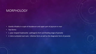
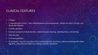
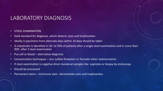
GIARDIASIS
INTRODUCTION
CAUSATIVE AGENT – Giardia lamblia [ also known as G . Intestinalis or G . Duodenalis ]
MORPHOLOGY
• Giardia inhabits in crypts of duodenum and upper part of jejunum in man
• Two forms:
• 1. pear shaped trophozoite : pathogenic form and feeding stage of parasite
• 2. tetra nucleated oval cysts : infective form as well as the diagnostic form of parasite
LIFE CYCLE
PATHOGENICITY
• Infective dose : 10 – 25 cysts
• Risk factors : children more commonly effected
• Other high risk group : elderly debilitated persons , poor hygiene and immune deficient individuals
• Pathogenic form : trophozoites , pathogenic form adhere to duodenal mucosa ,cause disruption of
intestinal epithelium , leads to increased permeability and malabsorption
• Malabsorption of fat :leads to foul smelling profuse frothy diarrhea
• Disaccharidase deficiency :leading to lactose intolerance
• Malabsorption of vitamin A , B12 and iron
• Protein loosing enteropathy
• Antigenic variation : Giardia undergoes frequent antigenic variation due to a cysteine rich protein on its
surface called variant surface protein [VSP].
CLINICAL FEATURES
• 3 Stages:
• 1. Asymptomatic carriers : most infected persons are asymptomatic , harbor the cysts in the gut and
spread the infection
• 2. Acute giardiasis
• Common symptoms include diarrhea , abdominal pain, bloating , belching ,flatus and vomiting
• Fatty diarrhea
• 3. Chronic giardiasis :
• GI symptoms : recurrent episodes of foul smelling diarrhea ,foul flatus ,sulfurous belching with rotten
egg taste, and profound weight loss leading to growth retardation
LABORATORY DIAGNOSIS
• STOOL EXAMINATION
• Gold standard for diagnosis ,which detects cysts and trophozoites
• Ideally 3 specimens from alternate days within 10 days should be taken
• G.intestinalis is identified in 50 to 70% of patients after a single stool examination and in more than
90% after 3 stool examination
• Pus cell or blood – alternative diagnosis
• Concentration techniques – zinc sulfate floatation or formalin ether sedimentation
• If stool examination is negative direct duodenal samples like aspirates or biopsy by endoscopy
• Should be processed
• Permanent stains – trichrome stain –demontrate cysts and trophozoites
ENTERO TEST [ STRING TEST ]
• Uses gelatin capsule attached to a thread containing a weight
• Capsule dissolved in stomachthread carried to duodenumgets unfoldedtakes up the duodenal
samples
• 4 hrs later thread withdrawn and shaken in saline to release trophozoites which can be detected
microscopically by wet mount or permanent stained smear
HISTOPATHOLOGY
• Endoscopy guided duodenal biopsy tissue
• Processed by touch preparation
• Stain – Giemsa stain
ANTIGEN DETECTION STAIN
• ELISA
• Direct fluorescent antibody [DFA]
• Rapid ICT
• Detect cyst wall antigens
• Highly specific and sensitive
ANTIBODY DETECTION
• ELISA
• IFA
• Drawback -Cannot differentiate recent and past infection
CULTURE
• Axenic media like Diamond’s media
• For research purpose
• To Prepare the antigens
MOLECULAR METHODS
• BIOFIRE FILM ARRAY
• MOLECULAR TYPING
RADIOLOGICAL FINDING
• FLUOROSCOPY May reveal hypermotility at the duodenal and jejunal levels
• XRAY AFTER BARIUM MEAL may reveal non specific irregular mucosal thickening with large dilated
loops of hypotonic bowel
TREATMENT
• TINIDAZOLE 2g once orally DOC
• Metronidazole for 5 days or Nitazoxanide for 3g given alternatively
• FurazolidoneChildren
• Auranofin, Paromomycin Pregnancy
• AIDS & Hypogammaglobulinemia prolonged therapy with metronidazole [21 days]
• Metronidazole resistance Auranofin
PREVENTION
• Impoved food and personal hygiene
• Boiling and filtering of potentially contaminated water
• Treatment of asymptomatic carriers
• No vaccine is currently available
THANK YOU

giardiasis.pptx

  • 1.
  • 2.
    INTRODUCTION CAUSATIVE AGENT –Giardia lamblia [ also known as G . Intestinalis or G . Duodenalis ]
  • 3.
    MORPHOLOGY • Giardia inhabitsin crypts of duodenum and upper part of jejunum in man • Two forms: • 1. pear shaped trophozoite : pathogenic form and feeding stage of parasite • 2. tetra nucleated oval cysts : infective form as well as the diagnostic form of parasite
  • 4.
  • 5.
    PATHOGENICITY • Infective dose: 10 – 25 cysts • Risk factors : children more commonly effected • Other high risk group : elderly debilitated persons , poor hygiene and immune deficient individuals • Pathogenic form : trophozoites , pathogenic form adhere to duodenal mucosa ,cause disruption of intestinal epithelium , leads to increased permeability and malabsorption
  • 6.
    • Malabsorption offat :leads to foul smelling profuse frothy diarrhea • Disaccharidase deficiency :leading to lactose intolerance • Malabsorption of vitamin A , B12 and iron • Protein loosing enteropathy
  • 7.
    • Antigenic variation: Giardia undergoes frequent antigenic variation due to a cysteine rich protein on its surface called variant surface protein [VSP].
  • 8.
    CLINICAL FEATURES • 3Stages: • 1. Asymptomatic carriers : most infected persons are asymptomatic , harbor the cysts in the gut and spread the infection • 2. Acute giardiasis • Common symptoms include diarrhea , abdominal pain, bloating , belching ,flatus and vomiting • Fatty diarrhea • 3. Chronic giardiasis : • GI symptoms : recurrent episodes of foul smelling diarrhea ,foul flatus ,sulfurous belching with rotten egg taste, and profound weight loss leading to growth retardation
  • 9.
    LABORATORY DIAGNOSIS • STOOLEXAMINATION • Gold standard for diagnosis ,which detects cysts and trophozoites • Ideally 3 specimens from alternate days within 10 days should be taken • G.intestinalis is identified in 50 to 70% of patients after a single stool examination and in more than 90% after 3 stool examination • Pus cell or blood – alternative diagnosis • Concentration techniques – zinc sulfate floatation or formalin ether sedimentation • If stool examination is negative direct duodenal samples like aspirates or biopsy by endoscopy • Should be processed • Permanent stains – trichrome stain –demontrate cysts and trophozoites
  • 11.
    ENTERO TEST [STRING TEST ] • Uses gelatin capsule attached to a thread containing a weight • Capsule dissolved in stomachthread carried to duodenumgets unfoldedtakes up the duodenal samples • 4 hrs later thread withdrawn and shaken in saline to release trophozoites which can be detected microscopically by wet mount or permanent stained smear
  • 12.
    HISTOPATHOLOGY • Endoscopy guidedduodenal biopsy tissue • Processed by touch preparation • Stain – Giemsa stain
  • 13.
    ANTIGEN DETECTION STAIN •ELISA • Direct fluorescent antibody [DFA] • Rapid ICT • Detect cyst wall antigens • Highly specific and sensitive
  • 14.
    ANTIBODY DETECTION • ELISA •IFA • Drawback -Cannot differentiate recent and past infection
  • 15.
    CULTURE • Axenic medialike Diamond’s media • For research purpose • To Prepare the antigens
  • 16.
    MOLECULAR METHODS • BIOFIREFILM ARRAY • MOLECULAR TYPING
  • 17.
    RADIOLOGICAL FINDING • FLUOROSCOPYMay reveal hypermotility at the duodenal and jejunal levels • XRAY AFTER BARIUM MEAL may reveal non specific irregular mucosal thickening with large dilated loops of hypotonic bowel
  • 18.
    TREATMENT • TINIDAZOLE 2gonce orally DOC • Metronidazole for 5 days or Nitazoxanide for 3g given alternatively • FurazolidoneChildren • Auranofin, Paromomycin Pregnancy • AIDS & Hypogammaglobulinemia prolonged therapy with metronidazole [21 days] • Metronidazole resistance Auranofin
  • 19.
    PREVENTION • Impoved foodand personal hygiene • Boiling and filtering of potentially contaminated water • Treatment of asymptomatic carriers • No vaccine is currently available
  • 20.