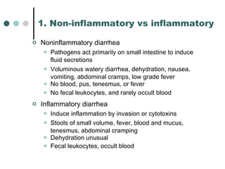
Gastro-intestinal Infections Dr Kamran Afzal Classified Microbiologist
Diarrhea Symptoms   Increase in stool frequency/ volume and/or decrease in consistency Signs Stool water excretion more than 150-300 ml above normal
Classification Of Diarrheas Duration acute chronic Affected organ  small intestine  large intestine Clinical description volume frequency consistency presence of blood Patho-physiologic inflammatory (infectious) non-inflammatory (non-infectious)
1. Non-inflammatory vs inflammatory Noninflammatory diarrhea Pathogens act primarily on small intestine to induce fluid secretions Voluminous watery diarrhea, dehydration, nausea, vomiting, abdominal cramps, low grade fever No blood, pus, tenesmus, or fever No fecal leukocytes, and rarely occult blood Inflammatory diarrhea Induce inflammation by invasion or cytotoxins Stools of small volume, fever, blood and mucus, tenesmus, abdominal cramping Dehydration unusual Fecal leukocytes, occult blood
Infectious Diarrhea - Possible Pathogens Bacteria Invasive and Cytotoxin-producing Microorganisms   Salmonella spp Shigella spp Campylobacter spp Vibrio cholerae Escherichia coli  (EPEC, EHEC, ETEC, EIEC) Clostridium difficile Aeromonas hydrophila Yersinia enterocolitica Vibrio parahaemolyticus Listeria monocytogenes
Bacteria (Food Poisoning-Enterotoxigenic) Staphylococcus aureus Clostridium perfringens Bacillus cereus
Protozoa Giardia lamblia  Entamoeba histolytica Balantidium coli Isospora belli Cryptosporidium Microsporidium Cyclospora Viruses Rota virus Enteric Adeno virus Norwalk virus Calici virus Astro virus
Infectious doses of enteric pathogens Shigella 10 1-2 Giardia lamblia 10 1-2 Entamoeba histolytica 10 1-2   Campylobacter jejuni 10 2-6 Salmonella 10 5  E. coli 10 8  Vibrio cholerae 10 8
Fecal leukocytes Shigellosis Enteroinvasive  E. coli Clostridium difficile Salmonella enteritidis Vibrio parahemolyticus Ulcerative colitis Ischemic colitis
Pathophysiology Toxin production Entero-adherence Mucosal invasion Antibiotic associated colitis
a. Toxin production Neurotoxins (preformed toxin) Bacillus cereus ,  Clostridium perfringens ,  Staphylococcus aureus Enterotoxin Intracellular mediators Increased cyclic AMP, GMP or Calcium Vibrio cholerae, Aeromonas  species, ETEC Cytotoxin Destruction of the mucosal cells Clostridium difficile, E. coli  0157:H7, Shigellae
b. Entero-adherence   Pili or fimbirae bind to specific receptors   Enteroadherent and enteropathogenic  E. coli Giardiasis ( Giardia lamblia )  Helminths Cryptosporidiosis ( Cryptosporidium parvum) Cyclospora  species (?)
c. Mucosal invasion Minimal invasion Norwalk virus, Rotavirus, other viruses Variable invasion Aeromonas  sp.,  Campylobacter  sp.,  Salmonella   sp.,  Vibrio parahemolyticus Severe invasion Intense inflammatory response   Entamoeba histolytica , enteroinvasive  E. coli , Salmonella and  Shigella   species
d. Antibiotic Associated Colitis Antibiotic exposure Intestinal endogenous flora Exposure to  C. difficile  in environment Colonic colonization -  C. difficile Elaboration of toxin(s) Diarrhea and colitis
Features Of Food Poisoning Agents ++ + +++ 8-72 hrs 8-24 hrs Cl. perfringens ++ +++ + 2-10 hrs 1-6 hrs Bacillus cereus ++ +++ + 12-24 hrs 1-8 hrs Staph aureus Enterotoxin Vomiting Diarrhea Duration Incubation period Organism
Salmonella  gastroenteritis   S. typhi  is confined to humans so spread is purely faecal-oral Non-typhoidal strains such as ( S. enterica ) serovars  S. typhimurium, S. enteritidis,  etc Found in GI tracts of mammals, birds, reptiles Acquired from ingestion of contaminated poultry, eggs, meat and exposure to pet reptiles Fever, abdominal cramping, nausea, vomiting, diarrhea with fecal leukocytes Untreated diarrhea lasts 4-10 days
Shigellosis Four species: S. dysenteriae, S. flexneri, S. boydi, S. sonnei Highly infectious: infectious dose < 200  Incubation period 1-7 days Symptoms can develop 12 hours after ingestion Presents as fever, abdominal pain, tenesmus, bloody diarrhea Anti-motility drugs have been associated with toxic megacolon Self-limited but treatment recommended to prevent secondary spread to contacts
E. coli  gastroenteritis Enterohemorrhagic ( E coli  0157:H7) Most common strain in developed countries Usually transmitted by beef, but many other foods Low infectious dose (as few as 100 bacteria) Shiga-toxin Crampy abdominal pain Copious bloody diarrhea sometimes with the hemolytic-uremic syndrome
Enterotoxigenic Most common cause of travelers diarrhea Acquired by ingestion of fecally contaminated food or water Occurs 3-14 days after ingestion Causes watery diarrhea, abdominal cramps occasionally nausea and vomiting Self-limiting lasts 1-5 days
Enteropathogenic Diarrhea in infants common in developing countries Enteroinvasive Dysentery with blood and mucus Enteroaggregative Dysentery with blood and mucus
Transmission of  E. coli  O157 Raw milk  “ Pasteurised milk” Yoghurt Cheese Cream Vegetables Salad vegetables Mayonnaise Apple cider (USA) Fruit
Vibrio cholerae 01 serotype:   (Cholera)  watery diarrhea, dehydration; associated with residence in endemic areas, Toxin causes host cells to secrete Cl – , HCO – , and water Non-01-serotypes:   (Noncholera vibrios)  diarrhea, fever, nausea, vomiting, blood in stool; often associated with traveler’s diarrhea V. parahaemolyticus V. vulnificus
Staphylococcal food poisoning Common food borne illness Infections originate from asymptomatic carriers of  Staphylococcus aureus Can contaminate processed meats, salad, ice-cream Preformed toxin (enterotoxin) in food rather than from direct effect of organism Incubation period about 4 hours Symptoms last < 24 hours No new toxin produced by ingested bacteria Severe nausea and vomiting along with abdominal pain and diarrhea No fever
Traveler’s diarrhea Bacterial Enterotoxigenic  E. coli  (ETEC) Shigella Salmonella Campylobacter Protozoa Giardia, Entamoeba, Cryptosporidium Virus Norwalk, rotavirus, enterovirus
Helicobacter pylori 70-90% of population in developing countries Associated with gastritis, duodenal ulcer, gastric ulcer and gastric adenocarcinoma Fecal-oral transmission
Intestinal tuberculosis Primary – ingestion of organism in unsensitised host Can cause severe ulcero-inflammatory disease with perforation Secondary – swallowing of infected sputum Most common in terminal ileum and jejunum Complications – obstruction, fistula
Clostridium difficile 1970s: Found to be the cause of enterocolitis related to the antibiotic clindamycin Pseudomembranous colitis with yellow-white plaques; can progress to toxic megacolon At least two toxins (A and B) cause necrosis of epithelium Nosocomial transmission Widespread contamination of hospital environments
Endoscopic view of multiple scattered, yellowish plaques consistent with pseudomembranous colitis
Abdominal radiograph demonstrating markedly dilated colon, wall edema, and loss of haustrations in a patient with  Clostridium difficile- associated pseudomembranous colitis complicated by toxic megacolon
Entamoeba histolytica  (amebiasis) Affects 10% of world’s population Order of involvement: caecum, ascending colon, rectum, sigmoid Flask-shaped ulcers Liver abscesses in up to 10%
Giardia lamblia  (giardiasis) Waterborne Can cause diarrhea by several mechanisms Weight loss, cramps, steatorrhea, flatulence, vomiting, belching, fever
Rotavirus  diarrhea Usually sporadic but can cause epidemics in institutions, including nursing homes Cytopathic effect on mature enterocytes, replaced by immature cells with loss of absorptive function Usually infants and young children Fecal-oral transmission
Norovirus  diarrhea Typically epidemic, often traced to a common source Throughout the year Often traced to contaminated food or water Usually adults and school-aged children
HIV associated diarrhea Diarrhoea is a big problem Opportunistic infection  Candida, cryptosporidia, cytomegalovirus, Mycobacterium avium-intracellulare, strongyloides, leishmaniasis HIV itself causes enteropathy
Dysentery Bacillary High frequency Blood and mucus Not sticky Alkaline Pus cells  >  RBCs Macrophages Amoebic Low frequency Blood and mucus, mixed with stools Sticky Acidic RBCs  >  Pus cells Trophozoites of  E. histolytica
Diagnosis - Stool Collection and Transport Appropriate container Proper instructions to the patient Representative portion of stool Fresh specimen Freshly collected rectal swab Transport as early as possible Use transport media – buffered glycerol saline or alkaline peptone water
Diagnostic  Evaluation
Protocol For Specimen Processing
Laboratory Diagnosis Stool Microscopy Saline preparation stool/rectal swab Pus cells RBCs Trophozoites Entamoeba histolytica Giardia lamblia Balantidium coli Cysts Entamoeba histolytica Giardia lamblia
Methylene Blue Preparation Pus cells Iodine Preparation Cysts of Entamoeba, Giardia Modified ZN Stain Cryptosporidium,  Isospora belli Fluorescent Staining Cryptosporidium parvum Gram’s Staining Food Poisoning cases
Special Cultures Campylobacters Skirrow’s or Preston medium Microaerophilic conditions Temp 42 o C Yersinia enterocolitica Yersinia Selective Agar Clostridium difficile Anaerobic Blood Agar
Other Investigations Stool Antigen Detection By ELISA Giardia, Entamoeba, Rota virus, Norwalk virus Clostridium difficile  Toxin Detection Latex agglutination ELISA, Neutralisation tests Electron Microscopy Enteric Adeno, Norwalk and Astro viruses Procto-sigmoidoscopy Pseudomembranous colitis, Amoebic colitis
Treatment Fluid Replacement Oral Intravascular Antimotility Agents Antisecretory Agents Antibiotics Extremes of age Toxic patient Profound deyhydration Probably having bacteremia Blood and mucus in stools Immunocompromised
Treatment Antibiotics Conventional Agents Ampicillin Co-trimoxazole Tetracycline Chloramphenicol Metronidazole Other Agents Nalidixic acid Norfloxacin Ciprofloxacin and other FQs Doxycylcline, Minocycline Cephalosporins Newer Macrolides
2. Duration of diarrhea ACUTE (symptoms    14 days) sudden onset, 90% are infectious in etiology Community-acquired:  Gram-negative bacterial pathogens Viral pathogens Norovirus Rotovirus Protozoal pathogens Entamoeba histolytica Nosocomial (> 3 days after hospitalization) Clostridium difficile Travelers: ETEC
PERSISTENT/CHRONIC (symptoms > 14 days) Most of the causes are non-infectious Parasitic Giardia lamblia, Cryptosporidium parvum, Cyclospora, Isospora belli Immunocompromised host Also consider  Microsporidium and CMV
3. Low-versus high-volume diarrhea Low volume  (low water): colonic High volume  (high water): small bowel  OSMOTIC DIARRHEA: high-volume diarrhea in which the measured fecal osmolality is less than 2 [Na + K] Sorbitol-containing liquid medications, tube feedings, lactose intolerance SECRETORY DIARRHEA: high-volume diarrhea in which the measured fecal osmolality equals 2 [Na + K] E. Coli , rotavirus, laxative abuse
4. Small- versus large-bowel diarrhea Small bowel Large volume, watery, less frequent, painless stools Blood and WBCs are rare Proctoscopy is normal Pain is mid-abdominal Large bowel Small volume, often mucoid, more frequent, painful stools Blood and WBCs are common Proctoscopy is abnormal Pain is lower-abdominal (left lower quadrant)
Case History A 40-year-old woman presented with diarrhea. She had had loose stools for 2 years, progressing over the past 6 months to fifteen large-volume watery stools daily.  What is your differential diagnosis? What diagnostic and management steps would you choose?
Differential Diagnosis Celiac disease   IBS Lactose intolerance Intestinal TB  Intestinal worms Cryptosporidium parvum  secondary to AIDS HUS  Cancer colon This case is a transition from semi-solid to watery diarrhea which is a classical hallmark of progressive ileal disease . When less than 50-60% ileum is involved, there is fat malabsorption and semisolid stools. But when more than 50% of ileum is involved, it will also lead ultimately to bile salt malabsorption
Investigations Stool analysis, C/S, serum electrolytes (Na, K, Ca), electrolytes, CBP Anti-gliadin/endomysial antibody testing to r/o celiac ELISA Barium swallow/enema, colonoscopy Treatment Fluid replacement Oral Antimotility agents

Gi infections

  • 1.
    Gastro-intestinal Infections DrKamran Afzal Classified Microbiologist
  • 2.
    Diarrhea Symptoms Increase in stool frequency/ volume and/or decrease in consistency Signs Stool water excretion more than 150-300 ml above normal
  • 3.
    Classification Of DiarrheasDuration acute chronic Affected organ small intestine large intestine Clinical description volume frequency consistency presence of blood Patho-physiologic inflammatory (infectious) non-inflammatory (non-infectious)
  • 4.
    1. Non-inflammatory vsinflammatory Noninflammatory diarrhea Pathogens act primarily on small intestine to induce fluid secretions Voluminous watery diarrhea, dehydration, nausea, vomiting, abdominal cramps, low grade fever No blood, pus, tenesmus, or fever No fecal leukocytes, and rarely occult blood Inflammatory diarrhea Induce inflammation by invasion or cytotoxins Stools of small volume, fever, blood and mucus, tenesmus, abdominal cramping Dehydration unusual Fecal leukocytes, occult blood
  • 5.
    Infectious Diarrhea -Possible Pathogens Bacteria Invasive and Cytotoxin-producing Microorganisms Salmonella spp Shigella spp Campylobacter spp Vibrio cholerae Escherichia coli (EPEC, EHEC, ETEC, EIEC) Clostridium difficile Aeromonas hydrophila Yersinia enterocolitica Vibrio parahaemolyticus Listeria monocytogenes
  • 6.
    Bacteria (Food Poisoning-Enterotoxigenic)Staphylococcus aureus Clostridium perfringens Bacillus cereus
  • 7.
    Protozoa Giardia lamblia Entamoeba histolytica Balantidium coli Isospora belli Cryptosporidium Microsporidium Cyclospora Viruses Rota virus Enteric Adeno virus Norwalk virus Calici virus Astro virus
  • 8.
    Infectious doses ofenteric pathogens Shigella 10 1-2 Giardia lamblia 10 1-2 Entamoeba histolytica 10 1-2 Campylobacter jejuni 10 2-6 Salmonella 10 5 E. coli 10 8 Vibrio cholerae 10 8
  • 9.
    Fecal leukocytes ShigellosisEnteroinvasive E. coli Clostridium difficile Salmonella enteritidis Vibrio parahemolyticus Ulcerative colitis Ischemic colitis
  • 10.
    Pathophysiology Toxin productionEntero-adherence Mucosal invasion Antibiotic associated colitis
  • 11.
    a. Toxin productionNeurotoxins (preformed toxin) Bacillus cereus , Clostridium perfringens , Staphylococcus aureus Enterotoxin Intracellular mediators Increased cyclic AMP, GMP or Calcium Vibrio cholerae, Aeromonas species, ETEC Cytotoxin Destruction of the mucosal cells Clostridium difficile, E. coli 0157:H7, Shigellae
  • 12.
    b. Entero-adherence Pili or fimbirae bind to specific receptors Enteroadherent and enteropathogenic E. coli Giardiasis ( Giardia lamblia ) Helminths Cryptosporidiosis ( Cryptosporidium parvum) Cyclospora species (?)
  • 13.
    c. Mucosal invasionMinimal invasion Norwalk virus, Rotavirus, other viruses Variable invasion Aeromonas sp., Campylobacter sp., Salmonella sp., Vibrio parahemolyticus Severe invasion Intense inflammatory response Entamoeba histolytica , enteroinvasive E. coli , Salmonella and Shigella species
  • 14.
    d. Antibiotic AssociatedColitis Antibiotic exposure Intestinal endogenous flora Exposure to C. difficile in environment Colonic colonization - C. difficile Elaboration of toxin(s) Diarrhea and colitis
  • 15.
    Features Of FoodPoisoning Agents ++ + +++ 8-72 hrs 8-24 hrs Cl. perfringens ++ +++ + 2-10 hrs 1-6 hrs Bacillus cereus ++ +++ + 12-24 hrs 1-8 hrs Staph aureus Enterotoxin Vomiting Diarrhea Duration Incubation period Organism
  • 16.
    Salmonella gastroenteritis S. typhi is confined to humans so spread is purely faecal-oral Non-typhoidal strains such as ( S. enterica ) serovars S. typhimurium, S. enteritidis, etc Found in GI tracts of mammals, birds, reptiles Acquired from ingestion of contaminated poultry, eggs, meat and exposure to pet reptiles Fever, abdominal cramping, nausea, vomiting, diarrhea with fecal leukocytes Untreated diarrhea lasts 4-10 days
  • 17.
    Shigellosis Four species:S. dysenteriae, S. flexneri, S. boydi, S. sonnei Highly infectious: infectious dose < 200 Incubation period 1-7 days Symptoms can develop 12 hours after ingestion Presents as fever, abdominal pain, tenesmus, bloody diarrhea Anti-motility drugs have been associated with toxic megacolon Self-limited but treatment recommended to prevent secondary spread to contacts
  • 18.
    E. coli gastroenteritis Enterohemorrhagic ( E coli 0157:H7) Most common strain in developed countries Usually transmitted by beef, but many other foods Low infectious dose (as few as 100 bacteria) Shiga-toxin Crampy abdominal pain Copious bloody diarrhea sometimes with the hemolytic-uremic syndrome
  • 19.
    Enterotoxigenic Most commoncause of travelers diarrhea Acquired by ingestion of fecally contaminated food or water Occurs 3-14 days after ingestion Causes watery diarrhea, abdominal cramps occasionally nausea and vomiting Self-limiting lasts 1-5 days
  • 20.
    Enteropathogenic Diarrhea ininfants common in developing countries Enteroinvasive Dysentery with blood and mucus Enteroaggregative Dysentery with blood and mucus
  • 21.
    Transmission of E. coli O157 Raw milk “ Pasteurised milk” Yoghurt Cheese Cream Vegetables Salad vegetables Mayonnaise Apple cider (USA) Fruit
  • 22.
    Vibrio cholerae 01serotype: (Cholera) watery diarrhea, dehydration; associated with residence in endemic areas, Toxin causes host cells to secrete Cl – , HCO – , and water Non-01-serotypes: (Noncholera vibrios) diarrhea, fever, nausea, vomiting, blood in stool; often associated with traveler’s diarrhea V. parahaemolyticus V. vulnificus
  • 23.
    Staphylococcal food poisoningCommon food borne illness Infections originate from asymptomatic carriers of Staphylococcus aureus Can contaminate processed meats, salad, ice-cream Preformed toxin (enterotoxin) in food rather than from direct effect of organism Incubation period about 4 hours Symptoms last < 24 hours No new toxin produced by ingested bacteria Severe nausea and vomiting along with abdominal pain and diarrhea No fever
  • 24.
    Traveler’s diarrhea BacterialEnterotoxigenic E. coli (ETEC) Shigella Salmonella Campylobacter Protozoa Giardia, Entamoeba, Cryptosporidium Virus Norwalk, rotavirus, enterovirus
  • 25.
    Helicobacter pylori 70-90%of population in developing countries Associated with gastritis, duodenal ulcer, gastric ulcer and gastric adenocarcinoma Fecal-oral transmission
  • 26.
    Intestinal tuberculosis Primary– ingestion of organism in unsensitised host Can cause severe ulcero-inflammatory disease with perforation Secondary – swallowing of infected sputum Most common in terminal ileum and jejunum Complications – obstruction, fistula
  • 27.
    Clostridium difficile 1970s:Found to be the cause of enterocolitis related to the antibiotic clindamycin Pseudomembranous colitis with yellow-white plaques; can progress to toxic megacolon At least two toxins (A and B) cause necrosis of epithelium Nosocomial transmission Widespread contamination of hospital environments
  • 28.
    Endoscopic view ofmultiple scattered, yellowish plaques consistent with pseudomembranous colitis
  • 29.
    Abdominal radiograph demonstratingmarkedly dilated colon, wall edema, and loss of haustrations in a patient with Clostridium difficile- associated pseudomembranous colitis complicated by toxic megacolon
  • 30.
    Entamoeba histolytica (amebiasis) Affects 10% of world’s population Order of involvement: caecum, ascending colon, rectum, sigmoid Flask-shaped ulcers Liver abscesses in up to 10%
  • 31.
    Giardia lamblia (giardiasis) Waterborne Can cause diarrhea by several mechanisms Weight loss, cramps, steatorrhea, flatulence, vomiting, belching, fever
  • 32.
    Rotavirus diarrheaUsually sporadic but can cause epidemics in institutions, including nursing homes Cytopathic effect on mature enterocytes, replaced by immature cells with loss of absorptive function Usually infants and young children Fecal-oral transmission
  • 33.
    Norovirus diarrheaTypically epidemic, often traced to a common source Throughout the year Often traced to contaminated food or water Usually adults and school-aged children
  • 34.
    HIV associated diarrheaDiarrhoea is a big problem Opportunistic infection Candida, cryptosporidia, cytomegalovirus, Mycobacterium avium-intracellulare, strongyloides, leishmaniasis HIV itself causes enteropathy
  • 35.
    Dysentery Bacillary Highfrequency Blood and mucus Not sticky Alkaline Pus cells > RBCs Macrophages Amoebic Low frequency Blood and mucus, mixed with stools Sticky Acidic RBCs > Pus cells Trophozoites of E. histolytica
  • 36.
    Diagnosis - StoolCollection and Transport Appropriate container Proper instructions to the patient Representative portion of stool Fresh specimen Freshly collected rectal swab Transport as early as possible Use transport media – buffered glycerol saline or alkaline peptone water
  • 37.
  • 38.
  • 39.
    Laboratory Diagnosis StoolMicroscopy Saline preparation stool/rectal swab Pus cells RBCs Trophozoites Entamoeba histolytica Giardia lamblia Balantidium coli Cysts Entamoeba histolytica Giardia lamblia
  • 40.
    Methylene Blue PreparationPus cells Iodine Preparation Cysts of Entamoeba, Giardia Modified ZN Stain Cryptosporidium, Isospora belli Fluorescent Staining Cryptosporidium parvum Gram’s Staining Food Poisoning cases
  • 41.
    Special Cultures CampylobactersSkirrow’s or Preston medium Microaerophilic conditions Temp 42 o C Yersinia enterocolitica Yersinia Selective Agar Clostridium difficile Anaerobic Blood Agar
  • 42.
    Other Investigations StoolAntigen Detection By ELISA Giardia, Entamoeba, Rota virus, Norwalk virus Clostridium difficile Toxin Detection Latex agglutination ELISA, Neutralisation tests Electron Microscopy Enteric Adeno, Norwalk and Astro viruses Procto-sigmoidoscopy Pseudomembranous colitis, Amoebic colitis
  • 43.
    Treatment Fluid ReplacementOral Intravascular Antimotility Agents Antisecretory Agents Antibiotics Extremes of age Toxic patient Profound deyhydration Probably having bacteremia Blood and mucus in stools Immunocompromised
  • 44.
    Treatment Antibiotics ConventionalAgents Ampicillin Co-trimoxazole Tetracycline Chloramphenicol Metronidazole Other Agents Nalidixic acid Norfloxacin Ciprofloxacin and other FQs Doxycylcline, Minocycline Cephalosporins Newer Macrolides
  • 45.
    2. Duration ofdiarrhea ACUTE (symptoms  14 days) sudden onset, 90% are infectious in etiology Community-acquired: Gram-negative bacterial pathogens Viral pathogens Norovirus Rotovirus Protozoal pathogens Entamoeba histolytica Nosocomial (> 3 days after hospitalization) Clostridium difficile Travelers: ETEC
  • 46.
    PERSISTENT/CHRONIC (symptoms >14 days) Most of the causes are non-infectious Parasitic Giardia lamblia, Cryptosporidium parvum, Cyclospora, Isospora belli Immunocompromised host Also consider Microsporidium and CMV
  • 47.
    3. Low-versus high-volumediarrhea Low volume (low water): colonic High volume (high water): small bowel OSMOTIC DIARRHEA: high-volume diarrhea in which the measured fecal osmolality is less than 2 [Na + K] Sorbitol-containing liquid medications, tube feedings, lactose intolerance SECRETORY DIARRHEA: high-volume diarrhea in which the measured fecal osmolality equals 2 [Na + K] E. Coli , rotavirus, laxative abuse
  • 48.
    4. Small- versuslarge-bowel diarrhea Small bowel Large volume, watery, less frequent, painless stools Blood and WBCs are rare Proctoscopy is normal Pain is mid-abdominal Large bowel Small volume, often mucoid, more frequent, painful stools Blood and WBCs are common Proctoscopy is abnormal Pain is lower-abdominal (left lower quadrant)
  • 49.
    Case History A40-year-old woman presented with diarrhea. She had had loose stools for 2 years, progressing over the past 6 months to fifteen large-volume watery stools daily. What is your differential diagnosis? What diagnostic and management steps would you choose?
  • 50.
    Differential Diagnosis Celiacdisease IBS Lactose intolerance Intestinal TB Intestinal worms Cryptosporidium parvum secondary to AIDS HUS Cancer colon This case is a transition from semi-solid to watery diarrhea which is a classical hallmark of progressive ileal disease . When less than 50-60% ileum is involved, there is fat malabsorption and semisolid stools. But when more than 50% of ileum is involved, it will also lead ultimately to bile salt malabsorption
  • 51.
    Investigations Stool analysis,C/S, serum electrolytes (Na, K, Ca), electrolytes, CBP Anti-gliadin/endomysial antibody testing to r/o celiac ELISA Barium swallow/enema, colonoscopy Treatment Fluid replacement Oral Antimotility agents

Editor's Notes

  • #10 Inflammatory diarrhea
  • #12 SA: n/v CNS, Botulinum on NMJ Enterotoxin: direct effect on intestinal mucosa to elicit net fluid secretion Cyto; mucosal destruction causing inflam colitis