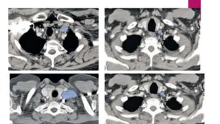
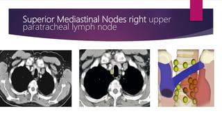
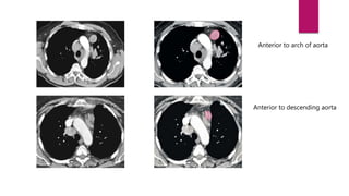
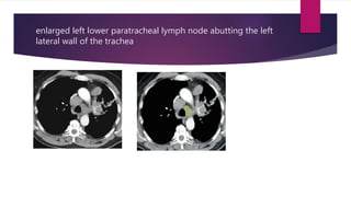
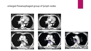
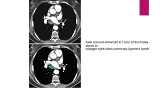
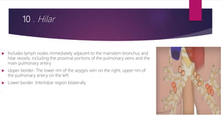
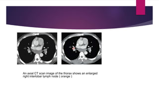
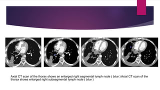
Mediastinal lymph nodes
DR VIPIN
 The body has approximately 600 lymph nodes, but only those in the
submandibular, axillary or inguinal regions may normally be palpable in healthy
people. Lymphadenopathy refers to nodes that are abnormal in either size,
consistency or number.
 In 2009, a new lung cancer lymph node map was proposed by the International
Association for the Study of Lung Cancer (IASLC) to reconcile the difference
between the Naruke and the Mountain–Dresler–American Thoracic Society (ATS)
maps and redefine the definitions of the anatomical boundaries of each lymph
node station
Supraclavicular Nodes
 1R and 1L . Low cervical, supraclavicular, and sternal notch nodes
 Upper border: Lower margin of cricoid cartilage.
 Lower border: Clavicles bilaterally and, in the midline, the upper border of the
manubrium;
 1R designates right-sided nodes;
 1L designates left-sided nodes in this region.
Supraclavicular Nodes
Superior Mediastinal Nodes
2-4
2R . Upper paratracheal
 Includes nodes extending to the left lateral border of the trachea.
 Upper border: Apex of the right lung and pleural space and in the midline, the upper border of
the manubrium.
 Lower border: Intersection of caudal margin of innominate vein with the trachea.
2L . Upper paratracheal.
 Upper border: Apex of the left lung and pleural space and in the midline, the
upper border of the manubrium.
 Lower border: Superior border of the aortic arch
Superior Mediastinal Nodes right upper
paratracheal lymph node
3A . Prevascular
 On the right:
 Upper border: Apex of chest.
 Lower border: Level of carina.
 Anterior border: Posterior aspect of sternum.
 Posterior border: Anterior border of superior vena cava.
 On the left:
 Upper border: Apex of chest.
 Lower border: Level of carina.
 Anterior border: Posterior aspect of sternum.
 Posterior border: Left carotid artery
Anterior to arch of aorta
Anterior to descending aorta
3P . Retrotracheal
 Upper border: Apex of chest.
 Lower border: Carina.
4R . Lower paratracheal.
 Includes right paratracheal nodes, and pretracheal nodes
extending to the left lateral border of trachea
 Upper border: Intersection of caudal margin of innominate veins with the
 trachea.
 Lower border: Lower border of azygos vein.
enlarged right-sided lower pretracheal lymph node
4L . Lower paratracheal.
 Includes nodes to the left of the left lateral border of the trachea, medial to the
ligamentum arteriosum.
 Upper border: Upper margins of the aortic arch.
 Lower border: Upper rim of the left main pulmonary artery
enlarged left lower paratracheal lymph node abutting the left
lateral wall of the trachea
Aortic Nodes 5–6
5 . Subaortic
 Lymph nodes lateral to the ligamentum arteriosum ( see Fig. 2.15 ).
 Upper border: The lower border of the aortic arch.
 Lower border: Upper rim of the left main pulmonary artery.
6 . Para-aortic.
 Lymph nodes anterior and lateral to the ascending aorta and aortic
 arch ( see Figs. 2.16 and 2.17 ).
 Upper border: A line tangential to the upper border of the aortic arch.
 Lower border: The lower border of the aortic arch.
Schematic illustration shows the anatomic location of
subaortic lymph nodes.
Axial contrast-enhanced CT scan image of the thorax shows an enlarged
Subaortic lymph node
Inferior Mediastinal Nodes
7 -9
7 Subcarinal
 Upper border: The carina of the trachea.
 Lower border: The upper border of the lower lobe bronchus on the left; the
lower
 border of the bronchus intermedius on the right.
Axial contrast-enhanced CT scan of the thorax shows
enlarged subcarinal group of lymph nodes
8 . Paraesophageal.
 Lymph nodes adjacent to the wall of the esophagus and to the
 right or left of the midline, excluding subcarinal nodes ( see Figs. 2.19 – 2.22 ).
 Upper border: The upper border of the lower lobe bronchus on the left; the
lower
 border of the bronchus intermedius on the right.
 Lower border: The diaphragm.
enlarged Paraesophageal group of lymph nodes
9 . Pulmonary ligament
Lymph nodes lying within the pulmonary ligament
 ( see Fig. 2.23 ).
 Upper border: The inferior pulmonary vein.
 Lower border: The diaphragm.
Axial contrast-enhanced CT scan of the thorax
shows an
enlarged right-sided pulmonary ligament lymph
Hilar, Lobar, and
(Sub)Segmental Nodes 10–
14
10 . Hilar
 Includes lymph nodes immediately adjacent to the mainstem bronchus and
hilar vessels, including the proximal portions of the pulmonary veins and the
main pulmonary artery
 Upper border: The lower rim of the azygos vein on the right; upper rim of
the pulmonary artery on the left.
 Lower border: Interlobar region bilaterally.
.
An axial CT scan image of the thorax shows an enlarged
right interlobar lymph node ( orange )
 11 . Interlobar. Between the origin of the lobar bronchi ( see Fig. 2.25 ).
 11s: Between the upper lobe bronchus and bronchus intermedius on the right.
 11i: Between the middle and lower lobe bronchus on the right.
 12 . Lobar. Adjacent to the lobar bronchi ( see Fig. 2.26 ).
 13 . Segmental. Adjacent to the segmental bronchi.
 14 . Subsegmental . Adjacent to the subsegmental bronchi
Axial CT scan of the thorax shows an enlarged right segmental lymph node ( blue ) Axial CT scan of the
thorax shows enlarged right subsegmental lymph node ( blue )

Mediastinal lymph nodes

  • 1.
  • 2.
     The bodyhas approximately 600 lymph nodes, but only those in the submandibular, axillary or inguinal regions may normally be palpable in healthy people. Lymphadenopathy refers to nodes that are abnormal in either size, consistency or number.  In 2009, a new lung cancer lymph node map was proposed by the International Association for the Study of Lung Cancer (IASLC) to reconcile the difference between the Naruke and the Mountain–Dresler–American Thoracic Society (ATS) maps and redefine the definitions of the anatomical boundaries of each lymph node station
  • 4.
  • 5.
    Supraclavicular Nodes  1Rand 1L . Low cervical, supraclavicular, and sternal notch nodes  Upper border: Lower margin of cricoid cartilage.  Lower border: Clavicles bilaterally and, in the midline, the upper border of the manubrium;  1R designates right-sided nodes;  1L designates left-sided nodes in this region.
  • 7.
  • 8.
  • 9.
    2R . Upperparatracheal  Includes nodes extending to the left lateral border of the trachea.  Upper border: Apex of the right lung and pleural space and in the midline, the upper border of the manubrium.  Lower border: Intersection of caudal margin of innominate vein with the trachea.
  • 10.
    2L . Upperparatracheal.  Upper border: Apex of the left lung and pleural space and in the midline, the upper border of the manubrium.  Lower border: Superior border of the aortic arch
  • 11.
    Superior Mediastinal Nodesright upper paratracheal lymph node
  • 12.
    3A . Prevascular On the right:  Upper border: Apex of chest.  Lower border: Level of carina.  Anterior border: Posterior aspect of sternum.  Posterior border: Anterior border of superior vena cava.  On the left:  Upper border: Apex of chest.  Lower border: Level of carina.  Anterior border: Posterior aspect of sternum.  Posterior border: Left carotid artery
  • 13.
    Anterior to archof aorta Anterior to descending aorta
  • 14.
    3P . Retrotracheal Upper border: Apex of chest.  Lower border: Carina.
  • 15.
    4R . Lowerparatracheal.  Includes right paratracheal nodes, and pretracheal nodes extending to the left lateral border of trachea  Upper border: Intersection of caudal margin of innominate veins with the  trachea.  Lower border: Lower border of azygos vein.
  • 16.
    enlarged right-sided lowerpretracheal lymph node
  • 17.
    4L . Lowerparatracheal.  Includes nodes to the left of the left lateral border of the trachea, medial to the ligamentum arteriosum.  Upper border: Upper margins of the aortic arch.  Lower border: Upper rim of the left main pulmonary artery
  • 18.
    enlarged left lowerparatracheal lymph node abutting the left lateral wall of the trachea
  • 19.
  • 20.
    5 . Subaortic Lymph nodes lateral to the ligamentum arteriosum ( see Fig. 2.15 ).  Upper border: The lower border of the aortic arch.  Lower border: Upper rim of the left main pulmonary artery.
  • 21.
    6 . Para-aortic. Lymph nodes anterior and lateral to the ascending aorta and aortic  arch ( see Figs. 2.16 and 2.17 ).  Upper border: A line tangential to the upper border of the aortic arch.  Lower border: The lower border of the aortic arch.
  • 22.
    Schematic illustration showsthe anatomic location of subaortic lymph nodes. Axial contrast-enhanced CT scan image of the thorax shows an enlarged Subaortic lymph node
  • 23.
  • 24.
    7 Subcarinal  Upperborder: The carina of the trachea.  Lower border: The upper border of the lower lobe bronchus on the left; the lower  border of the bronchus intermedius on the right.
  • 25.
    Axial contrast-enhanced CTscan of the thorax shows enlarged subcarinal group of lymph nodes
  • 26.
    8 . Paraesophageal. Lymph nodes adjacent to the wall of the esophagus and to the  right or left of the midline, excluding subcarinal nodes ( see Figs. 2.19 – 2.22 ).  Upper border: The upper border of the lower lobe bronchus on the left; the lower  border of the bronchus intermedius on the right.  Lower border: The diaphragm.
  • 27.
  • 28.
    9 . Pulmonaryligament Lymph nodes lying within the pulmonary ligament  ( see Fig. 2.23 ).  Upper border: The inferior pulmonary vein.  Lower border: The diaphragm.
  • 29.
    Axial contrast-enhanced CTscan of the thorax shows an enlarged right-sided pulmonary ligament lymph
  • 30.
  • 31.
    10 . Hilar Includes lymph nodes immediately adjacent to the mainstem bronchus and hilar vessels, including the proximal portions of the pulmonary veins and the main pulmonary artery  Upper border: The lower rim of the azygos vein on the right; upper rim of the pulmonary artery on the left.  Lower border: Interlobar region bilaterally. .
  • 32.
    An axial CTscan image of the thorax shows an enlarged right interlobar lymph node ( orange )
  • 33.
     11 .Interlobar. Between the origin of the lobar bronchi ( see Fig. 2.25 ).  11s: Between the upper lobe bronchus and bronchus intermedius on the right.  11i: Between the middle and lower lobe bronchus on the right.  12 . Lobar. Adjacent to the lobar bronchi ( see Fig. 2.26 ).  13 . Segmental. Adjacent to the segmental bronchi.  14 . Subsegmental . Adjacent to the subsegmental bronchi
  • 34.
    Axial CT scanof the thorax shows an enlarged right segmental lymph node ( blue ) Axial CT scan of the thorax shows enlarged right subsegmental lymph node ( blue )