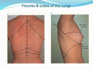
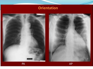
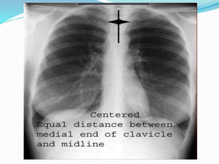
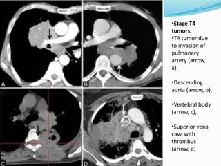
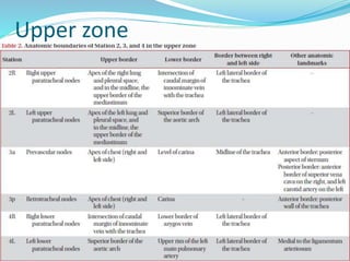
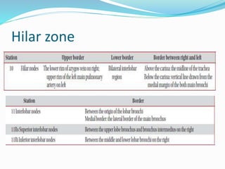
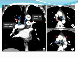
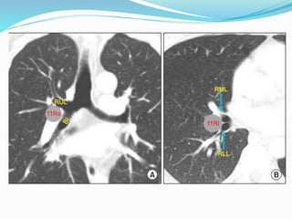
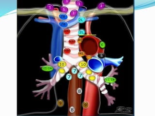
BORDERS OF THE LUNG
 The apex is about 2cm above the medial 1/3 of the clavicle
 The anterior border of both lungs run downwards & medially meeting each other
in the midline behind the angle of Louis.
 The anterior border of right lung continues running downwards till the 6th
costochondral junction.
 The anterior border of left lung continues running downwards till the 4th costal
cartilage then curves laterally ½ inch forming the cardiac notch then descends
downwards till the 6th costochondral junction.
 The lower border of the lungs represented by a line starting from 6th rib in the
MCL, 8th rib in the MAL & 10th rib in the mid scapular line.
Lung Fissures:
Oblique fissure (Right & Left):
It starts at the 3rd thoracic spine while the arms are elevated,
descends downwards, laterally & anteriorly along the medial
border of the scapula touching the inferior angle of the
scapula) cutting the mid axillary line in the 5th rib & ending at
the 6th costal cartilage 3 inches from the midline.
The transverse fissure (Right):
It arises at the 4th costal cartilage, runs horizontally to meet the
oblique fissure in the mid axillary line in the 5th rib.
Fissures & Lobes of the Lungs
Findings on Chest X-ray
 Nodule (< 3cm) vs. Mass (>= 3cm)
 Location:
 Peripheral (Adenocarcinoma) vs
 Central (Squamous)
 Single or multiple (metastases)
 Atelectasis of lobe or lung
 Pneumonia
 Hilar and mediastinal adenopathy
 Pleural effusions
CT Scan of Thorax
 Nodule details:
 Calcification, spiculation
 Evaluate extension into adjacent structures:
 Endobronchial, great vessels, pericardium etc
 Evaluation of adenopathy
 Upper abdominal pathology:
 Metastatic lesions in liver, adrenals, & kidneys.
•Common radiological
appearances of lung
cancer.
•Centrally located mass
with mediastinal
invasion (arrow, a)
•Peripherally situated
mass abutting the
pleura (arrow, B)
•Mass with smooth,
lobulated margins
(arrow, C)
•With spiculated,
irregular margins (arrow,
D)
•Squamous cell cancer
presenting as a cavitating
mass (arrow,a)
•Adenocarcinoma
presenting as dense
consolidation (arrow, B).
•Bronchoalveolar
carcinoma presenting as
ground-glass opacity
(arrow, c)
•Mixed density, solid
(arrow), and ground-glass
nodules (arrowhead) in d
•Stage T1 tumor
due to size <3 cm
(arrow, A).
•Stage T2
endobronchial
tumor
(arrowhead)
causing
pneumonitis
restricted to the
upper lobe
(arrow) in B.
•T2a tumor >3 cm
but <5 cm (arrow,
C).
•T2b tumor >5 cm
but <7 cm (arrow
in D)
•Stage T4
tumors.
•T4 tumor due
to invasion of
pulmonary
artery (arrow,
a),
•Descending
aorta (arrow, b),
•Vertebral body
(arrow, c),
•Superior vena
cava with
thrombus
(arrow, d)
The International Association for the
Study of Lung Cancer Lymph Node Map: A
Radiologic Atlas and Review
 14 LN stations reorganized into 7 zones
 Supraclavicular zone
 Upper zone
 AP zone
 Subcarinal zone
 Lower zone
 Hilar zone
 Peripheral zone
Station 1
 Low cervical, supraclavicular and sternal notch nodes
 Upper border : Lower margin of cricoid cartilage
 Lower border : Clavicle bilaterally and in midline upper
border of manubrium
Upper zone
AP zone
Subcarinal zone
Lower zone
Hilar zone
Peripheral Zone
 Station 12 : Node adjacent to lobar bronchi
 Station 13 : Node adjacent to segmental bronchi
 Station 14 : Node adjacent to subsegmental bronchi
Lymph nodal stations in ca lung

Lymph nodal stations in ca lung

  • 3.
    BORDERS OF THELUNG  The apex is about 2cm above the medial 1/3 of the clavicle  The anterior border of both lungs run downwards & medially meeting each other in the midline behind the angle of Louis.  The anterior border of right lung continues running downwards till the 6th costochondral junction.  The anterior border of left lung continues running downwards till the 4th costal cartilage then curves laterally ½ inch forming the cardiac notch then descends downwards till the 6th costochondral junction.  The lower border of the lungs represented by a line starting from 6th rib in the MCL, 8th rib in the MAL & 10th rib in the mid scapular line.
  • 4.
    Lung Fissures: Oblique fissure(Right & Left): It starts at the 3rd thoracic spine while the arms are elevated, descends downwards, laterally & anteriorly along the medial border of the scapula touching the inferior angle of the scapula) cutting the mid axillary line in the 5th rib & ending at the 6th costal cartilage 3 inches from the midline. The transverse fissure (Right): It arises at the 4th costal cartilage, runs horizontally to meet the oblique fissure in the mid axillary line in the 5th rib.
  • 5.
    Fissures & Lobesof the Lungs
  • 7.
    Findings on ChestX-ray  Nodule (< 3cm) vs. Mass (>= 3cm)  Location:  Peripheral (Adenocarcinoma) vs  Central (Squamous)  Single or multiple (metastases)  Atelectasis of lobe or lung  Pneumonia  Hilar and mediastinal adenopathy  Pleural effusions
  • 13.
    CT Scan ofThorax  Nodule details:  Calcification, spiculation  Evaluate extension into adjacent structures:  Endobronchial, great vessels, pericardium etc  Evaluation of adenopathy  Upper abdominal pathology:  Metastatic lesions in liver, adrenals, & kidneys.
  • 14.
    •Common radiological appearances oflung cancer. •Centrally located mass with mediastinal invasion (arrow, a) •Peripherally situated mass abutting the pleura (arrow, B) •Mass with smooth, lobulated margins (arrow, C) •With spiculated, irregular margins (arrow, D)
  • 15.
    •Squamous cell cancer presentingas a cavitating mass (arrow,a) •Adenocarcinoma presenting as dense consolidation (arrow, B). •Bronchoalveolar carcinoma presenting as ground-glass opacity (arrow, c) •Mixed density, solid (arrow), and ground-glass nodules (arrowhead) in d
  • 16.
    •Stage T1 tumor dueto size <3 cm (arrow, A). •Stage T2 endobronchial tumor (arrowhead) causing pneumonitis restricted to the upper lobe (arrow) in B. •T2a tumor >3 cm but <5 cm (arrow, C). •T2b tumor >5 cm but <7 cm (arrow in D)
  • 17.
    •Stage T4 tumors. •T4 tumordue to invasion of pulmonary artery (arrow, a), •Descending aorta (arrow, b), •Vertebral body (arrow, c), •Superior vena cava with thrombus (arrow, d)
  • 19.
    The International Associationfor the Study of Lung Cancer Lymph Node Map: A Radiologic Atlas and Review  14 LN stations reorganized into 7 zones  Supraclavicular zone  Upper zone  AP zone  Subcarinal zone  Lower zone  Hilar zone  Peripheral zone
  • 20.
    Station 1  Lowcervical, supraclavicular and sternal notch nodes  Upper border : Lower margin of cricoid cartilage  Lower border : Clavicle bilaterally and in midline upper border of manubrium
  • 22.
  • 25.
  • 27.
  • 28.
  • 30.
  • 33.
    Peripheral Zone  Station12 : Node adjacent to lobar bronchi  Station 13 : Node adjacent to segmental bronchi  Station 14 : Node adjacent to subsegmental bronchi

Editor's Notes