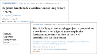
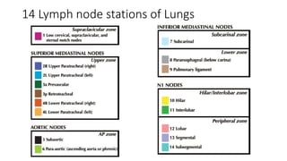
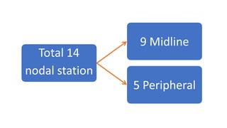
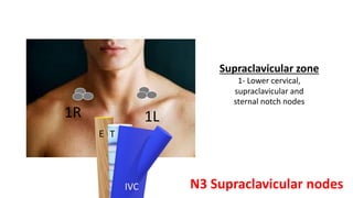
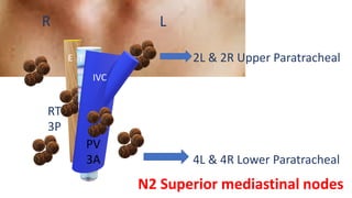
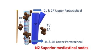
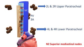
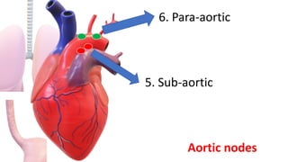
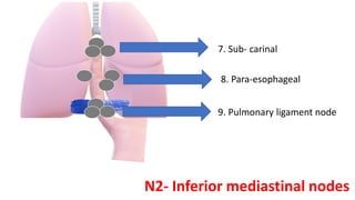
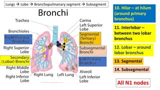
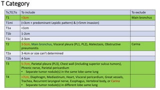
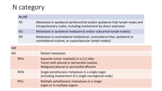
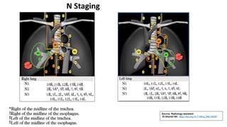
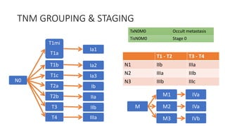
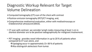
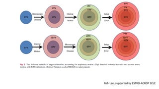
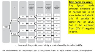
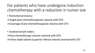
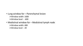
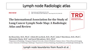
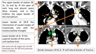
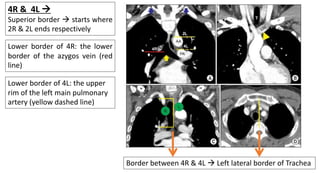
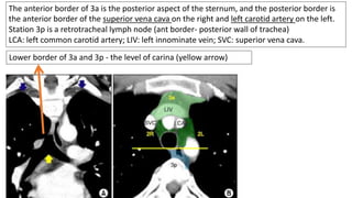
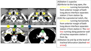
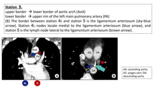
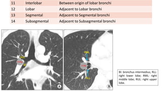
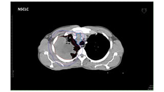
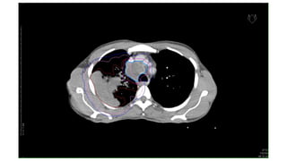
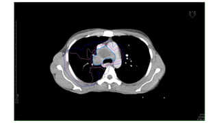
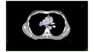
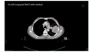
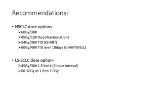
Radiotherapy techniques &
contouring guidelines for Radical
treatment of Carcinoma Lungs
Dr. Abani Kanta Nanda
Senior Resident
AHPGIC, Cuttack
Lymphnode stations in
Ca Lungs
Naruke Japanese Lymph node map
Naruke
Japanese
Lymph node
map
14 Lymph node stations of Lungs
Supraclavicular zone
1- Lower cervical,
supraclavicular and
sternal notch nodes
1R 1L
T
E
IVC N3 Supraclavicular nodes
R L
T
E
IVC
PV
3A
RT
3P
2L & 2R Upper Paratracheal
4L & 4R Lower Paratracheal
N2 Superior mediastinal nodes
T
E
IVC
PV
3A
RT
3P
2L & 2R Upper Paratracheal
4L & 4R Lower Paratracheal
N2 Superior mediastinal nodes
T
E
IVC
PV
3A
RT
3P
2L & 2R Upper Paratracheal
4L & 4R Lower Paratracheal
N2 Superior mediastinal nodes
5. Sub-aortic
6. Para-aortic
Aortic nodes
7. Sub- carinal
8. Para-esophageal
9. Pulmonary ligament node
N2- Inferior mediastinal nodes
10. Hilar – at hilum
(around primary
bronchus)
14. Subsegmental
13. Segmental
12. Lobar – around
lobar bronchus
11. Interlobar –
between two lobar
bronchus
Lungs → Lobe → Bronchopulmonary segment → Subsegment
All N1 nodes
AJCC Classification
T Category
Tx,T0,Tis To include To exclude
T1 <3cm Main bronchus
T1mi (<3cm + predominant Lepidic pattern) & (<5mm invasion)
T1a <1cm
T1b 1-2cm
T1c 2-3cm
T2 3-5cm, Main bronchus, Visceral pleura (PL1, PL2), Atelectasis, Obstructive
pneumonitis
Carina
T2a 3-4cm or size can’t determined
T2b 4-5cm
T3 5-7cm, Parietal pleura (PL3), Chest wall (including superior sulcus tumors),
Phrenic nerve, Parietal pericardium
• Separate tumor nodule(s) in the same lobe same lung
T4 >7cm, Diaphragm, Mediastinum, Heart, Visceral pericardium, Great vessels,
Trachea, Recurrent laryngeal nerve, Esophagus, Vertebral body, or Carina
• Separate tumor nodule(s) in different lobe same lung
N category
Nx,N0
N1 Metastasis in ipsilateral peribronchial and/or ipsilateral hilar lymph nodes and
intrapulmonary nodes, including involvement by direct extension
N2 Metastasis in ipsilateral mediastinal and/or subcarinal lymph node(s)
N3 Metastasis in contralateral mediastinal, contralateral hilar, ipsilateral or
contralateral scalene, or supraclavicular lymph node(s)
M0
M1 Distant metastasis
M1a Separate tumor nodule(s) in a C/L lobe;
Tumor with pleural or pericardial nodules
Malignant pleural or pericardial effusion
M1b Single extrathoracic metastasis in a single organ
(including involvement of a single nonregional node)
M1c Multiple extrathoracic metastases in a single
organ or in multiple organs
Source: Radiology assistant
El-Sherief AH. https://doi.org/10.1148/rg.346130097
N Staging
T1 - T2 T3 - T4
N1 IIb IIIa
N2 IIIa IIIb
N3 IIIb IIIc
N0
T1mi
T1a
Ia1
T1b Ia2
T1c Ia3
T2a Ib
T2b IIa
T3 IIb
T4 IIIa
M
M1 IVa
M2 IVa
M3 IVb
TNM GROUPING & STAGING
TxN0M0 Occult metastasis
TisN0M0 Stage 0
Contouring guidelines
Diagnostic Workup Relevant for Target
Volume Delineation
➢Computed tomography (CT) scan of the chest with contrast
➢Positron emission tomography (PET)/CT imaging, and,
➢Comprehensive mediastinal evaluation, either with mediastinoscopy or
Endobronchial ultrasound (EBUS).
• CT scan with contrast- we consider lymph nodes measuring at least 1 cm in
shortest diameter are to be positive radiographically for malignant involvement.
• PET imaging- provides novel information in up to 20 % of patients when
compared to CT scan alone, and
❖Upstaging occur in approximately 15–30 % of patients
❖Also distinguish atelectasis from tumor.
Simulation
• Patients immobilized in supine position with their arms over their head
• IV contrast to differentiate tumor involvement from mediastinal structures such as the
vasculature.
• Four-dimensional (4D) CT scans are acquired, to account for internal motion. When
the magnitude of respiratory motion is >1 cm and regular, patients are treated with a
free -breathing approach, in which they breathe regularly during treatment.
• If irregular or motion >1 cm, then respiratory management is considered, either deep
breath hold (inspiratory or expiratory) or respiratory gating, in which radiation is
delivered at specific periods of the breathing cycle.
• Both techniques have been shown to be beneficial in reducing target volumes for
tumors that have substantial motion.
• For patients to tolerate the deep breath hold technique, they need to be able to
maintain the appropriate position in the respiratory cycle for at least 15 s, which
is difficult for a significant percentage of patients with lung cancer.
• The 4D CT scan- typically extends from at least the the thoracic inlet to the
inferior portion of the diaphragm.
• Image fusion with PET/CT scans is recommended when feasible, particularly in
cases with atelectasis or when directly adjacent to critical structures.
• Image registration and fusion applications with MRI and PET scans should be used
to help in the delineation of target volumes, especially for regions of interest
encompassing the GTV, skull base, brainstem, and optic chiasm.
• The GTV and CTV and normal tissues should be outlined on all CT slices in which
the structures exist.
• Daily kV imaging and weekly CT scan alignment for localization (cone beam CT
scan or CT scan on rails).
• Daily CT scan localization is utilized when more precise localization is necessary
(e.g., tumors adjacent to the spinal cord), when the size of the tumor is rapidly
changing, or when bony landmarks are not representative of internal anatomy.
• Prior studies have shown that with daily kV imaging, interfractional variation is
approximately 5 mm and, with CBCT, can be reduced to approximately 3 mm. →
These are thus the PTV margins to be given. (Nelson et al. 2008 )
Target volume delineation
• GTV- Gross disease (primary tumor + lymph nodes)
• CTV- gross disease with margin of 0.5-1.0cm + region at risk of microscopic
spread.
• GTV to CTV margin- 8mm (6mm for SCC can be given) (SCLC- 5mm)
(To respect anatomic boundaries)
(This volume includes the remainder of involved lymph node stations)
• ITV- CTV + respiratory motion (to be determined during simulation)
• PTV- 5 or 3 mm margin
PET images – to identify the location of tumour
CT images – to identify the tumour edge
But in case of atelectasis & node(not seen on CT) , you can visualise boundary with PET
Respiratory motion management
Planning with
motion
unrestrained
Reducing and
controlling motion
Managing motion
Mitigation motion
• 4D CT scanning
• Slow CT scanning
• CT scans at
maximal inspiration
and expiration
• Fluoroscopy
• Abdominal
compression
• Active breathing
control
• DIBH/Coached
respiration
• Passive
respiratory
gating
• Tracking motion
Attempts must be made to control or mitigate
this if respiratory motion amplitude is >1cm
ITV • Conventional scanning: Just a guide
• ITV= CTV + 1cm S-I
+ 0.5cm laterally,
+ 0.5cm A-P
• Mediastinal movement: 5mm
Tumour motion
166 tumours from 152
lung cancer patients
S-I >5mm : 39.2%
>10mm : 10.8%
Lateral >5mm : 1.8%
A-P >5mm : 5.4%
Liu etal. IJROBP.2007;68:531-40
Encompass tumour motion in 90% of tumours
Ref- Lee, supported by ESTRO-ACROP SCLC
Take home message-
Any lymph node
whether enlarged or
of normal size in CT
scan, to be included in
GTV if positive in
either PET or EBUS.
But to be excluded
from GTV if negative
in both.
Ref- Radiother Oncol . 2018 Apr;127(1):1-5. doi: 10.1016/j.radonc.2018.02.023. Epub 2018 Mar 28; ESTRO ACROP guidelines
• In case of diagnostic uncertainty, a node should be included in GTV.
Elective nodal irradiation
• In general is not recommended for both SCLC & NSCLC.
• Inclusion of uninvolved areas between involved stations and primary
tumor volume is optional.
• In case of upper mediastinal lymph node involvement (station 2 or
3A) + no pretreatment PET scan → I/L Supraclavicular nodal region
can be considered to include in CTVn.
Ref- ESTRO-ACROP guideline SCLC 2020 & NSCLC 2018
Adaptive replanning
• In SCLC
• As responsive to RT → important volume changes >50% occur in first
week.
• Only for GTVp and not for GTVn
• Not to interrupt the treatment for adaptive planning → due to risk of
repopulation in SCLC.
ESTRO-ACROP guideline SCLC 2020
For patients who have undergone induction
chemotherapy with a reduction in tumor size
• Parenchymal lesions:
➢Target post-chemotherapeutic volume with GTV
➢Coverage of pre-chemotherapeutic volume with CTV
• Involved lymph nodes:
➢Post-chemotherapy volume covered with GTV
➢Entire nodal station (superior-inferior extent) covered with CTV.
Lee
• Lung window for – Parenchymal lesion
➢Window width: 1600
➢Window level : -600
• Medistinal window for – Medistinal lymph node
➢Window width: 400
➢Window level : 20
Lymph node Radiologic atlas
Lymph node boundaries from Rusch et al.
The upper border of station 2R,
2L, 3a and 3p → the apex of
each lung and pleural space
(blue arrows), and in the
midline, the upper border of
the manubrium.
Lower border of 2R→ the
intersection of caudal margin of
innominate vein with the
trachea (yellow triangle)
Lower border of 2L → the
superior border of the aortic
arch (yellow line)
Border between 2R & 2L → Left lateral border of Trachea
(AA: aortic arch; AZ: azygos vein; LIV: left
innominate vein; PA: left main pulmonary
artery; T: trachea)
4R & 4L →
Superior border → starts where
2R & 2L ends respectively
Lower border of 4R: the lower
border of the azygos vein (red
line)
Lower border of 4L: the upper
rim of the left main pulmonary
artery (yellow dashed line)
Border between 4R & 4L → Left lateral border of Trachea
Lower border of 3a and 3p - the level of carina (yellow arrow)
The anterior border of 3a is the posterior aspect of the sternum, and the posterior border is
the anterior border of the superior vena cava on the right and left carotid artery on the left.
Station 3p is a retrotracheal lymph node (ant border- posterior wall of trachea)
LCA: left common carotid artery; LIV: left innominate vein; SVC: superior vena cava.
(A)Station 1 supaclav
(B)Inferior to the lung apex, the
yellow line running horizontally
from anterior margin of both
lung pleura interface separates
station 1 from station 3p.
(C)At the suprasternal notch, the
yellow line running horizontally
from anterior margin of both
lung pleura interface separates
station 1 from station 2. The red
line running along posterior wall
of trachea separates station 2
from 3p.
(D)Station 3a and 3p at the level of
tracheal bifurcation (dashed red
arrow).
Station 5.
upper border → lower border of aortic arch (AoA)
lower border → upper rim of the left main pulmonary artery (PA)
(B) The border between station 4L and station 5 is the ligamentum arteriosum (sky-blue
arrow). Station 4L nodes locate medial to the ligamentum arteriosum (blue arrow), and
station 5 is the lymph node lateral to the ligamentum arteriosum (brown arrow).
AA: ascending aorta;
AZ: azygos vein; DA:
descending aorta.
Station 6. (A)
• upper border → a line tangential to the upper border of the aortic arch
• lower border → lower border of aortic arch (green lines).
(B) anterior border → the imaginary horizontal line extending from the anterior wall of the
aortic arch (yellow line), which discriminates station 6 from station 3a. AoA: aortic arch.
Subcarinal zone: Station 7. (A) Upper and lower borders of station 7 are well identified
on coronal image.
(B) Station 7 nodes are noted in the space between the medial margin of both main
bronchi (yellow lines) and
• nodes outside of the space are station 10 (Hilar nodes).
• Station 7 extends posteriorly around the esophagus.
AoA: aortic arch; BI:
bronchus
intermedius; LLL:
left lower lobe;
LMB: left main
bronchus; RMB:
right main
bronchus; RUL:
right upper lobe.
Lower zone: stations 8 and 9:
station 8→ adjacent to esophagus
➢ upper border→ lower border of station 7
• station 9
➢ Upper border→ inferior pulmonary vein.
• The lower border of station 8 and 9 is the diaphragm.
• The border between 8R and 8L is the midline (dashed
line)
(B) Station 4R and 4L nodes at the
level of azygos vein. Note the border
between station 4R and 4L is the left
lateral wall of the trachea (yellow line).
(C) Stations 10R and 10L below the
azygos vein.
The pleural reflection (pink
arrowhead) no longer serves as the
border between station 4 and 10.
• The border between station 10R
and 10L is the midline of tracheal
bifurcation (yellow line).
• Lower border- B/L inter lobar region
AoA: aortic arch; AZ: azygos vein; PA:
left pulmonary artery.
Station 10. (A) The border between station 4R
and 10R is the lower rim of the azygos vein (red
dashed arrow). The border between station 4L
and 10L is the upper rim of the left main
pulmonary artery (red line).
Means station 10 starts where 4 ends
BI: bronchus intermedius; RLL:
right lower lobe; RML: right
middle lobe; RUL: right upper
lobe.
11 Interlobar Between origin of lobar bronchi
12 Lobar Adjacent to Lobar bronchi
13 Segmental Adjacent to Segmental bronchi
14 Subsegmental Adjacent to Subsegmental bronchi
CT images of Ca Lungs
NSCLC
Ca Left Lung post NACT with residual
Dose
RTOG 0617
KQ2: What is the ideal external beam dose fractionation for the curative-intent treatment of
locally advanced non-small cell lung cancer with chemotherapy?
A. The standard thoracic radiation therapy dose fractionation for patients treatedwith
concurrent chemotherapy is 60 Gy given in 2 Gy once-daily fractions over 6 weeks
(MQE,recommendation rated as “strong”).
B. Dose escalation beyond 60 Gy with conventional fractionation has not been demonstrated
to be associated with any clinical benefits, including OS (MQE, recommendation rated as
“strong”).
Recommendations:
• NSCLC dose options-
➢60Gy/30#
➢45Gy/15# (hypofractionation)
➢54Gy/36# TID (CHART)
➢60Gy/40# TID over 18days (CHARTWELL)
• LS-SCLC dose option-
➢45Gy/30# 1.5 bid # (6 hour interval)
➢60-70Gy at 1.8 to 2.0Gy
Thank you

Lungs contouring Dr. Abani.pdf

  • 1.
    Radiotherapy techniques & contouringguidelines for Radical treatment of Carcinoma Lungs Dr. Abani Kanta Nanda Senior Resident AHPGIC, Cuttack
  • 2.
  • 3.
    Naruke Japanese Lymphnode map Naruke Japanese Lymph node map
  • 4.
    14 Lymph nodestations of Lungs
  • 5.
    14 Lymph nodestations of Lungs
  • 7.
    Supraclavicular zone 1- Lowercervical, supraclavicular and sternal notch nodes 1R 1L T E IVC N3 Supraclavicular nodes
  • 8.
    R L T E IVC PV 3A RT 3P 2L &2R Upper Paratracheal 4L & 4R Lower Paratracheal N2 Superior mediastinal nodes
  • 9.
    T E IVC PV 3A RT 3P 2L & 2RUpper Paratracheal 4L & 4R Lower Paratracheal N2 Superior mediastinal nodes
  • 10.
    T E IVC PV 3A RT 3P 2L & 2RUpper Paratracheal 4L & 4R Lower Paratracheal N2 Superior mediastinal nodes
  • 11.
  • 12.
    7. Sub- carinal 8.Para-esophageal 9. Pulmonary ligament node N2- Inferior mediastinal nodes
  • 13.
    10. Hilar –at hilum (around primary bronchus) 14. Subsegmental 13. Segmental 12. Lobar – around lobar bronchus 11. Interlobar – between two lobar bronchus Lungs → Lobe → Bronchopulmonary segment → Subsegment All N1 nodes
  • 14.
  • 15.
    T Category Tx,T0,Tis Toinclude To exclude T1 <3cm Main bronchus T1mi (<3cm + predominant Lepidic pattern) & (<5mm invasion) T1a <1cm T1b 1-2cm T1c 2-3cm T2 3-5cm, Main bronchus, Visceral pleura (PL1, PL2), Atelectasis, Obstructive pneumonitis Carina T2a 3-4cm or size can’t determined T2b 4-5cm T3 5-7cm, Parietal pleura (PL3), Chest wall (including superior sulcus tumors), Phrenic nerve, Parietal pericardium • Separate tumor nodule(s) in the same lobe same lung T4 >7cm, Diaphragm, Mediastinum, Heart, Visceral pericardium, Great vessels, Trachea, Recurrent laryngeal nerve, Esophagus, Vertebral body, or Carina • Separate tumor nodule(s) in different lobe same lung
  • 16.
    N category Nx,N0 N1 Metastasisin ipsilateral peribronchial and/or ipsilateral hilar lymph nodes and intrapulmonary nodes, including involvement by direct extension N2 Metastasis in ipsilateral mediastinal and/or subcarinal lymph node(s) N3 Metastasis in contralateral mediastinal, contralateral hilar, ipsilateral or contralateral scalene, or supraclavicular lymph node(s) M0 M1 Distant metastasis M1a Separate tumor nodule(s) in a C/L lobe; Tumor with pleural or pericardial nodules Malignant pleural or pericardial effusion M1b Single extrathoracic metastasis in a single organ (including involvement of a single nonregional node) M1c Multiple extrathoracic metastases in a single organ or in multiple organs
  • 17.
    Source: Radiology assistant El-SheriefAH. https://doi.org/10.1148/rg.346130097 N Staging
  • 18.
    T1 - T2T3 - T4 N1 IIb IIIa N2 IIIa IIIb N3 IIIb IIIc N0 T1mi T1a Ia1 T1b Ia2 T1c Ia3 T2a Ib T2b IIa T3 IIb T4 IIIa M M1 IVa M2 IVa M3 IVb TNM GROUPING & STAGING TxN0M0 Occult metastasis TisN0M0 Stage 0
  • 19.
  • 21.
    Diagnostic Workup Relevantfor Target Volume Delineation ➢Computed tomography (CT) scan of the chest with contrast ➢Positron emission tomography (PET)/CT imaging, and, ➢Comprehensive mediastinal evaluation, either with mediastinoscopy or Endobronchial ultrasound (EBUS). • CT scan with contrast- we consider lymph nodes measuring at least 1 cm in shortest diameter are to be positive radiographically for malignant involvement. • PET imaging- provides novel information in up to 20 % of patients when compared to CT scan alone, and ❖Upstaging occur in approximately 15–30 % of patients ❖Also distinguish atelectasis from tumor.
  • 22.
    Simulation • Patients immobilizedin supine position with their arms over their head • IV contrast to differentiate tumor involvement from mediastinal structures such as the vasculature. • Four-dimensional (4D) CT scans are acquired, to account for internal motion. When the magnitude of respiratory motion is >1 cm and regular, patients are treated with a free -breathing approach, in which they breathe regularly during treatment. • If irregular or motion >1 cm, then respiratory management is considered, either deep breath hold (inspiratory or expiratory) or respiratory gating, in which radiation is delivered at specific periods of the breathing cycle.
  • 23.
    • Both techniqueshave been shown to be beneficial in reducing target volumes for tumors that have substantial motion. • For patients to tolerate the deep breath hold technique, they need to be able to maintain the appropriate position in the respiratory cycle for at least 15 s, which is difficult for a significant percentage of patients with lung cancer. • The 4D CT scan- typically extends from at least the the thoracic inlet to the inferior portion of the diaphragm.
  • 24.
    • Image fusionwith PET/CT scans is recommended when feasible, particularly in cases with atelectasis or when directly adjacent to critical structures. • Image registration and fusion applications with MRI and PET scans should be used to help in the delineation of target volumes, especially for regions of interest encompassing the GTV, skull base, brainstem, and optic chiasm. • The GTV and CTV and normal tissues should be outlined on all CT slices in which the structures exist.
  • 25.
    • Daily kVimaging and weekly CT scan alignment for localization (cone beam CT scan or CT scan on rails). • Daily CT scan localization is utilized when more precise localization is necessary (e.g., tumors adjacent to the spinal cord), when the size of the tumor is rapidly changing, or when bony landmarks are not representative of internal anatomy. • Prior studies have shown that with daily kV imaging, interfractional variation is approximately 5 mm and, with CBCT, can be reduced to approximately 3 mm. → These are thus the PTV margins to be given. (Nelson et al. 2008 )
  • 26.
    Target volume delineation •GTV- Gross disease (primary tumor + lymph nodes) • CTV- gross disease with margin of 0.5-1.0cm + region at risk of microscopic spread. • GTV to CTV margin- 8mm (6mm for SCC can be given) (SCLC- 5mm) (To respect anatomic boundaries) (This volume includes the remainder of involved lymph node stations) • ITV- CTV + respiratory motion (to be determined during simulation) • PTV- 5 or 3 mm margin PET images – to identify the location of tumour CT images – to identify the tumour edge But in case of atelectasis & node(not seen on CT) , you can visualise boundary with PET
  • 28.
    Respiratory motion management Planningwith motion unrestrained Reducing and controlling motion Managing motion Mitigation motion • 4D CT scanning • Slow CT scanning • CT scans at maximal inspiration and expiration • Fluoroscopy • Abdominal compression • Active breathing control • DIBH/Coached respiration • Passive respiratory gating • Tracking motion Attempts must be made to control or mitigate this if respiratory motion amplitude is >1cm
  • 29.
    ITV • Conventionalscanning: Just a guide • ITV= CTV + 1cm S-I + 0.5cm laterally, + 0.5cm A-P • Mediastinal movement: 5mm Tumour motion 166 tumours from 152 lung cancer patients S-I >5mm : 39.2% >10mm : 10.8% Lateral >5mm : 1.8% A-P >5mm : 5.4% Liu etal. IJROBP.2007;68:531-40 Encompass tumour motion in 90% of tumours
  • 30.
    Ref- Lee, supportedby ESTRO-ACROP SCLC
  • 31.
    Take home message- Anylymph node whether enlarged or of normal size in CT scan, to be included in GTV if positive in either PET or EBUS. But to be excluded from GTV if negative in both. Ref- Radiother Oncol . 2018 Apr;127(1):1-5. doi: 10.1016/j.radonc.2018.02.023. Epub 2018 Mar 28; ESTRO ACROP guidelines • In case of diagnostic uncertainty, a node should be included in GTV.
  • 32.
    Elective nodal irradiation •In general is not recommended for both SCLC & NSCLC. • Inclusion of uninvolved areas between involved stations and primary tumor volume is optional. • In case of upper mediastinal lymph node involvement (station 2 or 3A) + no pretreatment PET scan → I/L Supraclavicular nodal region can be considered to include in CTVn. Ref- ESTRO-ACROP guideline SCLC 2020 & NSCLC 2018
  • 33.
    Adaptive replanning • InSCLC • As responsive to RT → important volume changes >50% occur in first week. • Only for GTVp and not for GTVn • Not to interrupt the treatment for adaptive planning → due to risk of repopulation in SCLC. ESTRO-ACROP guideline SCLC 2020
  • 34.
    For patients whohave undergone induction chemotherapy with a reduction in tumor size • Parenchymal lesions: ➢Target post-chemotherapeutic volume with GTV ➢Coverage of pre-chemotherapeutic volume with CTV • Involved lymph nodes: ➢Post-chemotherapy volume covered with GTV ➢Entire nodal station (superior-inferior extent) covered with CTV. Lee
  • 35.
    • Lung windowfor – Parenchymal lesion ➢Window width: 1600 ➢Window level : -600 • Medistinal window for – Medistinal lymph node ➢Window width: 400 ➢Window level : 20
  • 36.
    Lymph node Radiologicatlas Lymph node boundaries from Rusch et al.
  • 38.
    The upper borderof station 2R, 2L, 3a and 3p → the apex of each lung and pleural space (blue arrows), and in the midline, the upper border of the manubrium. Lower border of 2R→ the intersection of caudal margin of innominate vein with the trachea (yellow triangle) Lower border of 2L → the superior border of the aortic arch (yellow line) Border between 2R & 2L → Left lateral border of Trachea (AA: aortic arch; AZ: azygos vein; LIV: left innominate vein; PA: left main pulmonary artery; T: trachea)
  • 39.
    4R & 4L→ Superior border → starts where 2R & 2L ends respectively Lower border of 4R: the lower border of the azygos vein (red line) Lower border of 4L: the upper rim of the left main pulmonary artery (yellow dashed line) Border between 4R & 4L → Left lateral border of Trachea
  • 40.
    Lower border of3a and 3p - the level of carina (yellow arrow) The anterior border of 3a is the posterior aspect of the sternum, and the posterior border is the anterior border of the superior vena cava on the right and left carotid artery on the left. Station 3p is a retrotracheal lymph node (ant border- posterior wall of trachea) LCA: left common carotid artery; LIV: left innominate vein; SVC: superior vena cava.
  • 41.
    (A)Station 1 supaclav (B)Inferiorto the lung apex, the yellow line running horizontally from anterior margin of both lung pleura interface separates station 1 from station 3p. (C)At the suprasternal notch, the yellow line running horizontally from anterior margin of both lung pleura interface separates station 1 from station 2. The red line running along posterior wall of trachea separates station 2 from 3p. (D)Station 3a and 3p at the level of tracheal bifurcation (dashed red arrow).
  • 42.
    Station 5. upper border→ lower border of aortic arch (AoA) lower border → upper rim of the left main pulmonary artery (PA) (B) The border between station 4L and station 5 is the ligamentum arteriosum (sky-blue arrow). Station 4L nodes locate medial to the ligamentum arteriosum (blue arrow), and station 5 is the lymph node lateral to the ligamentum arteriosum (brown arrow). AA: ascending aorta; AZ: azygos vein; DA: descending aorta.
  • 43.
    Station 6. (A) •upper border → a line tangential to the upper border of the aortic arch • lower border → lower border of aortic arch (green lines). (B) anterior border → the imaginary horizontal line extending from the anterior wall of the aortic arch (yellow line), which discriminates station 6 from station 3a. AoA: aortic arch.
  • 44.
    Subcarinal zone: Station7. (A) Upper and lower borders of station 7 are well identified on coronal image. (B) Station 7 nodes are noted in the space between the medial margin of both main bronchi (yellow lines) and • nodes outside of the space are station 10 (Hilar nodes). • Station 7 extends posteriorly around the esophagus. AoA: aortic arch; BI: bronchus intermedius; LLL: left lower lobe; LMB: left main bronchus; RMB: right main bronchus; RUL: right upper lobe.
  • 45.
    Lower zone: stations8 and 9: station 8→ adjacent to esophagus ➢ upper border→ lower border of station 7 • station 9 ➢ Upper border→ inferior pulmonary vein. • The lower border of station 8 and 9 is the diaphragm. • The border between 8R and 8L is the midline (dashed line)
  • 46.
    (B) Station 4Rand 4L nodes at the level of azygos vein. Note the border between station 4R and 4L is the left lateral wall of the trachea (yellow line). (C) Stations 10R and 10L below the azygos vein. The pleural reflection (pink arrowhead) no longer serves as the border between station 4 and 10. • The border between station 10R and 10L is the midline of tracheal bifurcation (yellow line). • Lower border- B/L inter lobar region AoA: aortic arch; AZ: azygos vein; PA: left pulmonary artery. Station 10. (A) The border between station 4R and 10R is the lower rim of the azygos vein (red dashed arrow). The border between station 4L and 10L is the upper rim of the left main pulmonary artery (red line). Means station 10 starts where 4 ends
  • 47.
    BI: bronchus intermedius;RLL: right lower lobe; RML: right middle lobe; RUL: right upper lobe. 11 Interlobar Between origin of lobar bronchi 12 Lobar Adjacent to Lobar bronchi 13 Segmental Adjacent to Segmental bronchi 14 Subsegmental Adjacent to Subsegmental bronchi
  • 48.
    CT images ofCa Lungs
  • 49.
  • 53.
    Ca Left Lungpost NACT with residual
  • 54.
  • 55.
  • 57.
    KQ2: What isthe ideal external beam dose fractionation for the curative-intent treatment of locally advanced non-small cell lung cancer with chemotherapy? A. The standard thoracic radiation therapy dose fractionation for patients treatedwith concurrent chemotherapy is 60 Gy given in 2 Gy once-daily fractions over 6 weeks (MQE,recommendation rated as “strong”). B. Dose escalation beyond 60 Gy with conventional fractionation has not been demonstrated to be associated with any clinical benefits, including OS (MQE, recommendation rated as “strong”).
  • 58.
    Recommendations: • NSCLC doseoptions- ➢60Gy/30# ➢45Gy/15# (hypofractionation) ➢54Gy/36# TID (CHART) ➢60Gy/40# TID over 18days (CHARTWELL) • LS-SCLC dose option- ➢45Gy/30# 1.5 bid # (6 hour interval) ➢60-70Gy at 1.8 to 2.0Gy
  • 59.