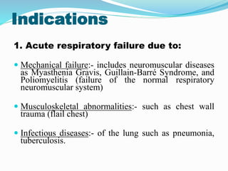
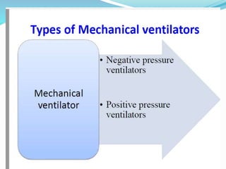
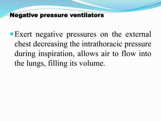
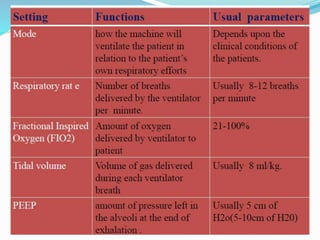
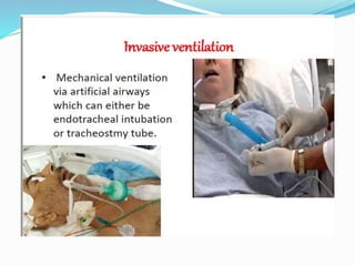
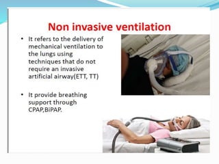
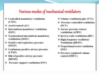
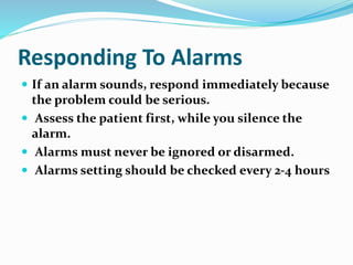
Overview
Introduction
The basic principles
Types of Mechanical Ventilation
Indication
Purpose
Modes
Initial settings
Alarms
Advantage and complications of ventilator
 Weaning from ventilator support
Nursing care of patient in ventilator
Mechanical Ventilation
 Delivery of mechanically generated breaths to get
oxygen in and carbon dioxide out.
 Mechanical Ventilation is ventilation of the lungs
by artificial means usually by a ventilator.
 A ventilator delivers gas to the lungs with either
negative or positive pressure.
 A mechanical ventilator is a machine that generates a
controlled flow of gas into a patient’s airways.
 Oxygen and air are received from cylinders or wall
outlets, the gas pressure is reduced and blended
according to the prescribed inspired oxygen tension
(FiO2), accumulated in a receptacle within the
machine, and delivered to the patient using one of
many available modes of ventilation.
Purposes
Improve and/or maintain oxygenation
Improve and/or maintain ventilation
Minimize ventilator induced lung
injury
Indications
1. Acute respiratory failure due to:
 Mechanical failure:- includes neuromuscular diseases
as Myasthenia Gravis, Guillain-Barré Syndrome, and
Poliomyelitis (failure of the normal respiratory
neuromuscular system)
 Musculoskeletal abnormalities:- such as chest wall
trauma (flail chest)
 Infectious diseases:- of the lung such as pneumonia,
tuberculosis.
2.Abnormalities of pulmonary gas exchange
as in:
 Obstructive lung disease in the form of asthma, chronic
bronchitis or emphysema.
 Conditions such as pulmonary edema, atelectasis,
pulmonary fibrosis.
 Patients who has received general anesthesia as well as
post cardiac arrest patients often require ventilator support
until they have recovered from the effects of the
anesthesia or the insult of an arrest.
Indication
S.N Components Values
1. Respiratory rate >35-40/min
2. Tidal capacity < 10-15 ml/kg
3. PaO2 <60-70 mmHg under
O2 therapy
4. PaCO2 >50-55mmHg
5. Inspiratory pressure >20-25 cm H2O
9
Principle
 Normal chest wall uses negative pressure
 Ventilators use positive pressure
 Gas is pumped into the patient during
inspiration and the patient passively expires
during expiration
Negative pressure ventilators
Exert negative pressures on the external
chest decreasing the intrathoracic pressure
during inspiration, allows air to flow into
the lungs, filling its volume.
 Rather than connecting to an artificial airway,
these ventilators enclosed the body from
outside.
 As gas pulled out of the ventilator chamber, the
resulting negative pressure caused the chest
wall to expand, which pulled air into the lungs
 Cessation of the negative pressure caused the
chest wall to fall and exhalation to occur
Positive pressure ventilation
 Inflate the lungs by exerting positive pressure on the
airway forcing the alveoli to expand during inspiration
 Expiration occurs passively
What variables can we control?
When to deliver each breath
How to deliver each breath
How big to make each breath
How much oxygen to deliver
Settings of mechanical ventilations
Respiratory Rate(f)
Tidal Volume(VT)
Oxygen concentration (fio2)
PEEP
I:E
Flow
Tidal Volume
 The tidal volume is the amount of air delivered with
each breath. The appropriate initial tidal volume
depends on numerous factors, most notably the
disease for which the patient requires mechanical
ventilation.
 Commonly 10-12 ml/kg, 6-8 ml/kg in acute lung
injury.
Respiratory Rate
 An optimal method for setting the respiratory rate
has not been established. For most patients, an
initial respiratory rate between 12 and 16 breaths
per minute is reasonable
Positive End-Expiratory Pressure (PEEP)
 Applied PEEP is generally added to mitigate
end-expiratory alveolar collapse. A typical initial
applied PEEP is 5 cm H2O. However, up to 20
cm H2O may be used in patients undergoing low
tidal volume ventilation for acute respiratory
distress syndrome (ARDS)
Inspiratory Time: Expiratory Time
Relationship (I:E Ratio)
 During spontaneous breathing, the normal I:E ratio is
1:2, indicating that for normal patients the exhalation
time is about twice as long as inhalation time.
 If exhalation time is too short “breath stacking” occurs
resulting in an increase in end-expiratory pressure also
called auto-PEEP.
 Depending on the disease process, such as in ARDS,
the I:E ratio can be changed to improve ventilation.
Fraction of Inspired Oxygen
 The lowest possible fraction of inspired oxygen (FiO2)
necessary to meet oxygenation goals should be used.
This will decrease the likelihood that adverse
consequences of supplemental oxygen will develop,
such as absorption atelectasis, airway injury, and
parenchyma injury.
Ventilator setting before use
 Fio2 - 50-70%
 Airway pressure 30-40 cm of H2O
 PEEP generally 5 cm of H2o
 Breath per minute-in adult 12-14 /min
in children 16-22/min
 Tidal volume 7-9 ml /kg body wt.
 Mandatory modes: ventilator decides when to
breathe
 Spontaneous modes: patient decides when to
breathe
 Assisted modes: a bit of both
Ventilator Modes
Types Of MECHANICAL Ventilation
Types Of MECHANICAL
Ventilation
1.INVASIVE
2. NON INVASIVE
Classification of positive-pressure
ventilators
They are classified as:
 Volume cycled ventilators
 Pressure cycled ventilators
Volume-cycled ventilator
 A predetermine tidal volume is delivered with each
inspiration .
 Inhalation proceeds until a set tidal volume (TV) is
delivered and is followed by passive exhalation
 Most commonly used in adult.
Pressure cycled ventilators
 With pressure ventilation the peak inspiratory
pressure is predetermined, and the Vt delivered to
the patient varies based on the selected pressure
and compliance and resistance factors of the patient-
ventilators system.
 Pressure ventilation was used in stable patients
being weaned from the ventilator.
 Today , pressure ventilation is frequently used to
treat critically ill patients
Modes of mechanical
ventilation
Mode refers to the manner in which ventilator breaths
are triggered, cycled, and limited. The trigger, either
an inspiratory effort or a time-based signal, defines
what the ventilator senses to initiate an assisted
breath. Cycle refers to the factors that determine the
end of inspiration
Various modes of mechanical
ventilators
 Controlled mandatory ventilation (CMV)
 Synchronized intermittent mandatory ventilation
(SIMV)
 Pressure support ventilation (PSV)
 Continuous positive airway pressure (CPAP)
 Bilevel positive airway pressure (BiPAP)
Controlled Mandatory
Ventilation
 Delivers a set tidal volume and respiratory rate. The
ventilator does ALL the work.
• Patient needs to be sedated or paralyzed.
Synchronized intermittent mandatory
ventilation
(SIMV)
 Synchronized intermittent mandatory ventilation
(SIMV) delivers a preset tidal volume and number of
breaths per minute.
 Between ventilator-delivered breaths, the patient can
breathe spontaneously with no assistance from the
ventilator on those extra breaths.
SIMV
 As the patient’s ability to breathe spontaneously
increases, the preset number of ventilator
breaths is decreased and the patient does more
of the work of breathing.
Pros Cons Clinical
Applicability
Patient can
exercise
respiratory
muscles. May
improve cardiac
output.
that it may
increase the
work of
breathing and
respiratory
muscle fatigue
• mode Patient
who require
partial
•ventilatoryUse
d as weaning
support.
Pressure Support Ventilation (PSV)
 Pressure support ventilation , is a spontaneous
mode of ventilation. The patient initiates every breath
and the ventilator delivers support with the preset
pressure value. With support from the ventilator, the
patient also regulates his own respiratory rate and
tidal volume.
PSV
 PSV is used for patients with a stable respiratory
status and is often used with SIMV to overcome the
resistance of breathing through ventilator circuits and
tubing.
Advantages OF PS
Ventilation
 Supports the patient's spontaneous tidal volume
 Decrease in work of breathing
 Facilitates weaning
 May be applied in any mode that allows
spontaneous breathing
 Improves patient comfort and reduces need for
sedation
NONINVASIVE FORMS OF MECHANICAL
VENTILATION??
NONINVASIVE FORMS OF MECHANICAL
VENTILATION
 Modality that supports breathing with out the
need for invasive artificial airway.
 2 types of NIPPV are commonly used
1.CPAP (continuous positive airway pressure )
2.BiPAP(Bilevel positive airway pressure )
Indications of nippv
 Obstructive sleep apnoea syndrome
 COPD with exacerbation
 Bilateral pneumonia
 Acute congestive heart
 failure with pulmonary oedema
 Neuromuscular disorder
 Acute lung injury
 Method of weaning
CPAP (Continuous Positive
Airway Pressure)
 CPAP is spontaneous breathing at a positive end
expiratory pressure.
 CPAP delivers a single, constant pressure
during both inhalation and exhalation.
 Requires only a source of oxygen and a face mask
with an expiratory valve that maintains a PEEP.
 Usually set at 5-10cm H2O.
BiPAP (Bi-level Positive Airway
Pressure)
 It delivers two pressures. It provides a combination
of both IPAP and EPAP.
 High pressure level is inspiratory positive
airway pressure (IPAP)
 And low pressure level is expiratory positive airway
pressure (EPAP).
High Pressure Alarm Low pressure alarm
•Secretions in ETT/airway or
•Kinking in tubing
•Patient biting on ETT
•Patient coughing, gagging, or
trying to talk
•Increased airway pressure from
bronchospasm or pneumothorax
•Vent tubing not connected
•Displaced ETT tube
High Respiratory Rate Low Exhaled Volume
•Patient anxiety or pain
•Secretions in
ETT/airway
•Hypoxia
•Hypercapnia
•Vent tubing not
connected
• Leak in cuff or
inadequate cuff seal
Responding to alarm
 Check client
 check circuit
 check ventilator setting and alarms limits
 Replace filter
 Remove water from tubing and check humidifier
setting
 Check cuff pressure
 Consider more ventilatory support for client
Complications of Mechanical ventilator
Pulmonary
1-Air way obstruction(thickened secretions,
mechanical problems with artificial airway or
ventilator circulatory.
2- Tracheal damage
3- pulmonary infection
4- Barotrauma (pneumothorax or tension
pneumothorax)
5- Decrease cardiac output.
6- Atelectasis.
7- Alteration in GI
8- Alteration in renal function.
9- Alteration in cognitive perceptual status
Weaning
Weaning from positive pressure
ventilation and extubation
 Weaning-The process of reducing ventilator support
and resuming spontaneous ventilation.
 Weaning is the word used to describe the process of
gradually removing the patient from ventilator and
restoring spontaneous breathing after a period of
mechanical ventilator.
Weaning can be view as 3 phase:
 Preweaning phase
 The weaning process
 Outcome phase
Weaning readiness
 Reversal of the underlying cause of respiratory
failure.
 Adequate oxygenation:
 Pao2/Fio2 > 150-200
 PEEP <_ 5-8 cm H2o
 Fio2 < 40-50 cm H2o
 pH > 7.25
 Haemodynamic stability:
 Absence of myocardial ishemia
 Absence of clinically significant hypotension
 Patient ability to initiate an respiratory efforts.
Weaning Assessment
Measurement s Normal value Indicates for weaning
Spontaneous Respiratory
Rate (f)
12-20 min <38 min
Spontaneous tidal volume 7-9 ml/kg >5 ml/kg
Minute ventilation 5-10 l/min <10 L/min
Negative inspiratory force
or pressure
-75 to -100 cm H2o > - 20 cm H2o
Positive Expiratory
pressure
60-85 cmH2o > 30 cm H2o
Vital capacity (VC) 65-75 ml/kg > 10-15 ml/kg
Weaning trials using a T-piece
Role of nurse before
weaning
1. Assess readiness for weaning
2. Ensure that the weaning criteria / parameters are met.
3. Explain the process of weaning to the patient and
offer reassurance to the patient.
4. Initiate weaning in the morning when the patient is
rested.
5. Elevate the head of the bed & Place the patient upright
6. Ensure a patent airway and suction if necessary
before a weaning trial,
7. Provide for rest period on ventilator for 15 – 20
minutes after suctioning
8. Ensure patient’s comfort & administer pharmacological
agents for comfort, such as bronchodilators or sedatives
as indicated.
9. Help the patient through some of the discomfort and
apprehension.
10. Support and reassurance help the patient through the
discomfort and apprehension as remains with the patient
after initiation of the weaning process.
11. Evaluate and document the patient’s response to
weaning.
Role of nurse during
weaning
1-Wean only during the day.
2- Remain with the patient during initiation of
weaning.
3- Instruct the patient to relax and breathe
normally.
4- Monitor the respiratory rate, vital signs, ABGs,
diaphoresis and use of accessory muscles
frequently.
If signs of fatigue or respiratory distress develop.
 Discontinue weaning trials and put the patient
on ventilatory support.
Role of nurse after
weaning
1- Ensure that extubation criteria are met .
2- Decanulate or extubat
3- Documentation
Extubation:
- Explain the procedure
- Ambu bag and mask ready in case ventilatory
assistance .
- Suction the tracheobonchial tree and
oropharynx
- Give oxygen for few breaths and insert new
nasal cannula or catheter .
NURSING DIAGNOSIS OF MECHANICAL
VENTILATION PATIENTS??
NURSING DIAGNOSIS
Impaired gas exchange and ineffective breathing pattern
related to underlying disease process and artificial
airways and ventilator system.
Ineffective airways clearance related to cough and
increased secretions formation in the lower
tracheobronchial tree from ET tube.
Anxiety related to dependence on Mechanical ventilation.
Risk for infection related to impaired primary defenses in
respiratory tact
Altered nutrition : Less than body requirements
related to lack ability to eat while on ventilator and
increased metabolic needs.
Impaired verbal communication related to ET tube .
Altered oral mucous membranes related to nothing
by mouth (NPO) status.
Knowledge deficit related to intubation and
mechanical ventilation
Potential for complications related to immobility
Nursing Intervention
1. Enhancing gas exchange
• The purpose of mechanical ventilator is to
optimize gas exchange by maintaining alveolar
ventilation and oxygen delivery.
• Continuously assessment: for adequate gas
exchange signs and symptoms of hypoxia
through arterial blod gas analysis, and response
to treatment
Nursing Intervention
(contd.)
 Administer analgesic to relieve pain .
 Monitor adequate fluid balance: presence of
peripheral edema, daily intake and output, daily
weighing
 Administer medication prescribed to control the
primary disease and monitor for their side effects
Nursing Intervention
(contd.)
2. Promoting effective airway clearance
 Assess patient for secretion by lung auscultation
at every 2 to 4 hours
 Clearing of airway secretion: suctioning, chest
physiotherapy, frequent position change,
increase mobility as soon as possible
Nursing Intervention (contd.)
Basic essential to be included for tracheal suctioning are:
i. Suction pressure not exceeding 120 mm Hg
i. Fill tracheostomy with sterile normal saline solution.
i. Ventilate the patient with manual resuscitation bag and high flow
oxygen.
i. Put sterile glove on dominant hand.
i. Pick up suction catheter in gloved hand and connect to suction.
Nursing Intervention
(contd.)
Hyper oxygenate the patient’s lungs for several deep breaths.
Instill normal saline solution into airway only if there are thick,
tenacious secretions.
Insert suction catheter at least as far as the end of the tube
without applying suction, just far enough to stimulate the
cough reflex.
Apply suction while withdrawing and gently rotating the
catheter 360° (no longer than 10 to 15 seconds, because
hypoxia and dysrhythmias may develop, leading to
cardiac arrest).
Nursing Intervention
(contd.)
 Re oxygenate and inflate the patient’s lungs for
several breaths.
 Repeat previous three steps until the airway is
clear. Rinse catheter with sterile normal saline
solution between suction attempts if necessary.
 Suction oropharyngeal cavity after completing
tracheal suctioning. Rinse suction tubing
Nursing Intervention
(contd.)
 Humidification of the airway through the
ventilation to liquefy secretions and remove
easily
 Administer bronchodilators to dilate the
bronchioles
 Administer mycolytic (eg acetylcysteine) if
ordered to liquefy the secretion
Nursing Intervention
(contd.)
3. Preventing trauma and infection
• Change the ventilator tubing and suction tube
periodically
• Perform oral care to the patient twice a day
• Elevate the head end of the patient above the
stomach
Nursing Intervention
(contd.)
4. Promoting optimal level of mobility
• Limited mobility when attached to ventilator
• Assist the patient to get out of bed when patient
improves and out of ventilator
• If possible encourage patient for active range of
motion while in ventilator or perform and assist
for passive exercises 8 hourly
Nursing Intervention
(contd.)
5. Promoting optimal communication
• Assess communication abilities
• Offer several communication approaches: lip
reading, pad and pencil or magic slate,
communication board, gesturing etc
• Ask simple yes/no questions which patient can
nod or shake head
Nursing Intervention
(contd.)
6. Promoting coping abilities
• Explain family members about ventilator, every
new procedures, participate in decision making
• Introduce stress reduction techniques eg back
rub, relaxation measures
Nursing Intervention
(contd.)
7. Monitoring and managing potential
complications
• The positive intrathoracic pressure during
inspiration may compress the heart and great
vessels and reduce venous return and cardiac
output
• Check for cardiac functions such as
hypoxia(confusion, restless, apprehension,
tachycardia, tachypnoea, labored breathing, pallor,
cynosis, diaphoresis, decreased urine output)
Nursing Intervention
(contd.)
Monitor the barotrauma and Pneumothorax
 Excessive positive pressure may cause
barotrauma and spontaneous pneumothorax
 Check for any sudden onset of changes in oxygen
saturation or respiratory distress
Pulmonary infection
 Report fever or a change in color or odor of
sputum to physician for follow up
Ventilation Care
 After disconnecting the ventilator from the
patient, the ventilator care is mandatory.
 Usually disposable tubes are disposed
 Reusable connecting tubes, sensor and valve
are cleaned in Virex and washed with soap
water. Then is send for autoclave.
Ventilation Care
(contd.)
 The equipments are either kept in cidex for high level
disinfection i.e. for 30 minutes or sterilize i.e. for 10
hours
 Reusable humidifier are usually cleaned and
autoclaved if
 Disposable humidifier are disposed
 Gas sterilization: usually used for equipments made
up of plastic or rubber and are more durable in such
method than autoclave
Responding To Alarms
 If an alarm sounds, respond immediately because
the problem could be serious.
 Assess the patient first, while you silence the
alarm.
 Alarms must never be ignored or disarmed.
 Alarms setting should be checked every 2-4 hours
mechanical ventilation.pptx

mechanical ventilation.pptx

  • 3.
    Overview Introduction The basic principles Typesof Mechanical Ventilation Indication Purpose Modes Initial settings Alarms Advantage and complications of ventilator  Weaning from ventilator support Nursing care of patient in ventilator
  • 4.
    Mechanical Ventilation  Deliveryof mechanically generated breaths to get oxygen in and carbon dioxide out.  Mechanical Ventilation is ventilation of the lungs by artificial means usually by a ventilator.  A ventilator delivers gas to the lungs with either negative or positive pressure.
  • 5.
     A mechanicalventilator is a machine that generates a controlled flow of gas into a patient’s airways.  Oxygen and air are received from cylinders or wall outlets, the gas pressure is reduced and blended according to the prescribed inspired oxygen tension (FiO2), accumulated in a receptacle within the machine, and delivered to the patient using one of many available modes of ventilation.
  • 6.
    Purposes Improve and/or maintainoxygenation Improve and/or maintain ventilation Minimize ventilator induced lung injury
  • 7.
    Indications 1. Acute respiratoryfailure due to:  Mechanical failure:- includes neuromuscular diseases as Myasthenia Gravis, Guillain-Barré Syndrome, and Poliomyelitis (failure of the normal respiratory neuromuscular system)  Musculoskeletal abnormalities:- such as chest wall trauma (flail chest)  Infectious diseases:- of the lung such as pneumonia, tuberculosis.
  • 8.
    2.Abnormalities of pulmonarygas exchange as in:  Obstructive lung disease in the form of asthma, chronic bronchitis or emphysema.  Conditions such as pulmonary edema, atelectasis, pulmonary fibrosis.  Patients who has received general anesthesia as well as post cardiac arrest patients often require ventilator support until they have recovered from the effects of the anesthesia or the insult of an arrest.
  • 9.
    Indication S.N Components Values 1.Respiratory rate >35-40/min 2. Tidal capacity < 10-15 ml/kg 3. PaO2 <60-70 mmHg under O2 therapy 4. PaCO2 >50-55mmHg 5. Inspiratory pressure >20-25 cm H2O 9
  • 10.
    Principle  Normal chestwall uses negative pressure  Ventilators use positive pressure  Gas is pumped into the patient during inspiration and the patient passively expires during expiration
  • 12.
    Negative pressure ventilators Exertnegative pressures on the external chest decreasing the intrathoracic pressure during inspiration, allows air to flow into the lungs, filling its volume.
  • 13.
     Rather thanconnecting to an artificial airway, these ventilators enclosed the body from outside.  As gas pulled out of the ventilator chamber, the resulting negative pressure caused the chest wall to expand, which pulled air into the lungs  Cessation of the negative pressure caused the chest wall to fall and exhalation to occur
  • 15.
    Positive pressure ventilation Inflate the lungs by exerting positive pressure on the airway forcing the alveoli to expand during inspiration  Expiration occurs passively
  • 17.
    What variables canwe control? When to deliver each breath How to deliver each breath How big to make each breath How much oxygen to deliver
  • 18.
    Settings of mechanicalventilations Respiratory Rate(f) Tidal Volume(VT) Oxygen concentration (fio2) PEEP I:E Flow
  • 20.
    Tidal Volume  Thetidal volume is the amount of air delivered with each breath. The appropriate initial tidal volume depends on numerous factors, most notably the disease for which the patient requires mechanical ventilation.  Commonly 10-12 ml/kg, 6-8 ml/kg in acute lung injury.
  • 21.
    Respiratory Rate  Anoptimal method for setting the respiratory rate has not been established. For most patients, an initial respiratory rate between 12 and 16 breaths per minute is reasonable
  • 22.
    Positive End-Expiratory Pressure(PEEP)  Applied PEEP is generally added to mitigate end-expiratory alveolar collapse. A typical initial applied PEEP is 5 cm H2O. However, up to 20 cm H2O may be used in patients undergoing low tidal volume ventilation for acute respiratory distress syndrome (ARDS)
  • 23.
    Inspiratory Time: ExpiratoryTime Relationship (I:E Ratio)  During spontaneous breathing, the normal I:E ratio is 1:2, indicating that for normal patients the exhalation time is about twice as long as inhalation time.  If exhalation time is too short “breath stacking” occurs resulting in an increase in end-expiratory pressure also called auto-PEEP.  Depending on the disease process, such as in ARDS, the I:E ratio can be changed to improve ventilation.
  • 24.
    Fraction of InspiredOxygen  The lowest possible fraction of inspired oxygen (FiO2) necessary to meet oxygenation goals should be used. This will decrease the likelihood that adverse consequences of supplemental oxygen will develop, such as absorption atelectasis, airway injury, and parenchyma injury.
  • 25.
    Ventilator setting beforeuse  Fio2 - 50-70%  Airway pressure 30-40 cm of H2O  PEEP generally 5 cm of H2o  Breath per minute-in adult 12-14 /min in children 16-22/min  Tidal volume 7-9 ml /kg body wt.
  • 31.
     Mandatory modes:ventilator decides when to breathe  Spontaneous modes: patient decides when to breathe  Assisted modes: a bit of both Ventilator Modes
  • 32.
  • 33.
  • 36.
    Classification of positive-pressure ventilators Theyare classified as:  Volume cycled ventilators  Pressure cycled ventilators
  • 37.
    Volume-cycled ventilator  Apredetermine tidal volume is delivered with each inspiration .  Inhalation proceeds until a set tidal volume (TV) is delivered and is followed by passive exhalation  Most commonly used in adult.
  • 38.
    Pressure cycled ventilators With pressure ventilation the peak inspiratory pressure is predetermined, and the Vt delivered to the patient varies based on the selected pressure and compliance and resistance factors of the patient- ventilators system.  Pressure ventilation was used in stable patients being weaned from the ventilator.  Today , pressure ventilation is frequently used to treat critically ill patients
  • 39.
    Modes of mechanical ventilation Moderefers to the manner in which ventilator breaths are triggered, cycled, and limited. The trigger, either an inspiratory effort or a time-based signal, defines what the ventilator senses to initiate an assisted breath. Cycle refers to the factors that determine the end of inspiration
  • 41.
    Various modes ofmechanical ventilators  Controlled mandatory ventilation (CMV)  Synchronized intermittent mandatory ventilation (SIMV)  Pressure support ventilation (PSV)  Continuous positive airway pressure (CPAP)  Bilevel positive airway pressure (BiPAP)
  • 42.
    Controlled Mandatory Ventilation  Deliversa set tidal volume and respiratory rate. The ventilator does ALL the work. • Patient needs to be sedated or paralyzed.
  • 44.
    Synchronized intermittent mandatory ventilation (SIMV) Synchronized intermittent mandatory ventilation (SIMV) delivers a preset tidal volume and number of breaths per minute.  Between ventilator-delivered breaths, the patient can breathe spontaneously with no assistance from the ventilator on those extra breaths.
  • 45.
    SIMV  As thepatient’s ability to breathe spontaneously increases, the preset number of ventilator breaths is decreased and the patient does more of the work of breathing.
  • 46.
    Pros Cons Clinical Applicability Patientcan exercise respiratory muscles. May improve cardiac output. that it may increase the work of breathing and respiratory muscle fatigue • mode Patient who require partial •ventilatoryUse d as weaning support.
  • 47.
    Pressure Support Ventilation(PSV)  Pressure support ventilation , is a spontaneous mode of ventilation. The patient initiates every breath and the ventilator delivers support with the preset pressure value. With support from the ventilator, the patient also regulates his own respiratory rate and tidal volume.
  • 48.
    PSV  PSV isused for patients with a stable respiratory status and is often used with SIMV to overcome the resistance of breathing through ventilator circuits and tubing.
  • 49.
    Advantages OF PS Ventilation Supports the patient's spontaneous tidal volume  Decrease in work of breathing  Facilitates weaning  May be applied in any mode that allows spontaneous breathing  Improves patient comfort and reduces need for sedation
  • 50.
    NONINVASIVE FORMS OFMECHANICAL VENTILATION??
  • 51.
    NONINVASIVE FORMS OFMECHANICAL VENTILATION  Modality that supports breathing with out the need for invasive artificial airway.  2 types of NIPPV are commonly used 1.CPAP (continuous positive airway pressure ) 2.BiPAP(Bilevel positive airway pressure )
  • 52.
    Indications of nippv Obstructive sleep apnoea syndrome  COPD with exacerbation  Bilateral pneumonia  Acute congestive heart  failure with pulmonary oedema  Neuromuscular disorder  Acute lung injury  Method of weaning
  • 55.
    CPAP (Continuous Positive AirwayPressure)  CPAP is spontaneous breathing at a positive end expiratory pressure.  CPAP delivers a single, constant pressure during both inhalation and exhalation.  Requires only a source of oxygen and a face mask with an expiratory valve that maintains a PEEP.  Usually set at 5-10cm H2O.
  • 56.
    BiPAP (Bi-level PositiveAirway Pressure)  It delivers two pressures. It provides a combination of both IPAP and EPAP.  High pressure level is inspiratory positive airway pressure (IPAP)  And low pressure level is expiratory positive airway pressure (EPAP).
  • 58.
    High Pressure AlarmLow pressure alarm •Secretions in ETT/airway or •Kinking in tubing •Patient biting on ETT •Patient coughing, gagging, or trying to talk •Increased airway pressure from bronchospasm or pneumothorax •Vent tubing not connected •Displaced ETT tube
  • 59.
    High Respiratory RateLow Exhaled Volume •Patient anxiety or pain •Secretions in ETT/airway •Hypoxia •Hypercapnia •Vent tubing not connected • Leak in cuff or inadequate cuff seal
  • 60.
    Responding to alarm Check client  check circuit  check ventilator setting and alarms limits  Replace filter  Remove water from tubing and check humidifier setting  Check cuff pressure  Consider more ventilatory support for client
  • 61.
    Complications of Mechanicalventilator Pulmonary 1-Air way obstruction(thickened secretions, mechanical problems with artificial airway or ventilator circulatory. 2- Tracheal damage 3- pulmonary infection 4- Barotrauma (pneumothorax or tension pneumothorax)
  • 62.
    5- Decrease cardiacoutput. 6- Atelectasis. 7- Alteration in GI 8- Alteration in renal function. 9- Alteration in cognitive perceptual status
  • 63.
  • 64.
    Weaning from positivepressure ventilation and extubation  Weaning-The process of reducing ventilator support and resuming spontaneous ventilation.  Weaning is the word used to describe the process of gradually removing the patient from ventilator and restoring spontaneous breathing after a period of mechanical ventilator.
  • 65.
    Weaning can beview as 3 phase:  Preweaning phase  The weaning process  Outcome phase
  • 66.
    Weaning readiness  Reversalof the underlying cause of respiratory failure.  Adequate oxygenation:  Pao2/Fio2 > 150-200  PEEP <_ 5-8 cm H2o  Fio2 < 40-50 cm H2o  pH > 7.25  Haemodynamic stability:  Absence of myocardial ishemia  Absence of clinically significant hypotension  Patient ability to initiate an respiratory efforts.
  • 67.
    Weaning Assessment Measurement sNormal value Indicates for weaning Spontaneous Respiratory Rate (f) 12-20 min <38 min Spontaneous tidal volume 7-9 ml/kg >5 ml/kg Minute ventilation 5-10 l/min <10 L/min Negative inspiratory force or pressure -75 to -100 cm H2o > - 20 cm H2o Positive Expiratory pressure 60-85 cmH2o > 30 cm H2o Vital capacity (VC) 65-75 ml/kg > 10-15 ml/kg
  • 68.
  • 69.
    Role of nursebefore weaning
  • 70.
    1. Assess readinessfor weaning 2. Ensure that the weaning criteria / parameters are met. 3. Explain the process of weaning to the patient and offer reassurance to the patient. 4. Initiate weaning in the morning when the patient is rested. 5. Elevate the head of the bed & Place the patient upright 6. Ensure a patent airway and suction if necessary before a weaning trial, 7. Provide for rest period on ventilator for 15 – 20 minutes after suctioning
  • 71.
    8. Ensure patient’scomfort & administer pharmacological agents for comfort, such as bronchodilators or sedatives as indicated. 9. Help the patient through some of the discomfort and apprehension. 10. Support and reassurance help the patient through the discomfort and apprehension as remains with the patient after initiation of the weaning process. 11. Evaluate and document the patient’s response to weaning.
  • 72.
    Role of nurseduring weaning 1-Wean only during the day. 2- Remain with the patient during initiation of weaning. 3- Instruct the patient to relax and breathe normally. 4- Monitor the respiratory rate, vital signs, ABGs, diaphoresis and use of accessory muscles frequently.
  • 73.
    If signs offatigue or respiratory distress develop.  Discontinue weaning trials and put the patient on ventilatory support.
  • 74.
    Role of nurseafter weaning 1- Ensure that extubation criteria are met . 2- Decanulate or extubat 3- Documentation
  • 75.
    Extubation: - Explain theprocedure - Ambu bag and mask ready in case ventilatory assistance . - Suction the tracheobonchial tree and oropharynx - Give oxygen for few breaths and insert new nasal cannula or catheter .
  • 76.
    NURSING DIAGNOSIS OFMECHANICAL VENTILATION PATIENTS??
  • 77.
    NURSING DIAGNOSIS Impaired gasexchange and ineffective breathing pattern related to underlying disease process and artificial airways and ventilator system. Ineffective airways clearance related to cough and increased secretions formation in the lower tracheobronchial tree from ET tube. Anxiety related to dependence on Mechanical ventilation. Risk for infection related to impaired primary defenses in respiratory tact
  • 78.
    Altered nutrition :Less than body requirements related to lack ability to eat while on ventilator and increased metabolic needs. Impaired verbal communication related to ET tube . Altered oral mucous membranes related to nothing by mouth (NPO) status. Knowledge deficit related to intubation and mechanical ventilation Potential for complications related to immobility
  • 79.
    Nursing Intervention 1. Enhancinggas exchange • The purpose of mechanical ventilator is to optimize gas exchange by maintaining alveolar ventilation and oxygen delivery. • Continuously assessment: for adequate gas exchange signs and symptoms of hypoxia through arterial blod gas analysis, and response to treatment
  • 80.
    Nursing Intervention (contd.)  Administeranalgesic to relieve pain .  Monitor adequate fluid balance: presence of peripheral edema, daily intake and output, daily weighing  Administer medication prescribed to control the primary disease and monitor for their side effects
  • 81.
    Nursing Intervention (contd.) 2. Promotingeffective airway clearance  Assess patient for secretion by lung auscultation at every 2 to 4 hours  Clearing of airway secretion: suctioning, chest physiotherapy, frequent position change, increase mobility as soon as possible
  • 82.
    Nursing Intervention (contd.) Basicessential to be included for tracheal suctioning are: i. Suction pressure not exceeding 120 mm Hg i. Fill tracheostomy with sterile normal saline solution. i. Ventilate the patient with manual resuscitation bag and high flow oxygen. i. Put sterile glove on dominant hand. i. Pick up suction catheter in gloved hand and connect to suction.
  • 83.
    Nursing Intervention (contd.) Hyper oxygenatethe patient’s lungs for several deep breaths. Instill normal saline solution into airway only if there are thick, tenacious secretions. Insert suction catheter at least as far as the end of the tube without applying suction, just far enough to stimulate the cough reflex. Apply suction while withdrawing and gently rotating the catheter 360° (no longer than 10 to 15 seconds, because hypoxia and dysrhythmias may develop, leading to cardiac arrest).
  • 84.
    Nursing Intervention (contd.)  Reoxygenate and inflate the patient’s lungs for several breaths.  Repeat previous three steps until the airway is clear. Rinse catheter with sterile normal saline solution between suction attempts if necessary.  Suction oropharyngeal cavity after completing tracheal suctioning. Rinse suction tubing
  • 85.
    Nursing Intervention (contd.)  Humidificationof the airway through the ventilation to liquefy secretions and remove easily  Administer bronchodilators to dilate the bronchioles  Administer mycolytic (eg acetylcysteine) if ordered to liquefy the secretion
  • 86.
    Nursing Intervention (contd.) 3. Preventingtrauma and infection • Change the ventilator tubing and suction tube periodically • Perform oral care to the patient twice a day • Elevate the head end of the patient above the stomach
  • 87.
    Nursing Intervention (contd.) 4. Promotingoptimal level of mobility • Limited mobility when attached to ventilator • Assist the patient to get out of bed when patient improves and out of ventilator • If possible encourage patient for active range of motion while in ventilator or perform and assist for passive exercises 8 hourly
  • 88.
    Nursing Intervention (contd.) 5. Promotingoptimal communication • Assess communication abilities • Offer several communication approaches: lip reading, pad and pencil or magic slate, communication board, gesturing etc • Ask simple yes/no questions which patient can nod or shake head
  • 89.
    Nursing Intervention (contd.) 6. Promotingcoping abilities • Explain family members about ventilator, every new procedures, participate in decision making • Introduce stress reduction techniques eg back rub, relaxation measures
  • 90.
    Nursing Intervention (contd.) 7. Monitoringand managing potential complications • The positive intrathoracic pressure during inspiration may compress the heart and great vessels and reduce venous return and cardiac output • Check for cardiac functions such as hypoxia(confusion, restless, apprehension, tachycardia, tachypnoea, labored breathing, pallor, cynosis, diaphoresis, decreased urine output)
  • 91.
    Nursing Intervention (contd.) Monitor thebarotrauma and Pneumothorax  Excessive positive pressure may cause barotrauma and spontaneous pneumothorax  Check for any sudden onset of changes in oxygen saturation or respiratory distress Pulmonary infection  Report fever or a change in color or odor of sputum to physician for follow up
  • 92.
    Ventilation Care  Afterdisconnecting the ventilator from the patient, the ventilator care is mandatory.  Usually disposable tubes are disposed  Reusable connecting tubes, sensor and valve are cleaned in Virex and washed with soap water. Then is send for autoclave.
  • 93.
    Ventilation Care (contd.)  Theequipments are either kept in cidex for high level disinfection i.e. for 30 minutes or sterilize i.e. for 10 hours  Reusable humidifier are usually cleaned and autoclaved if  Disposable humidifier are disposed  Gas sterilization: usually used for equipments made up of plastic or rubber and are more durable in such method than autoclave
  • 94.
    Responding To Alarms If an alarm sounds, respond immediately because the problem could be serious.  Assess the patient first, while you silence the alarm.  Alarms must never be ignored or disarmed.  Alarms setting should be checked every 2-4 hours