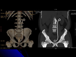
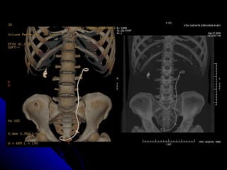
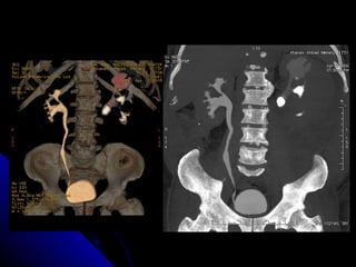
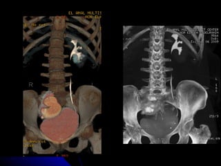
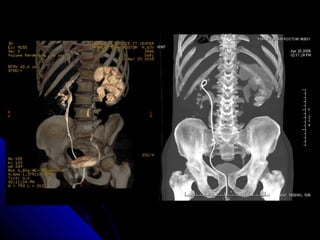
MMaaxxiimmuumm iinntteennssiittyy pprroojjeeccttiioonn ((MMIIPP)) 
iimmaaggeess ooff mmuullttiiddeetteeccttoorr CCTT ((MMDDCCTT)) 
eennhhaannccee tthhee vviissuuaalliizzaattiioonn ooff uurriinnaarryy 
ccaallccuullii iinn tthhee pprreesseennccee ooff ccoonnttrraasstt mmeeddiiaa 
oorr uurreetteerriicc sstteennttss.. 
HHaazzeemm AA.. YYoouusssseeff**,, MMoohhaammmmaadd KK.. OOmmaarr**,, HHoossssaamm AA.. 
YYoouusssseeff**,, aanndd HHaassssaann AA.. AAbboolleellllaa**** 
**RRaaddiioollooggyy DDeeppaarrttmmeenntt,, FFaaccuullttyy ooff MMeeddiicciinnee,, AAssssiiuutt UUnniivveerrssiittyy.. 
****UUrroollooggyy DDeeppaarrttmmeenntt,, FFaaccuullttyy ooff MMeeddiicciinnee,, AAssssiiuutt UUnniivveerrssiittyy..
IINNTTRROODDUUCCTTIIOONN 
With the introduction of multidetector technology, 
CT urography, to date, has emerged as the initial heir 
apparent to intravenous urography; many years of 
experience have now clearly demonstrated that CT is 
the test of choice for many urologic problems, 
including urolithiasis, renal masses, urinary tract 
infection, trauma, and obstructive uropathy.
CT urography provides a detailed anatomic depiction of 
each of the major portions of the urinary tract—the 
kidneys, intrarenal collecting systems, ureters, and 
bladder—and thus allows patients with hematuria to be 
evaluated comprehensively ©RSNA, 2009.
Because it provides fast results, unenhanced MDCT 
has largely supplanted intravenous urography, and, 
in the United States, MSCT generally prevails as 
the standard examination for detecting ureteral 
calculi (Wehrschuetz et. al., 2008).
OOBBJJEECCTTIIVVEE 
The objective of the current study is highlighting the 
rule of MIP images among the various image 
processing techniques in the special situation when 
ureteric stents are implemented or when the urinary 
tract was unintentionally opacified by IV contrast.
PPAATTIIEENNTTSS AANNDD MMEETTHHOODDSS 
Study Population 
A total of 23 consecutive patients (18 males and 5 
females, median age 41.2 years, range 2-65) with 
suspected urinary calculi were included in this 
retropective pilot study from April through September 
2009. 
Twelve patients were referred for detection of urinary 
calculi after inconclusive intravenous uroengraphy 
(IVU) and 11 patients were referred after placement of 
ureteric stents. In the former group IV contrast was 
administered 1-24 hours prior to CT examination.
Imaging protocol 
Unenhanced MDCT images were acquired with a 64 
detector row CT scanner VCT GE, Milwaukee, WI (18 
patients) or a 16 detector row CT scanner Brightspeed 
S GE, Milwaukee, WI (5 patients). 
Five-millimeter contiguous unenhanced axial CT 
images were obtained in a cephalocaudal direction 
from the diaphragm to the symphysis pubis. No oral or 
IV contrast was administered.
Scanning protocol 
For the 64-slice scanner, scans were obtained with tube 
rotation time 0.8sec, pitch 0.984:1, table feed/gantry 
rotation 39.75mm, a tube voltage of 140 kVp and 
effective tube current-time product of 548mAs. 
For the 16-slice scanner, scans were obtained with tube 
rotation time 0.8sec, pitch 1.375:1, table feed/gantry 
rotation 27.5mm, a tube voltage of 120 kVp and 
effective tube current-time product of 248mAs. 
The images were then reconstructed at 0.625 mm thick 
slices.
Image processing 
All datasets were sent to a commercially available 
workstation (ADW4.4 or ADW4.3). In all patients 
5mm MPR images were obtained in addition to the 
following: 
1) Thick slab MIP. 
2)Volume rendering 
3)Curved reformats.
Image evaluation 
Two independent radiologists and a urologist analyzed 
the images. The observers were asked to separately 
determine whether pyelocalyceal and ureteral calculi 
were present. They were instructed to document the 
location and size of each calculus using standard 
measurement devices.
RREESSUULLTTSS 
Urinary calculi were detected in 10 patients referred 
after inconclusive IVU and in 8 patients with ureteric 
stents. In 2 patients with inconclusive IVU ureteric 
stricture was the alternative diagnosis.
NNoo.. ooff ssttoonneess// ppaattiieenntt NNoo.. ooff ppaattiieennttss 
00 55 
11 22 
22 33 
33 33 
44 11 
55 33 
66 11 
77 22 
77--1155 22 
>>1155 11 
TToottaall 2233
Distinction between the urinary calculi and/or contrast 
and ureteric stents was not possible in the standard 
window for viewing the abdomen. In the VR images 
calculi can be demonstrated against ureteric stent by 
their morphology despite of having the same density 
but they could not be demonstrated within the 
opacified collecting system. All the stones were clearly 
depicted against the contrasted lumina (by having 
higher density) or the ureteric stents (by having lower 
density) in thick slab MIP at bone window setting.
Bone windowing also facilitates visualization of stones 
against the pelvic bones or the spine. 
The major weakness of MIP images was the inability 
to measure the size of the stones. VR or MPR were 
then reviewed for that context.
RREEPPRREESSEENNTTAATTIIVVEE CCAASSEESS
DDIISSCCUUSSSSIIOONN 
The use of maximum-intensity projection (MIP) and 
images in CT urography was initially described by 
McNicholas et al (1998). To our knowledge it was 
applied in the to date puplished literatures describing 
various techniques of CT urography. Since the 
majority of these studies were dedicated to detection of 
urothelial abnormalities they reported lower sensitivity 
of MIP images than thin section MPR images (Caoili 
et al. 2005), (Chow and Sommer 2001) in detection of 
similar lesions.
In our study, however, higher sensitivity of MIP 
images was evident. This is due to higher density of 
the stones compared with the low density uroepithelial 
lesions that may be masked by the contrast 
concentrated within the collecting system. Moreover 
the low contrast density in the collecting system 
caused by poor renal contrast concentration helped 
detection of the calculi.
The use of bone window for distinguishing stones from 
stent was reported by Tarnikut et al. (2004). In a series 
of a 3 patients they used bone window to distinguish 
stones from nephrostomy tubes in axial CT images 
after measuring the in vitro density values for stents.
The higher sensitivity of MIP images compared with 
that of VR images is related to the rendering technique 
as stated by Fishman et al (2006). VR images are 
formed by binary technique; voxels in volume are 
considered as either containing the specified densi 
Inaccurate size measurement of the recorded 
abnormality at MIP images is explained by lack of 
depth (or Z-axis perception) in the 2D image that 
represent the MIP slab.
CONCLUSION AANNDD RREECCOOMMMMEENNDDAATTIIOONNSS 
The CTU has become the radiologists most robust 
imaging tool for the evaluation of the kidneys, upper 
urinary tracts, and UB. 
Complete imaging of the kidneys and urinary tracts can 
be performed with MDCT. Studies are tailored to the 
clinical question and may be performed as noncontrast, 
combined non-contrast and post-contrast or post-contrast 
imaging studies only.
Finally radiologists need to educate emergency room 
and referring physicians about the limitations of 
unenhanced CT scans, as it does not detect infarcts, 
pyelonephritis, small renal cell carcinomas, or small 
ureteral tumors.
TTHHAANNKK YYOOUU

Maximum intensity projection (mip) (2)

  • 1.
    MMaaxxiimmuumm iinntteennssiittyy pprroojjeeccttiioonn((MMIIPP)) iimmaaggeess ooff mmuullttiiddeetteeccttoorr CCTT ((MMDDCCTT)) eennhhaannccee tthhee vviissuuaalliizzaattiioonn ooff uurriinnaarryy ccaallccuullii iinn tthhee pprreesseennccee ooff ccoonnttrraasstt mmeeddiiaa oorr uurreetteerriicc sstteennttss.. HHaazzeemm AA.. YYoouusssseeff**,, MMoohhaammmmaadd KK.. OOmmaarr**,, HHoossssaamm AA.. YYoouusssseeff**,, aanndd HHaassssaann AA.. AAbboolleellllaa**** **RRaaddiioollooggyy DDeeppaarrttmmeenntt,, FFaaccuullttyy ooff MMeeddiicciinnee,, AAssssiiuutt UUnniivveerrssiittyy.. ****UUrroollooggyy DDeeppaarrttmmeenntt,, FFaaccuullttyy ooff MMeeddiicciinnee,, AAssssiiuutt UUnniivveerrssiittyy..
  • 2.
    IINNTTRROODDUUCCTTIIOONN With theintroduction of multidetector technology, CT urography, to date, has emerged as the initial heir apparent to intravenous urography; many years of experience have now clearly demonstrated that CT is the test of choice for many urologic problems, including urolithiasis, renal masses, urinary tract infection, trauma, and obstructive uropathy.
  • 3.
    CT urography providesa detailed anatomic depiction of each of the major portions of the urinary tract—the kidneys, intrarenal collecting systems, ureters, and bladder—and thus allows patients with hematuria to be evaluated comprehensively ©RSNA, 2009.
  • 4.
    Because it providesfast results, unenhanced MDCT has largely supplanted intravenous urography, and, in the United States, MSCT generally prevails as the standard examination for detecting ureteral calculi (Wehrschuetz et. al., 2008).
  • 5.
    OOBBJJEECCTTIIVVEE The objectiveof the current study is highlighting the rule of MIP images among the various image processing techniques in the special situation when ureteric stents are implemented or when the urinary tract was unintentionally opacified by IV contrast.
  • 6.
    PPAATTIIEENNTTSS AANNDD MMEETTHHOODDSS Study Population A total of 23 consecutive patients (18 males and 5 females, median age 41.2 years, range 2-65) with suspected urinary calculi were included in this retropective pilot study from April through September 2009. Twelve patients were referred for detection of urinary calculi after inconclusive intravenous uroengraphy (IVU) and 11 patients were referred after placement of ureteric stents. In the former group IV contrast was administered 1-24 hours prior to CT examination.
  • 7.
    Imaging protocol UnenhancedMDCT images were acquired with a 64 detector row CT scanner VCT GE, Milwaukee, WI (18 patients) or a 16 detector row CT scanner Brightspeed S GE, Milwaukee, WI (5 patients). Five-millimeter contiguous unenhanced axial CT images were obtained in a cephalocaudal direction from the diaphragm to the symphysis pubis. No oral or IV contrast was administered.
  • 8.
    Scanning protocol Forthe 64-slice scanner, scans were obtained with tube rotation time 0.8sec, pitch 0.984:1, table feed/gantry rotation 39.75mm, a tube voltage of 140 kVp and effective tube current-time product of 548mAs. For the 16-slice scanner, scans were obtained with tube rotation time 0.8sec, pitch 1.375:1, table feed/gantry rotation 27.5mm, a tube voltage of 120 kVp and effective tube current-time product of 248mAs. The images were then reconstructed at 0.625 mm thick slices.
  • 9.
    Image processing Alldatasets were sent to a commercially available workstation (ADW4.4 or ADW4.3). In all patients 5mm MPR images were obtained in addition to the following: 1) Thick slab MIP. 2)Volume rendering 3)Curved reformats.
  • 10.
    Image evaluation Twoindependent radiologists and a urologist analyzed the images. The observers were asked to separately determine whether pyelocalyceal and ureteral calculi were present. They were instructed to document the location and size of each calculus using standard measurement devices.
  • 11.
    RREESSUULLTTSS Urinary calculiwere detected in 10 patients referred after inconclusive IVU and in 8 patients with ureteric stents. In 2 patients with inconclusive IVU ureteric stricture was the alternative diagnosis.
  • 12.
    NNoo.. ooff ssttoonneess//ppaattiieenntt NNoo.. ooff ppaattiieennttss 00 55 11 22 22 33 33 33 44 11 55 33 66 11 77 22 77--1155 22 >>1155 11 TToottaall 2233
  • 13.
    Distinction between theurinary calculi and/or contrast and ureteric stents was not possible in the standard window for viewing the abdomen. In the VR images calculi can be demonstrated against ureteric stent by their morphology despite of having the same density but they could not be demonstrated within the opacified collecting system. All the stones were clearly depicted against the contrasted lumina (by having higher density) or the ureteric stents (by having lower density) in thick slab MIP at bone window setting.
  • 14.
    Bone windowing alsofacilitates visualization of stones against the pelvic bones or the spine. The major weakness of MIP images was the inability to measure the size of the stones. VR or MPR were then reviewed for that context.
  • 15.
  • 23.
    DDIISSCCUUSSSSIIOONN The useof maximum-intensity projection (MIP) and images in CT urography was initially described by McNicholas et al (1998). To our knowledge it was applied in the to date puplished literatures describing various techniques of CT urography. Since the majority of these studies were dedicated to detection of urothelial abnormalities they reported lower sensitivity of MIP images than thin section MPR images (Caoili et al. 2005), (Chow and Sommer 2001) in detection of similar lesions.
  • 24.
    In our study,however, higher sensitivity of MIP images was evident. This is due to higher density of the stones compared with the low density uroepithelial lesions that may be masked by the contrast concentrated within the collecting system. Moreover the low contrast density in the collecting system caused by poor renal contrast concentration helped detection of the calculi.
  • 25.
    The use ofbone window for distinguishing stones from stent was reported by Tarnikut et al. (2004). In a series of a 3 patients they used bone window to distinguish stones from nephrostomy tubes in axial CT images after measuring the in vitro density values for stents.
  • 26.
    The higher sensitivityof MIP images compared with that of VR images is related to the rendering technique as stated by Fishman et al (2006). VR images are formed by binary technique; voxels in volume are considered as either containing the specified densi Inaccurate size measurement of the recorded abnormality at MIP images is explained by lack of depth (or Z-axis perception) in the 2D image that represent the MIP slab.
  • 27.
    CONCLUSION AANNDD RREECCOOMMMMEENNDDAATTIIOONNSS The CTU has become the radiologists most robust imaging tool for the evaluation of the kidneys, upper urinary tracts, and UB. Complete imaging of the kidneys and urinary tracts can be performed with MDCT. Studies are tailored to the clinical question and may be performed as noncontrast, combined non-contrast and post-contrast or post-contrast imaging studies only.
  • 28.
    Finally radiologists needto educate emergency room and referring physicians about the limitations of unenhanced CT scans, as it does not detect infarcts, pyelonephritis, small renal cell carcinomas, or small ureteral tumors.
  • 29.