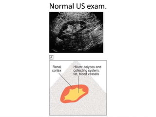
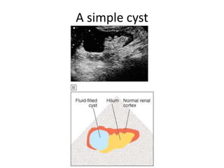
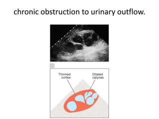
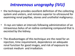
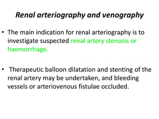
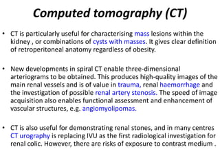
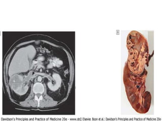
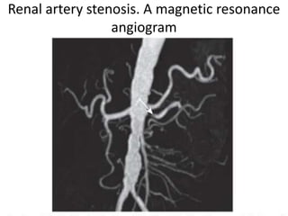
IMAGING TECHNIQUES
 Plain X-rays ( KUB)
• may show the renal outlines ,
• opaque calculi and calcification within the
  renal tract.
Ultrasound
• It can show renal size and position
• detect dilatation of the collecting system (suggesting
  obstruction),
• distinguish tumours and cysts,
• show other abdominal, pelvic and retroperitoneal
  pathology.
• it can image the prostate and bladder, and estimate
  completeness of emptying in suspected bladder outflow
  obstruction.

 Images are often less clear in obese individuals.
Ultrasound
In chronic renal disease(signs of chronicity):

1- ultrasonographic density of the renal cortex is
  increased (increase echogenicity)
2- corticomedullary differentiation is lost.
3- Thin cortical thickness.
4- small size kidneys.

Except in ( MM, DM, Amyloidosis)
Ultrasound (Doppler)
• used to show blood flow in extrarenal and larger intrarenal
   vessels.
• The resistivity index (RI) is the ratio of peak systolic and
   diastolic velocities, and is influenced by the resistance to
   flow through small intrarenal arteries. It may be elevated in
   various diseases, including
  1- acute glomerulonephritis and
  2- rejection of a renal transplant.

Severe renal artery stenosis causes damping of flow in
  intrarenal vessels with high peak velocities.
Normal US exam.
A simple cyst
chronic obstruction to urinary outflow.
Intravenous urography (IVU)
• the technique provides excellent definition of the collecting
  system and ureters, and remains superior to ultrasound for
  examining renal papillae, stones and urothelial malignancy .

• X-rays are taken at intervals following administration of an
  intravenous bolus of an iodine-containing compound that is
  excreted by the kidney.

• The disadvantages of this technique are the need for an
  injection, time requirement, dependence on adequate
  renal function for good images, and risk of exposure to
  contrast medium and irradiation.
5 min, IVU
Pyelography
• Pyelography (direct injection of contrast medium into the
  collecting system from above or below) offers the best
  views of the collecting system and upper tract, and is
  commonly used to identify the cause of urinary tract
  obstruction .
• Antegrade pyelography requires the insertion of a fine
  needle into the pelvicalyceal system under ultrasound or
  radiographic control. Contrast is injected to outline the
  collecting system, and particularly to localise the site of
  obstruction.

• Retrograde pyelography can be performed by inserting
  catheters into the ureteric orifices at cystoscopy .
Retrograde pyelography
Renal arteriography and venography

• The main indication for renal arteriography is to
  investigate suspected renal artery stenosis or
  haemorrhage.

• Therapeutic balloon dilatation and stenting of the
  renal artery may be undertaken, and bleeding
  vessels or arteriovenous fistulae occluded.
Computed tomography (CT)
• CT is particularly useful for characterising mass lesions within the
  kidney , or combinations of cysts with masses. It gives clear definition
  of retroperitoneal anatomy regardless of obesity.

• New developments in spiral CT enable three-dimensional
  arteriograms to be obtained. This produces high-quality images of the
  main renal vessels and is of value in trauma, renal haemorrhage and
  the investigation of possible renal artery stenosis. The speed of image
  acquisition also enables functional assessment and enhancement of
  vascular structures, e.g. angiomyolipomas.

• CT is also useful for demonstrating renal stones, and in many centres
  CT urography is replacing IVU as the first radiological investigation for
  renal colic. However, there are risks of exposure to contrast medium .
17.2 RENAL COMPLICATIONS OF RADIOLOGICAL
                     INVESTIGATIONS
• A. Contrast nephrotoxicity
 An acute deterioration in renal function, sometimes life-
  threatening, commencing < 48 hrs after administration of
  i.v. radiographic contrast media

    Risk factors
•    Pre-existing renal impairment
•    Use of high-osmolality, ionic contrast media and repetitive
     dosing in short time periods
•    Diabetes mellitus
•    Myeloma
PREVENTION

• Hydration-e.g. free oral fluids plus i.v. isotonic saline
  500 ml then 250 ml/hr during procedure
• Avoid nephrotoxic drugs and diuretics ; withhold
  non-steroidal anti-inflammatory drugs (NSAIDs);
  omit metformin for 48 hrs befor and after
  procedure
• N-acetyl cysteine , Aminophyline,sodium
  bicarbonate ( may provide protection).

• If the risks are high, consider alternative methods of
  imaging
B. Cholesterol atheroembolism
 Typically follows days to weeks after intra-
 arterial investigations .
The foot of a patient who suffered extensive
 atheroembolism following coronary artery
                   stenting
Magnetic resonance imaging (MRI)
• MRI offers excellent resolution and distinction between
  different tissues.
• Magnetic resonance angiography (MRA) uses gadolinium-
  based contrast media which are non-nephrotoxic, and
  avoids the risk of atheroemboli which accompany invasive
  angiography. It can produce good images of main renal
  vessels, although the resolution is not yet sufficient to
  exclude branch artery stenosis.

• These techniques are likely to develop further and are
  finding an important role in non-invasive screening for
  renal artery stenosis.
Renal artery stenosis. A magnetic resonance
                 angiogram
Radionuclide studies
• These are functional studies requiring the injection
  of gamma ray-emitting radiopharmaceuticals which
  are taken up and excreted by the kidney,

• A process which can be monitored by an external
  gamma camera.
99mTc-DTPA

• Diethylene triamine-penta acetic acid labelled
  with technetium (99mTc-DTPA) is excreted by
  glomerular filtration.
• DTPA injection and analysis of uptake and
  excretion provides information regarding the
  arterial perfusion of each kidney. A typical pattern
  of prolonged transit time, delayed peak activity
  and reduced excretion is seen in renal artery
  stenosis.
• In patients with significant obstruction of the
  outflow tract, persistence of the nuclide in the
  renal pelvis is seen , and a loop diuretic fails to
  accelerate its disappearance.
99mTc-DMSA

• Di mercapto succinic acid labelled with
  technetium (99mTc-DMSA) is filtered by glomeruli
  and partially bound to proximal tubular cells.
• Following intravenous injection, images of the
  renal cortex show the shape, size and function of
  each kidney.
• This is a sensitive method of demonstrating early
  cortical scarring that is of particular value in
  children with vesico-ureteric reflux and
  pyelonephritis.
• It is also possible to assess the relative
  contribution of each kidney to total function.
DMSA isotope renogram
RENAL BIOPSY
 Indications
• Acute renal failure that is not adequately explained

• Chronic renal failure with normal-sized kidneys

• Nephrotic syndrome or glomerular proteinuria in adults

• Nephrotic syndrome in children that has atypical features
  or is not responding to treatment
• Isolated haematuria or proteinuria with renal
  characteristics or associated abnormalities
Before the Bx.
• BP normal
• Cooperative pt.

•   Ix.
•   CBC
•   PT , PTT,
•   Bleeding time
•   Viral markers (HIV, HBs Ag. Anti HCV Ab.)
RENAL BIOPSY
 Contraindications
• Disordered coagulation or thrombocytopenia. Aspirin and
  other agents causing platelet dysfunction should be omitted
  for elective biopsies

• Uncontrolled hypertension

• Kidneys < 60% predicted size

• Solitary kidney (except transplants) (relative
  contraindication)
Complications

• Pain, usually mild

• Bleeding into urine, usually minor but may
  produce clot colic and obstruction

• Bleeding around the kidney, occasionally massive
  and requiring angiography with intervention, or
  surgery

• Arteriovenous fistula, rarely significant clinically

medicine.Kidney 2.(dr.ala)

  • 1.
    IMAGING TECHNIQUES PlainX-rays ( KUB) • may show the renal outlines , • opaque calculi and calcification within the renal tract.
  • 2.
    Ultrasound • It canshow renal size and position • detect dilatation of the collecting system (suggesting obstruction), • distinguish tumours and cysts, • show other abdominal, pelvic and retroperitoneal pathology. • it can image the prostate and bladder, and estimate completeness of emptying in suspected bladder outflow obstruction. Images are often less clear in obese individuals.
  • 3.
    Ultrasound In chronic renaldisease(signs of chronicity): 1- ultrasonographic density of the renal cortex is increased (increase echogenicity) 2- corticomedullary differentiation is lost. 3- Thin cortical thickness. 4- small size kidneys. Except in ( MM, DM, Amyloidosis)
  • 4.
    Ultrasound (Doppler) • usedto show blood flow in extrarenal and larger intrarenal vessels. • The resistivity index (RI) is the ratio of peak systolic and diastolic velocities, and is influenced by the resistance to flow through small intrarenal arteries. It may be elevated in various diseases, including 1- acute glomerulonephritis and 2- rejection of a renal transplant. Severe renal artery stenosis causes damping of flow in intrarenal vessels with high peak velocities.
  • 5.
  • 6.
  • 7.
    chronic obstruction tourinary outflow.
  • 8.
    Intravenous urography (IVU) •the technique provides excellent definition of the collecting system and ureters, and remains superior to ultrasound for examining renal papillae, stones and urothelial malignancy . • X-rays are taken at intervals following administration of an intravenous bolus of an iodine-containing compound that is excreted by the kidney. • The disadvantages of this technique are the need for an injection, time requirement, dependence on adequate renal function for good images, and risk of exposure to contrast medium and irradiation.
  • 9.
  • 10.
    Pyelography • Pyelography (directinjection of contrast medium into the collecting system from above or below) offers the best views of the collecting system and upper tract, and is commonly used to identify the cause of urinary tract obstruction . • Antegrade pyelography requires the insertion of a fine needle into the pelvicalyceal system under ultrasound or radiographic control. Contrast is injected to outline the collecting system, and particularly to localise the site of obstruction. • Retrograde pyelography can be performed by inserting catheters into the ureteric orifices at cystoscopy .
  • 11.
  • 12.
    Renal arteriography andvenography • The main indication for renal arteriography is to investigate suspected renal artery stenosis or haemorrhage. • Therapeutic balloon dilatation and stenting of the renal artery may be undertaken, and bleeding vessels or arteriovenous fistulae occluded.
  • 13.
    Computed tomography (CT) •CT is particularly useful for characterising mass lesions within the kidney , or combinations of cysts with masses. It gives clear definition of retroperitoneal anatomy regardless of obesity. • New developments in spiral CT enable three-dimensional arteriograms to be obtained. This produces high-quality images of the main renal vessels and is of value in trauma, renal haemorrhage and the investigation of possible renal artery stenosis. The speed of image acquisition also enables functional assessment and enhancement of vascular structures, e.g. angiomyolipomas. • CT is also useful for demonstrating renal stones, and in many centres CT urography is replacing IVU as the first radiological investigation for renal colic. However, there are risks of exposure to contrast medium .
  • 15.
    17.2 RENAL COMPLICATIONSOF RADIOLOGICAL INVESTIGATIONS • A. Contrast nephrotoxicity An acute deterioration in renal function, sometimes life- threatening, commencing < 48 hrs after administration of i.v. radiographic contrast media Risk factors • Pre-existing renal impairment • Use of high-osmolality, ionic contrast media and repetitive dosing in short time periods • Diabetes mellitus • Myeloma
  • 16.
    PREVENTION • Hydration-e.g. freeoral fluids plus i.v. isotonic saline 500 ml then 250 ml/hr during procedure • Avoid nephrotoxic drugs and diuretics ; withhold non-steroidal anti-inflammatory drugs (NSAIDs); omit metformin for 48 hrs befor and after procedure • N-acetyl cysteine , Aminophyline,sodium bicarbonate ( may provide protection). • If the risks are high, consider alternative methods of imaging
  • 17.
    B. Cholesterol atheroembolism Typically follows days to weeks after intra- arterial investigations .
  • 18.
    The foot ofa patient who suffered extensive atheroembolism following coronary artery stenting
  • 19.
    Magnetic resonance imaging(MRI) • MRI offers excellent resolution and distinction between different tissues. • Magnetic resonance angiography (MRA) uses gadolinium- based contrast media which are non-nephrotoxic, and avoids the risk of atheroemboli which accompany invasive angiography. It can produce good images of main renal vessels, although the resolution is not yet sufficient to exclude branch artery stenosis. • These techniques are likely to develop further and are finding an important role in non-invasive screening for renal artery stenosis.
  • 20.
    Renal artery stenosis.A magnetic resonance angiogram
  • 21.
    Radionuclide studies • Theseare functional studies requiring the injection of gamma ray-emitting radiopharmaceuticals which are taken up and excreted by the kidney, • A process which can be monitored by an external gamma camera.
  • 22.
    99mTc-DTPA • Diethylene triamine-pentaacetic acid labelled with technetium (99mTc-DTPA) is excreted by glomerular filtration. • DTPA injection and analysis of uptake and excretion provides information regarding the arterial perfusion of each kidney. A typical pattern of prolonged transit time, delayed peak activity and reduced excretion is seen in renal artery stenosis. • In patients with significant obstruction of the outflow tract, persistence of the nuclide in the renal pelvis is seen , and a loop diuretic fails to accelerate its disappearance.
  • 23.
    99mTc-DMSA • Di mercaptosuccinic acid labelled with technetium (99mTc-DMSA) is filtered by glomeruli and partially bound to proximal tubular cells. • Following intravenous injection, images of the renal cortex show the shape, size and function of each kidney. • This is a sensitive method of demonstrating early cortical scarring that is of particular value in children with vesico-ureteric reflux and pyelonephritis. • It is also possible to assess the relative contribution of each kidney to total function.
  • 24.
  • 25.
    RENAL BIOPSY Indications •Acute renal failure that is not adequately explained • Chronic renal failure with normal-sized kidneys • Nephrotic syndrome or glomerular proteinuria in adults • Nephrotic syndrome in children that has atypical features or is not responding to treatment • Isolated haematuria or proteinuria with renal characteristics or associated abnormalities
  • 26.
    Before the Bx. •BP normal • Cooperative pt. • Ix. • CBC • PT , PTT, • Bleeding time • Viral markers (HIV, HBs Ag. Anti HCV Ab.)
  • 27.
    RENAL BIOPSY Contraindications •Disordered coagulation or thrombocytopenia. Aspirin and other agents causing platelet dysfunction should be omitted for elective biopsies • Uncontrolled hypertension • Kidneys < 60% predicted size • Solitary kidney (except transplants) (relative contraindication)
  • 28.
    Complications • Pain, usuallymild • Bleeding into urine, usually minor but may produce clot colic and obstruction • Bleeding around the kidney, occasionally massive and requiring angiography with intervention, or surgery • Arteriovenous fistula, rarely significant clinically