TOMOSYNTHESIS
PRESENTED BY
SAHEB JANA
BWU/BRI/21/091
Breast Tomosynthesis
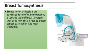
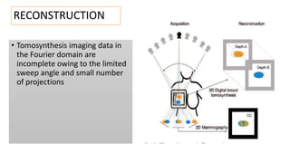
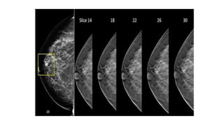
• Breast tomosynthesis is an
advanced form of mammography,
a specific type of breast imaging
that uses low-dose x-rays to detect
cancer early when it is most
treatable.
How does the procedure work?
 Breast tomosynthesis uses a low-dose x-ray system, electronics and a
computer to convert x-ray images of the breast to a three-
dimensional image set.
 Multiple x-ray images of the breast are digitized using systems similar
to those found in digital cameras and then transferred to a computer
where they are reconstructed or "synthesized" into a three-
dimensional image set.
 In this way, 3-D breast imaging is similar to computed tomography
(CT) imaging in which a series of thin high-resolution "slices" are
assembled together to create a 3-D reconstruction of the body.
TUBE MOTION
• DBT tube motion refers to the mode of x-ray
tube emission with respect to motion of the
gantry, with the use of either a continuous or
step-and-shoot technique.
SWEEP ANGLE
-Similar to CT images, tomosynthesis images are obtained
with a moving x-ray source. However, tomosynthesis images
are obtained over a limited angular range, or sweep angle.
The sweep angle for DBT is 15°–50°
RECONSTRUCTION
• Tomosynthesis imaging data in
the Fourier domain are
incomplete owing to the limited
sweep angle and small number
of projections
BENEFIT
Imaging of the breast allows detection of small tumors. When cancers
are small, the woman has more treatment options.
The use of screening mammography increases the detection of small
abnormal tissue growths confined to the milk ducts in the breast, called
ductal carcinoma in situ (DCIS).
Greater accuracy in pinpointing the size, shape and location of breast
abnormalities .
Fewer unnecessary biopsies or additional tests.
Greater likelihood of detecting multiple breast tumors.
Clearer images of abnormalities within dense breast tissue.
RISK
Exposure to a low level of radiation. A 3D mammogram uses X-rays to
create an image of the breast, which exposes you to a low level of
radiation. ...
The test may find something that turns out to not be cancer. ...
The test can't detect all cancers.
TOMOSYNTHESIS.pptx

TOMOSYNTHESIS.pptx

  • 1.
  • 2.
    Breast Tomosynthesis • Breasttomosynthesis is an advanced form of mammography, a specific type of breast imaging that uses low-dose x-rays to detect cancer early when it is most treatable.
  • 3.
    How does theprocedure work?  Breast tomosynthesis uses a low-dose x-ray system, electronics and a computer to convert x-ray images of the breast to a three- dimensional image set.  Multiple x-ray images of the breast are digitized using systems similar to those found in digital cameras and then transferred to a computer where they are reconstructed or "synthesized" into a three- dimensional image set.  In this way, 3-D breast imaging is similar to computed tomography (CT) imaging in which a series of thin high-resolution "slices" are assembled together to create a 3-D reconstruction of the body.
  • 4.
    TUBE MOTION • DBTtube motion refers to the mode of x-ray tube emission with respect to motion of the gantry, with the use of either a continuous or step-and-shoot technique. SWEEP ANGLE -Similar to CT images, tomosynthesis images are obtained with a moving x-ray source. However, tomosynthesis images are obtained over a limited angular range, or sweep angle. The sweep angle for DBT is 15°–50°
  • 5.
    RECONSTRUCTION • Tomosynthesis imagingdata in the Fourier domain are incomplete owing to the limited sweep angle and small number of projections
  • 7.
    BENEFIT Imaging of thebreast allows detection of small tumors. When cancers are small, the woman has more treatment options. The use of screening mammography increases the detection of small abnormal tissue growths confined to the milk ducts in the breast, called ductal carcinoma in situ (DCIS). Greater accuracy in pinpointing the size, shape and location of breast abnormalities . Fewer unnecessary biopsies or additional tests. Greater likelihood of detecting multiple breast tumors. Clearer images of abnormalities within dense breast tissue.
  • 8.
    RISK Exposure to alow level of radiation. A 3D mammogram uses X-rays to create an image of the breast, which exposes you to a low level of radiation. ... The test may find something that turns out to not be cancer. ... The test can't detect all cancers.