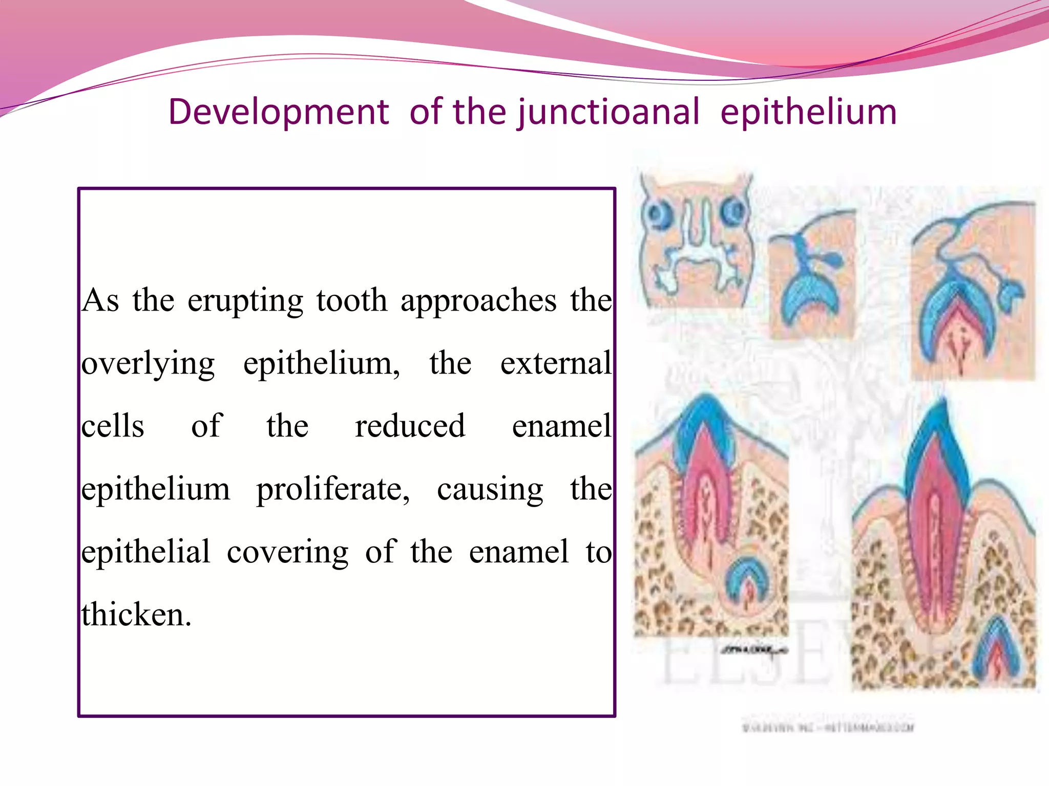
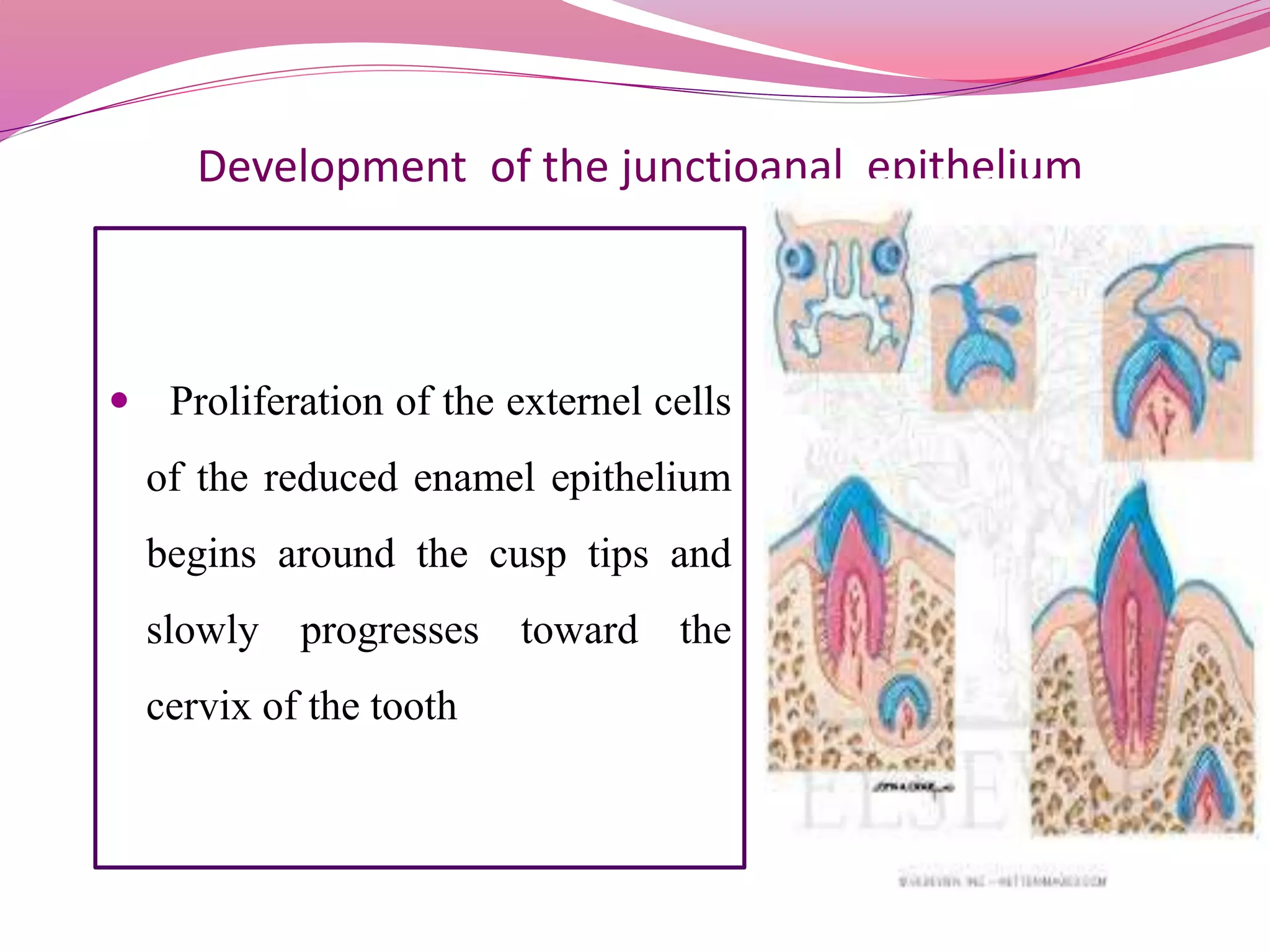
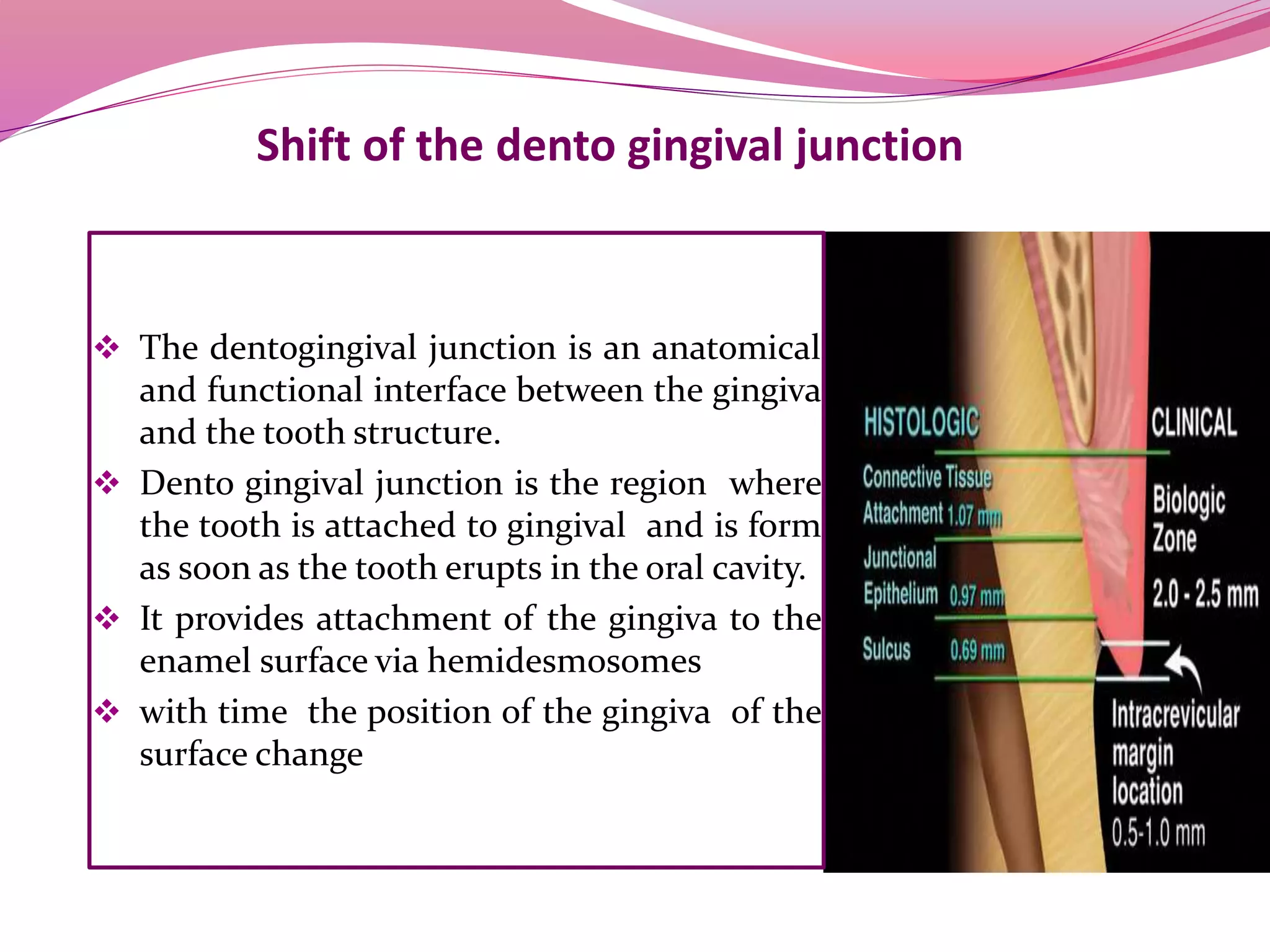
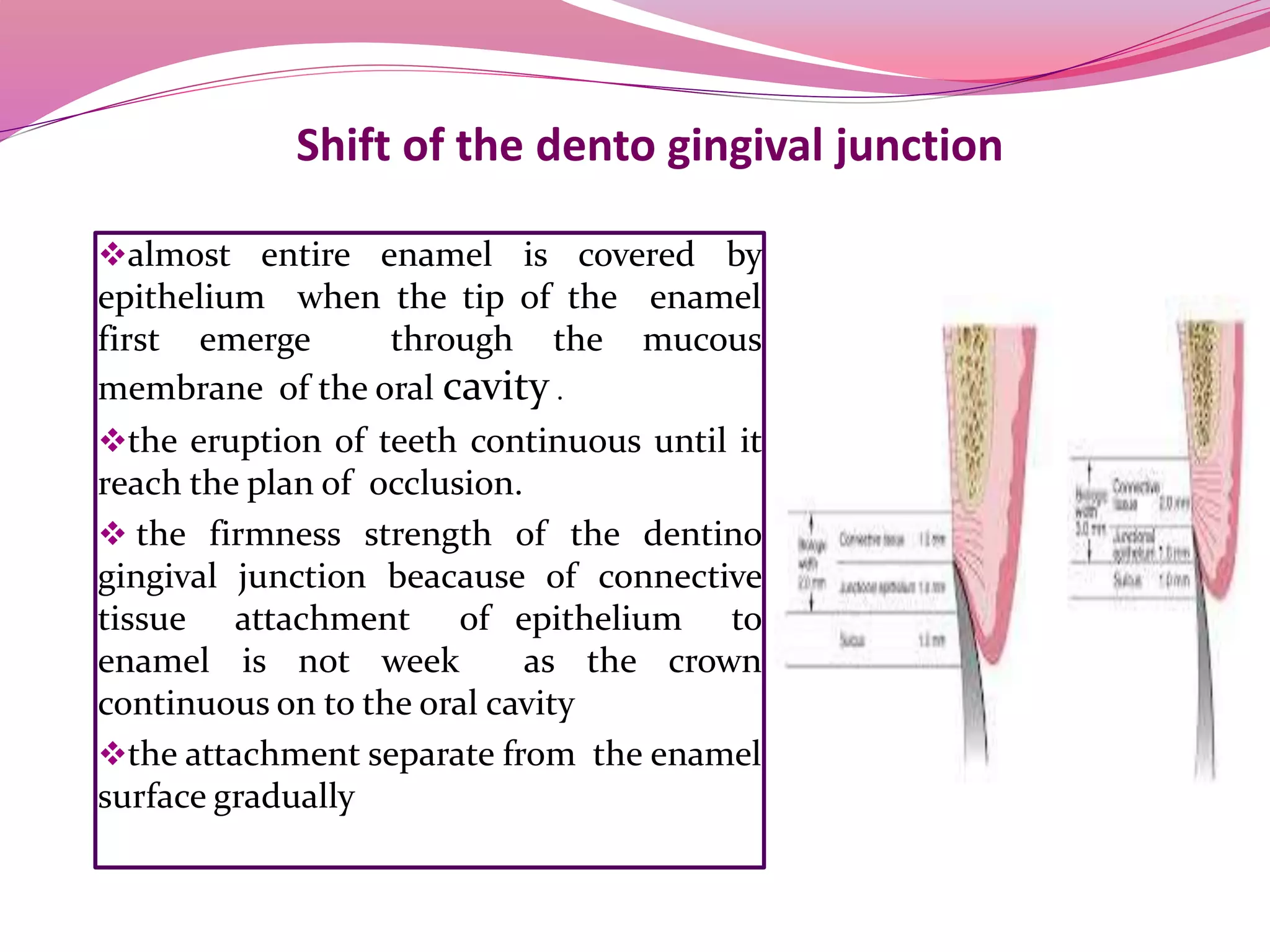
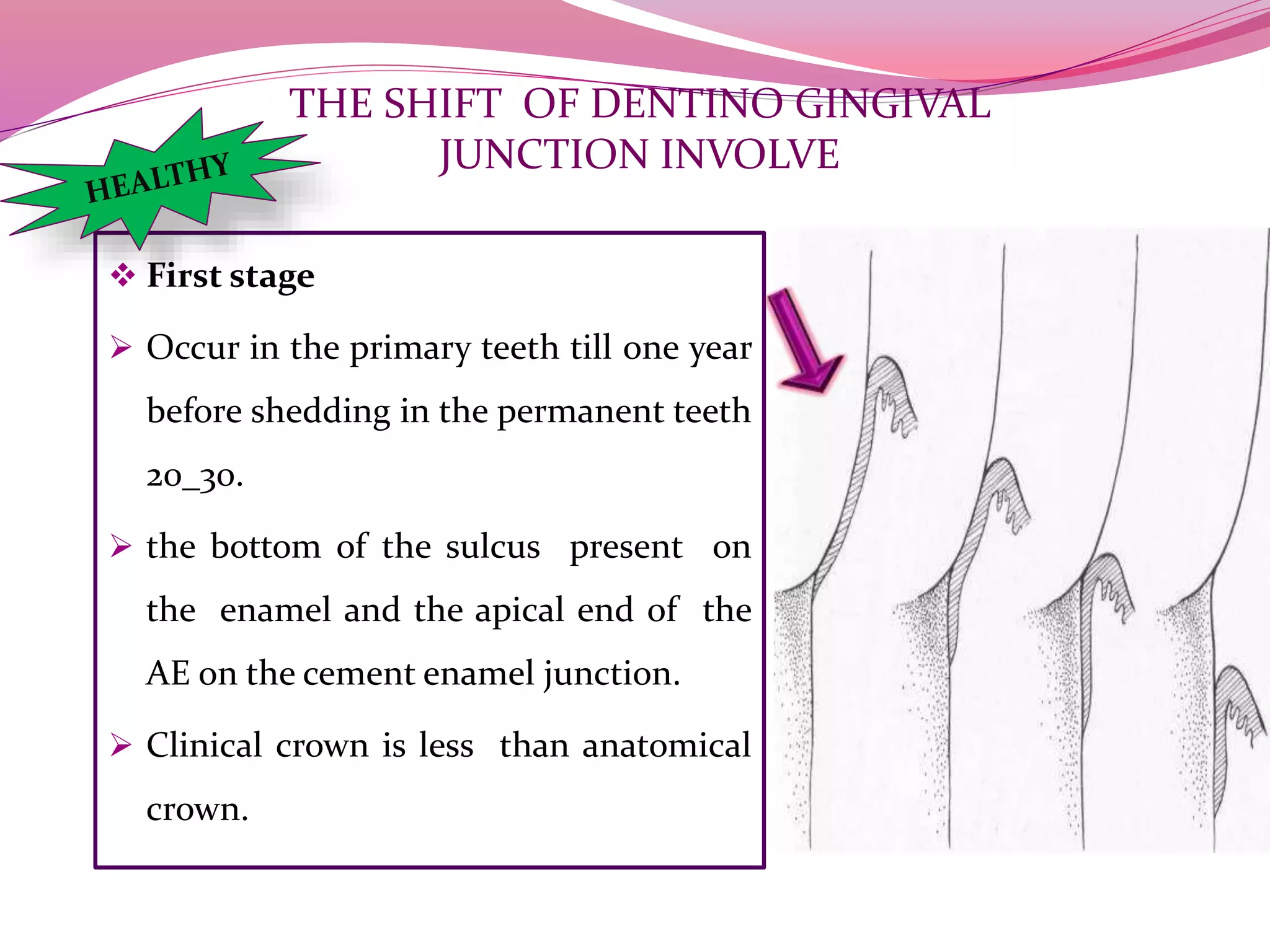
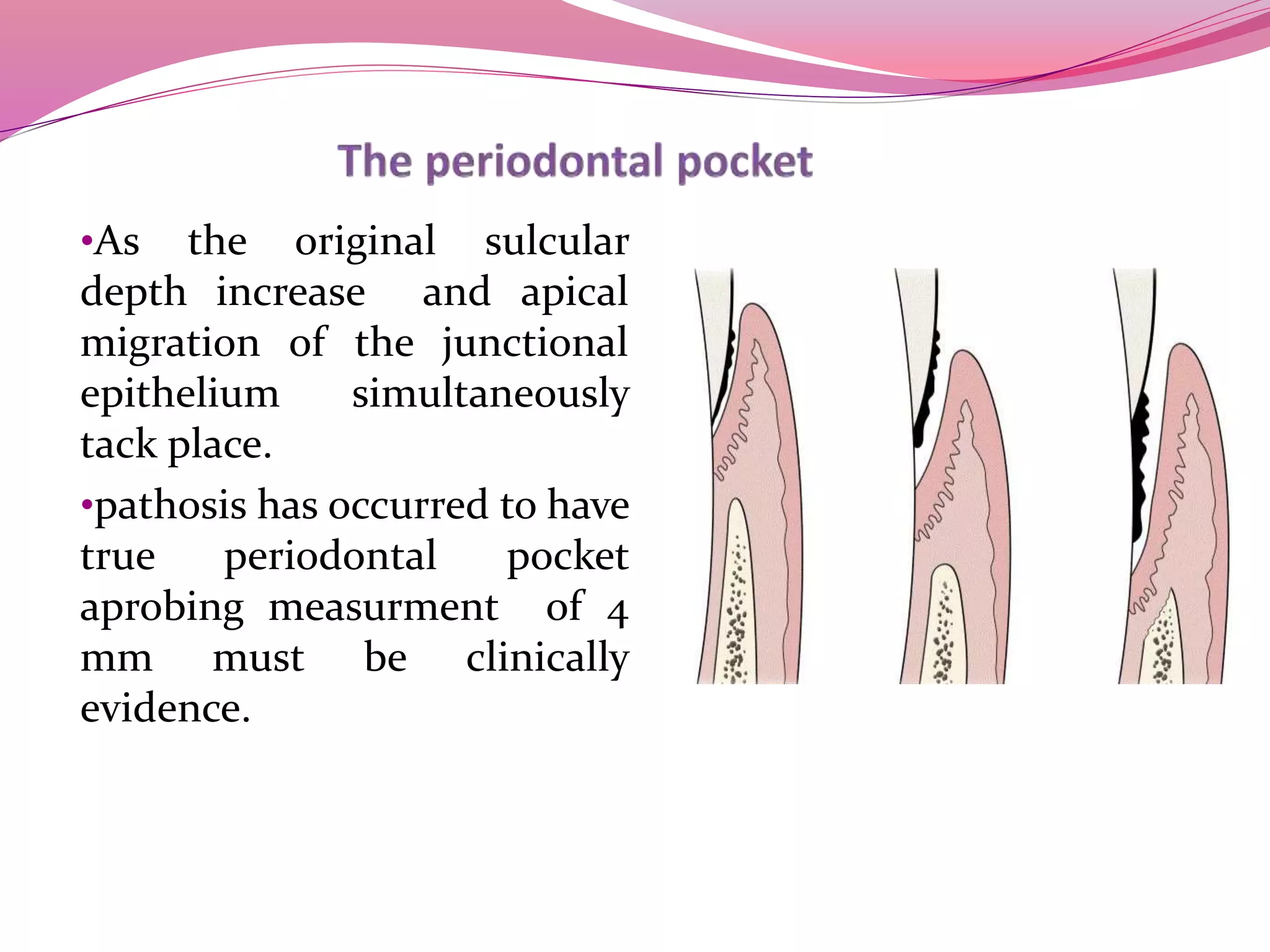
âOh GOSH! Reflecting on Hackteria's Collaborative Practices in a Global Do-It...
Â
Dentinogingival junction
1.
2.
3. The gingival sulcus
īļis an area of potential space between
a tooth and the surrounding gingival
tissue.
īļIt is lined by two entities:
-Apically by the gingival fibers of
connective tissue attachment .
_Coronally by free gingival margin .
4. The gingival sulcus
īļUnder normal conditions the depth
of the sulcus is variable.45% of all
measured sulci are below 0.5 mm
,the average sulcus is 1.8mm
īļthe shallower sulcus is the more
likely that the gingival margin is
not inflamed
5.
6. THE SULCULAR EPITHELIUM
īļIts stratified squamous
epithelium ,non keratinized .
Para keratinized that is
continuous with the oral
epithelium and lines the
lateral surface of the sulcus .
īļIts lacks epithelial ridges so
forms a smooth contact with
lamina properia
7. THE SULCULAR
EPITHELIUM
īļApically it overlaps the
coronal border of the
junctional epithelium
īļthis epithelium shares many
of the characteristics of oral
epithelium including good
resistance to mechanical
forces and relative
impermeability to fluid and
cells
8.
9. the junctional epithelium
īļIts the stratified non keratinizing
epithelium that surround the tooth like
a collar .
īļits attached by one broad surface to
the tooth and by the other to the
gingival connective tissue
īļThe junctional epithelium has two
basal lamina:
oOne that faces the tooth (internal basal
lamina )
oThe other faces connective tissue
(external basal lamina)
10. the junctional epithelium
īļThe desquamative shedding
surface of the junctional epithelium
is located at its coronal end which
also forms the bottom of the
gingival sulcus
īļThe junctional epithelium is more
permeable than oral or sulcular
epithelium It serves as the
perferential route for the passage of
bacterial products from the sulcus in
to connective tissue into the sulcus
11.
12. THE SULCULAR EPITHELIUM THE JUNCTION EPITHELIUM
1. It is epithelium that surround
the tooth like a collar .
2. It is more permeable than oral or
sulcular epithelium It serves as
the perferential route for the
passage of bacterial products
from the sulcus in to connective
tissue and fluid and cells from C.T
into the sulcus
1. It is continuous with the oral
epithelium and lines the lateral
surface of the sulcus .
2. It characterized by good
resistance to mechanical forces
and relative impermeability to
fluid and cells
13. Development of the junctioanal epithelium
As the erupting tooth approaches the
overlying epithelium, the external
cells of the reduced enamel
epithelium proliferate, causing the
epithelial covering of the enamel to
thicken.
14. Development of the junctioanal epithelium
ī Proliferation of the externel cells
of the reduced enamel epithelium
begins around the cusp tips and
slowly progresses toward the
cervix of the tooth
15. Development of the junctioanal epithelium
proliferating cells eventually displace
any remaining reduced ameloblasts, thus
replacing the relatively inert reduced
enamel epithelium with an epithelial
collar of cells with a high turnover
rate. This collar of cells with a high
turnover rate is the early junctional
epithelium.
Eventually, the entire reduced enamel
epithelium will become replaced by
junctional epithelium.
16.
17. Development of the junctioanal epithelium
īļAfter accomplishing of amelogenesis
(protective stage) the ameloblast secrete
or leave structure less material on enamel
surface primary enamel cuticle .
īļThe enamel organ becomes reduced in
thickness (reduced enamel epithelium )
and function as aprotection against
contact with connective tissue ,to inhibit
cementum deposition or enamel
resorption
The reduced enamel epi thelium
consists of two layers
1-the reduced ameloblast
2-remnant of the dental organ
18. Development of the junctioanal epithelium
īļThe ameloblast develop hemidesmosomes
to attach the reduced enamel epithelium to
the surface of the enamel at the time of
eruption the reduced enamel epithelium
secrete desmolytic enzymes causing
degenerating of the of the c.t present
between it and the oral mucosa (desmolytic
stage of amelo blasts )
īļThe outer layer of the reduced enamel
epithelium and the cells of the oral
epithelium proliferate into the degenerated
c.t. to form amass of cells over the erupting
tooth the epithelial plug.
19.
20. Development of the junctioanal epithelium
īļCell death in th middle portion of plug cause formation of epithelial lined canal
through which tooth will erupt with out hemorrhage
īļOnce the tip of the crown appear in the oral cavity reduced enamel epithelium will
be called primary attached epithelium
īļAnd the the shallow groove present between the tooth and the gingiva is called
GINGIVAL SULCUS
īļThe cells of primary attached epithelium (PAE) originate from reduced enamel
epithelium and attached to tooth by primary enamel cuticle
īļWhen primary attachment epithelium is replaced by oral epithelium its called
secondary attached epithelium that attaches by secondary enamel cuticle.
21. Primary enamel cuticle Secondary enamel cuticle
1. Binds secondary attached
epithelium to the enamel surface
2. It is formed finally when the teeth
erupt
1. Binds primary attached epithelium
to the enamel surface
2. It is formed initially at the
protective stage
Primary attached epi. Secondary attached epi.
1. It is Primary attachment
epithelium replaced by oral
epithelium
2. attached by secondary enamel
cuticle
1. originate from reduced enamel
epithelium
2. attached to tooth by primary
enamel cuticle
22. Shift of the dento gingival junction
īļ The dentogingival junction is an anatomical
and functional interface between the gingiva
and the tooth structure.
īļ Dento gingival junction is the region where
the tooth is attached to gingival and is form
as soon as the tooth erupts in the oral cavity.
īļ It provides attachment of the gingiva to the
enamel surface via hemidesmosomes
īļ with time the position of the gingiva of the
surface change
23. Shift of the dento gingival junction
īļ Components
1. Epithelial component is derived from
reduced dental (enamel) epithelium
and oral epithelium.
2. The connective tissue component is
derived from the lamina propria of the
oral mucosa.
īļ The attachment of the functional
epithelium to the tooth is reinforced with
the gingival fibers, which brace the gingival
against the tooth surface.
25. Shift of the dento gingival junction
īļalmost entire enamel is covered by
epithelium when the tip of the enamel
first emerge through the mucous
membrane of the oral cavity .
īļthe eruption of teeth continuous until it
reach the plan of occlusion.
īļ the firmness strength of the dentino
gingival junction beacause of connective
tissue attachment of epithelium to
enamel is not week as the crown
continuous on to the oral cavity
īļthe attachment separate from the enamel
surface gradually
26.
27. Shift of the dento gingival junction
ī when the tip of the enamel first emerge through the mucous membrane of
the oral cavity . one third to one fourth of enamel is still covered by the gingiva
īThe actual movement of teeth towards occlusal plane called acitve
eruption
īThe separation of primary attached epithelium from the enamel
surface called passive eruption
28. THE SHIFT OF DENTINO GINGIVAL
JUNCTION INVOLVE
īļ First stage
ī Occur in the primary teeth till one year
before shedding in the permanent teeth
20_30.
ī the bottom of the sulcus present on
the enamel and the apical end of the
AE on the cement enamel junction.
ī Clinical crown is less than anatomical
crown.
29. THE SHIFT OF DENTINO GINGIVAL
JUNCTION INVOLVE
īļ Second stage
ī Occur till age of the 40 or even later.
ī The bottom of the sulcus still present
on the enamel and the apical end of the
AE on cementum.
ī the clinical crown is less than
anatomical crown.
30. THE SHIFT OF DENTINO GINGIVAL
JUNCTION INVOLVE
īļ Third stage
ī It is unhealthy condition.
ī The bottom of the sulcus present at the
CEJ and the apical end at cementum.
ī The clinical crown equal to the
anatomical crown.
31. THE SHIFT OF DENTINO GINGIVAL
JUNCTION INVOLVE
īļ Fourth stage
ī It is unhealthy condition.
ī From 60 years later .
ī The bottom of the sulcucs and apical
end on the cementum.
ī The clinical crown is longer than
anatomical crown
32. âĸAs the original sulcular
depth increase and apical
migration of the junctional
epithelium simultaneously
tack place.
âĸpathosis has occurred to have
true periodontal pocket
aprobing measurment of 4
mm must be clinically
evidence.
33. The periodontal pocket
īļ It results in:
1. A bluish-red vertical zone from
the gingival margin to the
alveolar mucosa.
2. Gingival bleeding.
3. Suppuration & tooth mobility
4. And diastema formation.
īļ symptoms such as localized
pain or pain "deep in the bone"
are suggestive of the presence
of periodontal pockets
The enamel organ becomes reduced in thickness (reduced enamel epithelium ) and function as aprotection against contact with connective tissue ,to inhibit cementum deposition or enamel resorption