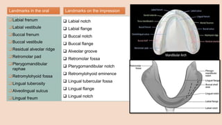
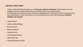
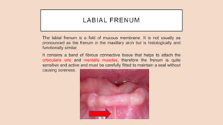
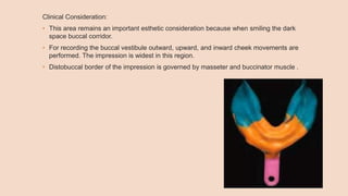
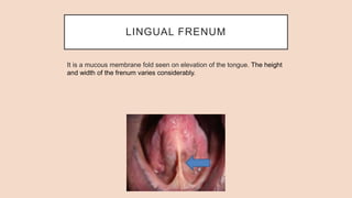
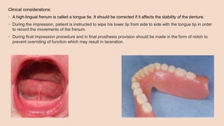
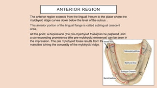
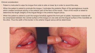
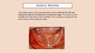
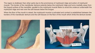
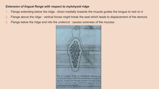
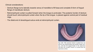
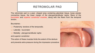
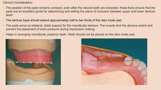
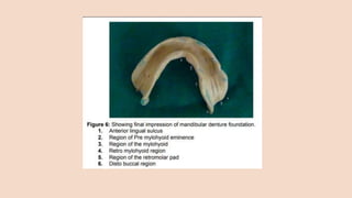
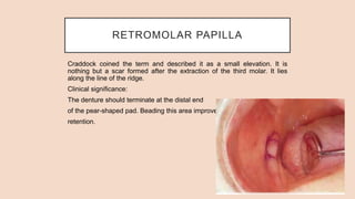
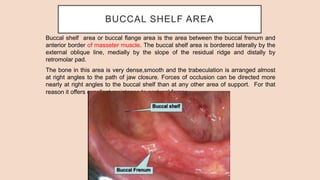
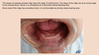
ANATOMICAL LANDMARKS OF
EDENTULOUS MANDIBULAR
ARCH
Nishu Priya
1st year PGT
CONTENTS
⮚ Introduction
⮚ Anatomical landmarks in Mandibular arch
❑ Limiting structures
❑ Supporting structures
❑ Relief areas
INTRODUCTION
Knowledge of the orofacial anatomy is necessary for making impressions,
recording jaw relations, adjusting dentures and fabrication of dentures. It is
necessary to review important structures that are directly related to
impression making. It is also important to know their function and to be
aware of anatomical variations.
Complete denture must function in harmony with the remaining natural
tissues so for the success, a thorough knowledge of anatomy is a must.
Labial frenum
Labial vestibule
Buccal frenum
Buccal vestibule
Residual alveolar ridge
Retromolar pad
Pterygomandibular
raphae
Retromylohyoid fossa
Lingual tuberosity
Alveolingual sulcus
Lingual freum
 Labial notch
 Labial flange
 Buccal notch
 Buccal flange
 Alveolar groove
 Retromolar fossa
 Pterygomandibular notch
 Retromylohyoid eminence
 Lingual tubercular fossa
 Lingual flange
 Lingual notch
Landmarks in the oral
cavity
Landmarks on the impression
Denture
bearing
surface
Limiting
structures
Supporting
areas
Relief areas
LIMITING STRUCTURES
LIMITING STRUCTURES
• These are the sites that will guide us in having an optimum extension of the denture so as to
engage maximum surface area without encroaching upon the muscle actions.
• Encroaching upon these structures will lead to dislodgement of the denture and/or soreness of the
area while failure to cover the areas up to the limiting structure will imply decreased retention
stability and support.
 Labial frenum
 Labial vestibule/flange
 Buccal frenum
 Buccal vestibule
 Lingual frenum
 Alveololingual sulcus
 Retromolar pad
 Pterygomandibular raphe
LABIAL FRENUM
The labial frenum is a fold of mucous membrane. It is not usually as
pronounced as the frenum in the maxillary arch but is histologically and
functionally similar.
It contains a band of fibrous connective tissue that helps to attach the
orbicularis oris and mentalis muscles, therefore the frenum is quite
sensitive and active and must be carefully fitted to maintain a seal without
causing soreness.
Clinical Consideration:
• Sufficient relief should be given during final impression procedure and in completed prosthesis because
overriding of function of frenum will cause pain and dislodgement of denture.
• During final impression this frenum is recorded as LABIAL NOTCH.
• If frenum is attached close to the crest frenectomy is done, failure of which will lead to the denture
border being placed on the bone tissue which will cause decreased border seal.
LABIAL VESTIBULE
The labial flange space extending from the labial frenum to the buccal
frenum is limited inferiorly by the mucous membrane reflection, internally
by the residual ridge, and labially by the lip.
The fibers of the orbicularis oris and the incisive labi inferioris run fairly to
the crest of the ridge thus producing an effect on the labial flange of the
denture base.
Mentalis is the active muscle which lies here.
Clinical Consideration:
• For effective border contact between denture and tissue, vestibule should be completely filled with
impression material. The length and the thickness of the labial flange of the denture occupying this
space is crucial in influencing lip support and retention.
• Muscle attachment close to the crest of the ridge –limits the denture flange extension.
BUCCAL FRENUM
The buccal frenum is a fold or folds of mucous membrane extending from
the buccal mucous membrane reflection to or toward the slope or crest of
the residual ridge in the region just distal to the cuspid eminence.
This membrane may be single or double, broad U-shaped, or sharp V-
shaped. It overlies the depressor anguli oris.
Clinical consideration:
• During impression procedure the cheek should be reflected laterally and posteriorly to
simulate the frenum movements.
• During final impression procedure and in final prosthesis sufficient relief should be given for
the movement of frenum because overriding of function of frenum will cause pain and
dislodgement of denture.
BUCCAL VESTIBULE
The buccal vestibule extends from the buccal frenum posteriorly to the
outside back corner of the retromolar pad and from the crest of the residual
ridge to the cheek.
The buccinator muscle in the cheek extends from the modiolus (anteriorly)
to the pterygomandibular raphe (posteriorly). It’s lower side attaches in the
molar region in the buccal shelf area of the mandible.
The buccinator muscle action occurs in horizontal direction, and so it cannot lift the lower denture, even
though the buccal flange of a properly extended denture will rest on it’s inferior attachment.
This space is also influenced by the action of masseter. When the masseter contracts, it pushes inwards
against buccinator, producing a bulge into the mouth. This bulge can be recorded only when the masseter
contracts. It is reproduced as a notch in the denture flange called the masseteric notch.
Clinical Consideration:
• This area remains an important esthetic consideration because when smiling the dark
space buccal corridor.
• For recording the buccal vestibule outward, upward, and inward cheek movements are
performed. The impression is widest in this region.
• Distobuccal border of the impression is governed by masseter and buccinator muscle .
LINGUAL FRENUM
It is a mucous membrane fold seen on elevation of the tongue. The height
and width of the frenum varies considerably.
Clinical considerations:
• A high-lingual frenum is called a tongue tie. It should be corrected if it affects the stability of the denture.
• During the impression, patient is instructed to wipe his lower lip from side to side with the tongue tip in order
to record the movements of the frenum.
• During final impression procedure and in final prosthesis provision should be made in the form of notch to
prevent overriding of function which may result in laceration.
ALVEOLO-LINGUAL SULCUS
The alveolo-lingual sulcus (the space between the residual ridge and the
tongue) extends posteriorly from the lingual frenum to the retro-mylohyoid
curtain. Part of this sulcus is available for the lingual flange of the denture.
The alveolo-lingual sulcus can be considered in three regions:
• Anterior region
• Middle region
• Posterior region
ANTERIOR REGION
The anterior region extends from the lingual frenum to the place where the
mylohyoid ridge curves down below the level of the sulcus. .
This anterior portion of the lingual flange is called sublingual crescent
area.
At this point, a depression (the pre-mylohyoid fossa)can be palpated ,and
a corresponding prominence (the pre-mylohyoid eminence) can be seen in
the impression. The pre mylohyoid fossa results from the concavity of the
mandible joining the convexity of the mylohyoid ridge.
Clinical consideration:
• Patient is instructed to wipe his tongue from side to side on lower lip in order to record this area.
• When the patient is asked to protrude the tongue, it activates the posterior fibers of the genioglossus muscle
which creates functional activity in the anterior part of the floor of the mouth. Floor of the mouth is raised to
determine the length (height) of the lingual flange in the anterior lingual sulcus.
• When the patient is asked to push the tongue forcefully against the front part of palate. Impression material will
be compressed between the ventral surface of the tongue on one side and the lingual surface of the mandible on
the other. Thus the width of the border in the anterior lingual sulcus will be determined.
MIDDLE REGION
The middle region of the alveolo-lingual sulcus extends from the pre
mylohyoid fossa to the distal end of mylohyoid ridge. The sulcus curves
medially from the body of the mandible. The curvature is caused by the
prominence of the mylohyoid ridge.
The region is shallower than other parts due to the prominence of mylohyoid ridge and action of mylohyoid
muscle. The border of the mandibular denture extends below the mylohyoid ridge and turns medially away from
the lingual surface of the mandible; parallel to the mylohyoid muscle fibers to avoid the undercut underneath the
mylohyoid ridge and rest over the soft tissues below the tongue.
When the floor of the mouth is raised, the mylohyoid muscle is activated and contact is established between the
borders of the mandibular denture and the soft tissues on the floor of the mouth which limits the denture border.
Extension of lingual flange with respect to mylohyoid ridge
1. Flange extending below the ridge : direct medially towards the muscle guides the tongue to rest on it
2. Flange above the ridge : vertical forces might break the seal which leads to displacement of the denture.
3. Flange below the ridge and into the undercut : causes soreness of the mucosa
The flange of the mandibular denture should provide adequate space for the sublingual gland. This is achieved by
the flange sloping inwards, medially away from the lingual surface of the mandible.
Lingual flanges are the only stabilizing feature against lateral displacement of the mandibular denture during
function.
Clinical considerations:
• The patient is asked to protrude the tongue, followed by swallowing action which activates mylohyoid muscle
and the floor of the mouth is raised to contact the material.
• Dental flange is shallower here due to prominence of mylohyoid ridge and action of mylohyoid muscle. Lingual
flange should slope medially towards tongue.
• The border should rest on the floor of the mouth below the tongue to accommodate the sublingual gland.
• Sloping helps in three ways: tongue rests over flange thus stabilizing denture, provides space for raising floor of
mouth without displacing denture, & peripheral seal maintained during function
POSTERIOR REGION
The third and the most posterior region of the alveolo-lingual sulcus is the
retro-mylohyoid space or fossa it extends from the end of the mylohyoid ridge
to the retro-mylohyoid curtain. It is also called lateral throat form.
It is bounded on the lingual side by the anterior tonsillar pillar, at the distal end
by the retro-mylohyoid superior constrictor muscles on the buccal side by the
mylohyoid muscles, the ramus and the retromolar pad.
Retro-mylohyoid fossa
It lies posterior to the mylohyoid muscle. This fossa is bounded anteriorly by retro-mylohyoid curtain, posteriorly
by superior constrictor of pharynx (lateral) and palatoglossus and the tongue (medial). Inferior border is sub-
mandibular gland.
The region of the retromylohyoid curtain influences the disto-lingual flange of the mandibular denture. The
superior support for the retro-mylohyoid curtain is provided by the superior pharyngeal constrictor muscle. Two
muscles that influence the denture border in the region of the retro mylohyoid curtain are- superior constrictor of
pharynx and medial pterygoid.
Clinical considerations:
• Denture flange turns laterally towards ramus of mandible to fill fossa and complete S-form of lingual
flange of mandibular denture.
• Retromylohyoid curtain is pulled forward when the tongue is protruded. The posterior border of denture
should touch retromylohyoid curtain when the tip of the tongue is placed against central part of residual
ridge.
• The distal end of alveolingual sulcus ends at retromylohyoid curtain.
RETROMOLAR PAD
The retromolar pad is a pear- shaped area containing glandular tissue, loose areolar
connective tissue, the lower margin of the pterygomandibular raphe, fibers of the
buccinator and superior constrictor muscles, along with the fibers from the temporal
tendon.
Boundaries :
❑ Posteriorly: tendons of the temporalis.
❑ Laterally : buccinator
❑ Medially : pterygomandibular raphe
and superior constrictor
The action of these muscles limits the extent of the denture
and prevents extra pressure during the impression procedure
Clinical Consideration:
• The position of the pads remains constant, even after the natural teeth are extracted. these facts ensure that the
pads are an excellent guide for determining and setting the plane of occlusion between upper and lower denture
teeth.
• The denture base should extend approximately half to two thirds of the retro molar pad.
• The pads serve as bilateral, distal support for the mandibular denture. The muscle limit the denture extent and
prevent the placement of extra pressure during impression making.
• Helps in arranging mandibular posterior teeth. Teeth should not be placed on the retro molar pad.
RETROMOLAR PAPILLA
Craddock coined the term and described it as a small elevation. It is
nothing but a scar formed after the extraction of the third molar. It lies
along the line of the ridge.
Clinical significance:
The denture should terminate at the distal end
of the pear-shaped pad. Beading this area improves
retention.
PTREYGOMANDIBULAR RAPHE
Pterygomandibular raphe arises from the hamular process of the medial pterygoid plate
and gets attached to the mylohyoid ridge.
A raphe is a tendinous insertion of the two muscles. In this case, the superior constrictor
is inserted postero-medially and the buccinator is inserted antero-laterally.
Clinical Significance:
• Its is very prominent in some patients. Most patients do not require any clearance. A simple wide-
open visual and digital inspection is sufficient to determine the need for clearance.
SUPPORTING STRUCTURES
SUPPORTING STRUCTURES/ STRESS BEARING AREAS
• Supporting structures are the load bearing areas. The denture should be designed such
that most of the load is concentrated on these areas.
• Support is the resistance to the displacement towards the basal tissue or underlying
structures.
 Buccal shelf area (primary)
 Alveolar ridge (secondary)
BUCCAL SHELF AREA
Buccal shelf area or buccal flange area is the area between the buccal frenum and
anterior border of masseter muscle. The buccal shelf area is bordered laterally by the
external oblique line, medially by the slope of the residual ridge and distally by
retromolar pad.
The bone in this area is very dense,smooth and the trabeculation is arranged almost
at right angles to the path of jaw closure. Forces of occlusion can be directed more
nearly at right angles to the buccal shelf than at any other area of support. For that
reason it offers excellent resistance to occlusal forces.
Clinical Significance:
• Some fibers of buccinator muscle are located under the buccal flange which
run anterioposteriorly paralleling the bone and the denture. For this reason
contraction of this muscle does not lift the lower denture.
RESIDUAL ALVEOLAR RIDGE
The bony ridge that contains the alveoli is known as the alveolar ridge.
The part of alveolar ridge that remains after the alveolar process has
disappeared post extraction of teeth is called residual alveolar ridge.
The crest is covered by fibrous connective tissue which is closely attached
to the bone. This makes it favorable for resisting externally applied forces.
The slopes of residual alveolar ridge have thin plate of cortical bone. The slope of the ridge are at an acute angle
to the occlusal force. Hence, it is considered as a Secondary Stress Bearing Area.
Since crest of the ridge has cancellous bone, it is not favorable as primary stress bearing area.
Clinical consideration:
• Residual ridge resorption in the mandible after tooth loss may lead to
worsening of complete denture stability and to various subjective
complaints.
• In some people, the submucosa is loosely attached to the bone over
the entire crest of the residual ridge, and the soft tissue is quite
movable. In others, the submucosa is firmly attached to the bone on
both the crest and the slopes of the lower residual ridge.
• Flat mandibular ridges- proximity of muscle attachment in anterior
region accounts for short flanges.
• Crest of alveolar ridge acts as a relief area when it is sharp, thin and
unfavourable.
RELIEF AREAS
RELIEF AREAS
• Relief areas are areas where they are either resorb under constant load, having fragile structures
within or covered by thin mucosa which can be easily traumatized.
• It should be designed in such away that the masticatory load is not concentrated over these area
 Mental foramen
 Mylohyoid ridge
 Genial tubercles
 Torus mandibularis
MENTAL FORAMEN
Located in the buccal surface of the premolar region. Bony margins of
mental foramen are usually more dense and resistant to the resorption
Clinical consideration:
• Severe resorption of bone results in mental foramen lying close to or
at the crest of the ridge results in compression of the mental nerves
and blood vessels, if relief is not provided in the denture base.
Pressure on the mental nerve can cause numbness of the lower lip.
Hence as a part of pre-prosthetic procedure it is necessary to
smoothen the bony margin of foramen and increase the foramen
diameter on its lower border, thereby relieving the nerve from excess
pressure
MYLOHYOID RIDGE
The mylohyoid ridge is a bony prominence along the
lingual aspect of the mandible. Soft tissue usually hides
the sharpness of mylohyoid ridge. Anteriorly, this ridge
with mylohyoid muscle is close to inferior surface of
mandible. Posteriorly, after resorption, it often flushes
with the residual ridge.
Clinical consideration:
• The mucous membrane overlying the sharp or
irregular
mylohyoid ridge needs to be relieved.
GENIAL TUBERCLE
Pair of bony tubercles found anteriorly on lingual side
of body of mandible.
Clinical consideration:
• Sometimes, the genial tubercles are extremely
prominent as a result of advanced ridge resorption
in the anterior part of the body of the mandible. the
genial tubercles are relieved and if it is clinically
necessary to deepen the alveololingual sulcus in
this area, the genioglossus muscle is sutured to the
geniohyoid muscle below it.
TORUS MANDIBULARIS
Abnormal bony prominence usually found bilaterally &
lingually near the 1st & 2nd premolar.
Clinical consideration:
• Covered by extremely thin mucosa which is easily
traumatized. Relief is provided for small tori but the
ones larger in size need to be surgically excised.
REFERENCES
1. Bolender Z. Prosthodontic treatment for edentulous patients .12thed.
Pg232-251.
2. Winkler S. Essentials of complete denture prosthodontics. 2nd. Pg134-
138.
3. Deepak Nallaswamy. Textbook of Prosthodontics edition 1
4. Anatomic landmarks in a maxillary and mandibular ridge - A clinical
perspective IJADS 2017; 3(2): 26-29 2017 IJADS
5. Anatomy of the Lingual Vestibule and its Influence on Denture Borders
Anat Physiol 2013, 3:2 DOI: 10.4172/2161-0940.1000122
THANK YOU

Mandibular anatomical landmarks

  • 1.
    ANATOMICAL LANDMARKS OF EDENTULOUSMANDIBULAR ARCH Nishu Priya 1st year PGT
  • 2.
    CONTENTS ⮚ Introduction ⮚ Anatomicallandmarks in Mandibular arch ❑ Limiting structures ❑ Supporting structures ❑ Relief areas
  • 3.
    INTRODUCTION Knowledge of theorofacial anatomy is necessary for making impressions, recording jaw relations, adjusting dentures and fabrication of dentures. It is necessary to review important structures that are directly related to impression making. It is also important to know their function and to be aware of anatomical variations. Complete denture must function in harmony with the remaining natural tissues so for the success, a thorough knowledge of anatomy is a must.
  • 4.
    Labial frenum Labial vestibule Buccalfrenum Buccal vestibule Residual alveolar ridge Retromolar pad Pterygomandibular raphae Retromylohyoid fossa Lingual tuberosity Alveolingual sulcus Lingual freum  Labial notch  Labial flange  Buccal notch  Buccal flange  Alveolar groove  Retromolar fossa  Pterygomandibular notch  Retromylohyoid eminence  Lingual tubercular fossa  Lingual flange  Lingual notch Landmarks in the oral cavity Landmarks on the impression
  • 5.
  • 6.
  • 7.
    LIMITING STRUCTURES • Theseare the sites that will guide us in having an optimum extension of the denture so as to engage maximum surface area without encroaching upon the muscle actions. • Encroaching upon these structures will lead to dislodgement of the denture and/or soreness of the area while failure to cover the areas up to the limiting structure will imply decreased retention stability and support.  Labial frenum  Labial vestibule/flange  Buccal frenum  Buccal vestibule  Lingual frenum  Alveololingual sulcus  Retromolar pad  Pterygomandibular raphe
  • 8.
    LABIAL FRENUM The labialfrenum is a fold of mucous membrane. It is not usually as pronounced as the frenum in the maxillary arch but is histologically and functionally similar. It contains a band of fibrous connective tissue that helps to attach the orbicularis oris and mentalis muscles, therefore the frenum is quite sensitive and active and must be carefully fitted to maintain a seal without causing soreness.
  • 9.
    Clinical Consideration: • Sufficientrelief should be given during final impression procedure and in completed prosthesis because overriding of function of frenum will cause pain and dislodgement of denture. • During final impression this frenum is recorded as LABIAL NOTCH. • If frenum is attached close to the crest frenectomy is done, failure of which will lead to the denture border being placed on the bone tissue which will cause decreased border seal.
  • 10.
    LABIAL VESTIBULE The labialflange space extending from the labial frenum to the buccal frenum is limited inferiorly by the mucous membrane reflection, internally by the residual ridge, and labially by the lip. The fibers of the orbicularis oris and the incisive labi inferioris run fairly to the crest of the ridge thus producing an effect on the labial flange of the denture base. Mentalis is the active muscle which lies here.
  • 11.
    Clinical Consideration: • Foreffective border contact between denture and tissue, vestibule should be completely filled with impression material. The length and the thickness of the labial flange of the denture occupying this space is crucial in influencing lip support and retention. • Muscle attachment close to the crest of the ridge –limits the denture flange extension.
  • 12.
    BUCCAL FRENUM The buccalfrenum is a fold or folds of mucous membrane extending from the buccal mucous membrane reflection to or toward the slope or crest of the residual ridge in the region just distal to the cuspid eminence. This membrane may be single or double, broad U-shaped, or sharp V- shaped. It overlies the depressor anguli oris.
  • 13.
    Clinical consideration: • Duringimpression procedure the cheek should be reflected laterally and posteriorly to simulate the frenum movements. • During final impression procedure and in final prosthesis sufficient relief should be given for the movement of frenum because overriding of function of frenum will cause pain and dislodgement of denture.
  • 14.
    BUCCAL VESTIBULE The buccalvestibule extends from the buccal frenum posteriorly to the outside back corner of the retromolar pad and from the crest of the residual ridge to the cheek. The buccinator muscle in the cheek extends from the modiolus (anteriorly) to the pterygomandibular raphe (posteriorly). It’s lower side attaches in the molar region in the buccal shelf area of the mandible.
  • 15.
    The buccinator muscleaction occurs in horizontal direction, and so it cannot lift the lower denture, even though the buccal flange of a properly extended denture will rest on it’s inferior attachment. This space is also influenced by the action of masseter. When the masseter contracts, it pushes inwards against buccinator, producing a bulge into the mouth. This bulge can be recorded only when the masseter contracts. It is reproduced as a notch in the denture flange called the masseteric notch.
  • 16.
    Clinical Consideration: • Thisarea remains an important esthetic consideration because when smiling the dark space buccal corridor. • For recording the buccal vestibule outward, upward, and inward cheek movements are performed. The impression is widest in this region. • Distobuccal border of the impression is governed by masseter and buccinator muscle .
  • 17.
    LINGUAL FRENUM It isa mucous membrane fold seen on elevation of the tongue. The height and width of the frenum varies considerably.
  • 18.
    Clinical considerations: • Ahigh-lingual frenum is called a tongue tie. It should be corrected if it affects the stability of the denture. • During the impression, patient is instructed to wipe his lower lip from side to side with the tongue tip in order to record the movements of the frenum. • During final impression procedure and in final prosthesis provision should be made in the form of notch to prevent overriding of function which may result in laceration.
  • 19.
    ALVEOLO-LINGUAL SULCUS The alveolo-lingualsulcus (the space between the residual ridge and the tongue) extends posteriorly from the lingual frenum to the retro-mylohyoid curtain. Part of this sulcus is available for the lingual flange of the denture. The alveolo-lingual sulcus can be considered in three regions: • Anterior region • Middle region • Posterior region
  • 20.
    ANTERIOR REGION The anteriorregion extends from the lingual frenum to the place where the mylohyoid ridge curves down below the level of the sulcus. . This anterior portion of the lingual flange is called sublingual crescent area. At this point, a depression (the pre-mylohyoid fossa)can be palpated ,and a corresponding prominence (the pre-mylohyoid eminence) can be seen in the impression. The pre mylohyoid fossa results from the concavity of the mandible joining the convexity of the mylohyoid ridge.
  • 21.
    Clinical consideration: • Patientis instructed to wipe his tongue from side to side on lower lip in order to record this area. • When the patient is asked to protrude the tongue, it activates the posterior fibers of the genioglossus muscle which creates functional activity in the anterior part of the floor of the mouth. Floor of the mouth is raised to determine the length (height) of the lingual flange in the anterior lingual sulcus. • When the patient is asked to push the tongue forcefully against the front part of palate. Impression material will be compressed between the ventral surface of the tongue on one side and the lingual surface of the mandible on the other. Thus the width of the border in the anterior lingual sulcus will be determined.
  • 22.
    MIDDLE REGION The middleregion of the alveolo-lingual sulcus extends from the pre mylohyoid fossa to the distal end of mylohyoid ridge. The sulcus curves medially from the body of the mandible. The curvature is caused by the prominence of the mylohyoid ridge.
  • 23.
    The region isshallower than other parts due to the prominence of mylohyoid ridge and action of mylohyoid muscle. The border of the mandibular denture extends below the mylohyoid ridge and turns medially away from the lingual surface of the mandible; parallel to the mylohyoid muscle fibers to avoid the undercut underneath the mylohyoid ridge and rest over the soft tissues below the tongue. When the floor of the mouth is raised, the mylohyoid muscle is activated and contact is established between the borders of the mandibular denture and the soft tissues on the floor of the mouth which limits the denture border.
  • 24.
    Extension of lingualflange with respect to mylohyoid ridge 1. Flange extending below the ridge : direct medially towards the muscle guides the tongue to rest on it 2. Flange above the ridge : vertical forces might break the seal which leads to displacement of the denture. 3. Flange below the ridge and into the undercut : causes soreness of the mucosa
  • 25.
    The flange ofthe mandibular denture should provide adequate space for the sublingual gland. This is achieved by the flange sloping inwards, medially away from the lingual surface of the mandible. Lingual flanges are the only stabilizing feature against lateral displacement of the mandibular denture during function.
  • 26.
    Clinical considerations: • Thepatient is asked to protrude the tongue, followed by swallowing action which activates mylohyoid muscle and the floor of the mouth is raised to contact the material. • Dental flange is shallower here due to prominence of mylohyoid ridge and action of mylohyoid muscle. Lingual flange should slope medially towards tongue. • The border should rest on the floor of the mouth below the tongue to accommodate the sublingual gland. • Sloping helps in three ways: tongue rests over flange thus stabilizing denture, provides space for raising floor of mouth without displacing denture, & peripheral seal maintained during function
  • 27.
    POSTERIOR REGION The thirdand the most posterior region of the alveolo-lingual sulcus is the retro-mylohyoid space or fossa it extends from the end of the mylohyoid ridge to the retro-mylohyoid curtain. It is also called lateral throat form. It is bounded on the lingual side by the anterior tonsillar pillar, at the distal end by the retro-mylohyoid superior constrictor muscles on the buccal side by the mylohyoid muscles, the ramus and the retromolar pad.
  • 28.
    Retro-mylohyoid fossa It liesposterior to the mylohyoid muscle. This fossa is bounded anteriorly by retro-mylohyoid curtain, posteriorly by superior constrictor of pharynx (lateral) and palatoglossus and the tongue (medial). Inferior border is sub- mandibular gland. The region of the retromylohyoid curtain influences the disto-lingual flange of the mandibular denture. The superior support for the retro-mylohyoid curtain is provided by the superior pharyngeal constrictor muscle. Two muscles that influence the denture border in the region of the retro mylohyoid curtain are- superior constrictor of pharynx and medial pterygoid.
  • 29.
    Clinical considerations: • Dentureflange turns laterally towards ramus of mandible to fill fossa and complete S-form of lingual flange of mandibular denture. • Retromylohyoid curtain is pulled forward when the tongue is protruded. The posterior border of denture should touch retromylohyoid curtain when the tip of the tongue is placed against central part of residual ridge. • The distal end of alveolingual sulcus ends at retromylohyoid curtain.
  • 30.
    RETROMOLAR PAD The retromolarpad is a pear- shaped area containing glandular tissue, loose areolar connective tissue, the lower margin of the pterygomandibular raphe, fibers of the buccinator and superior constrictor muscles, along with the fibers from the temporal tendon. Boundaries : ❑ Posteriorly: tendons of the temporalis. ❑ Laterally : buccinator ❑ Medially : pterygomandibular raphe and superior constrictor The action of these muscles limits the extent of the denture and prevents extra pressure during the impression procedure
  • 31.
    Clinical Consideration: • Theposition of the pads remains constant, even after the natural teeth are extracted. these facts ensure that the pads are an excellent guide for determining and setting the plane of occlusion between upper and lower denture teeth. • The denture base should extend approximately half to two thirds of the retro molar pad. • The pads serve as bilateral, distal support for the mandibular denture. The muscle limit the denture extent and prevent the placement of extra pressure during impression making. • Helps in arranging mandibular posterior teeth. Teeth should not be placed on the retro molar pad.
  • 33.
    RETROMOLAR PAPILLA Craddock coinedthe term and described it as a small elevation. It is nothing but a scar formed after the extraction of the third molar. It lies along the line of the ridge. Clinical significance: The denture should terminate at the distal end of the pear-shaped pad. Beading this area improves retention.
  • 34.
    PTREYGOMANDIBULAR RAPHE Pterygomandibular raphearises from the hamular process of the medial pterygoid plate and gets attached to the mylohyoid ridge. A raphe is a tendinous insertion of the two muscles. In this case, the superior constrictor is inserted postero-medially and the buccinator is inserted antero-laterally.
  • 35.
    Clinical Significance: • Itsis very prominent in some patients. Most patients do not require any clearance. A simple wide- open visual and digital inspection is sufficient to determine the need for clearance.
  • 36.
  • 37.
    SUPPORTING STRUCTURES/ STRESSBEARING AREAS • Supporting structures are the load bearing areas. The denture should be designed such that most of the load is concentrated on these areas. • Support is the resistance to the displacement towards the basal tissue or underlying structures.  Buccal shelf area (primary)  Alveolar ridge (secondary)
  • 38.
    BUCCAL SHELF AREA Buccalshelf area or buccal flange area is the area between the buccal frenum and anterior border of masseter muscle. The buccal shelf area is bordered laterally by the external oblique line, medially by the slope of the residual ridge and distally by retromolar pad. The bone in this area is very dense,smooth and the trabeculation is arranged almost at right angles to the path of jaw closure. Forces of occlusion can be directed more nearly at right angles to the buccal shelf than at any other area of support. For that reason it offers excellent resistance to occlusal forces.
  • 40.
    Clinical Significance: • Somefibers of buccinator muscle are located under the buccal flange which run anterioposteriorly paralleling the bone and the denture. For this reason contraction of this muscle does not lift the lower denture.
  • 41.
    RESIDUAL ALVEOLAR RIDGE Thebony ridge that contains the alveoli is known as the alveolar ridge. The part of alveolar ridge that remains after the alveolar process has disappeared post extraction of teeth is called residual alveolar ridge. The crest is covered by fibrous connective tissue which is closely attached to the bone. This makes it favorable for resisting externally applied forces.
  • 42.
    The slopes ofresidual alveolar ridge have thin plate of cortical bone. The slope of the ridge are at an acute angle to the occlusal force. Hence, it is considered as a Secondary Stress Bearing Area. Since crest of the ridge has cancellous bone, it is not favorable as primary stress bearing area.
  • 43.
    Clinical consideration: • Residualridge resorption in the mandible after tooth loss may lead to worsening of complete denture stability and to various subjective complaints. • In some people, the submucosa is loosely attached to the bone over the entire crest of the residual ridge, and the soft tissue is quite movable. In others, the submucosa is firmly attached to the bone on both the crest and the slopes of the lower residual ridge. • Flat mandibular ridges- proximity of muscle attachment in anterior region accounts for short flanges. • Crest of alveolar ridge acts as a relief area when it is sharp, thin and unfavourable.
  • 44.
  • 45.
    RELIEF AREAS • Reliefareas are areas where they are either resorb under constant load, having fragile structures within or covered by thin mucosa which can be easily traumatized. • It should be designed in such away that the masticatory load is not concentrated over these area  Mental foramen  Mylohyoid ridge  Genial tubercles  Torus mandibularis
  • 46.
    MENTAL FORAMEN Located inthe buccal surface of the premolar region. Bony margins of mental foramen are usually more dense and resistant to the resorption Clinical consideration: • Severe resorption of bone results in mental foramen lying close to or at the crest of the ridge results in compression of the mental nerves and blood vessels, if relief is not provided in the denture base. Pressure on the mental nerve can cause numbness of the lower lip. Hence as a part of pre-prosthetic procedure it is necessary to smoothen the bony margin of foramen and increase the foramen diameter on its lower border, thereby relieving the nerve from excess pressure
  • 47.
    MYLOHYOID RIDGE The mylohyoidridge is a bony prominence along the lingual aspect of the mandible. Soft tissue usually hides the sharpness of mylohyoid ridge. Anteriorly, this ridge with mylohyoid muscle is close to inferior surface of mandible. Posteriorly, after resorption, it often flushes with the residual ridge. Clinical consideration: • The mucous membrane overlying the sharp or irregular mylohyoid ridge needs to be relieved.
  • 48.
    GENIAL TUBERCLE Pair ofbony tubercles found anteriorly on lingual side of body of mandible. Clinical consideration: • Sometimes, the genial tubercles are extremely prominent as a result of advanced ridge resorption in the anterior part of the body of the mandible. the genial tubercles are relieved and if it is clinically necessary to deepen the alveololingual sulcus in this area, the genioglossus muscle is sutured to the geniohyoid muscle below it.
  • 49.
    TORUS MANDIBULARIS Abnormal bonyprominence usually found bilaterally & lingually near the 1st & 2nd premolar. Clinical consideration: • Covered by extremely thin mucosa which is easily traumatized. Relief is provided for small tori but the ones larger in size need to be surgically excised.
  • 50.
    REFERENCES 1. Bolender Z.Prosthodontic treatment for edentulous patients .12thed. Pg232-251. 2. Winkler S. Essentials of complete denture prosthodontics. 2nd. Pg134- 138. 3. Deepak Nallaswamy. Textbook of Prosthodontics edition 1 4. Anatomic landmarks in a maxillary and mandibular ridge - A clinical perspective IJADS 2017; 3(2): 26-29 2017 IJADS 5. Anatomy of the Lingual Vestibule and its Influence on Denture Borders Anat Physiol 2013, 3:2 DOI: 10.4172/2161-0940.1000122
  • 51.