Download to read offline


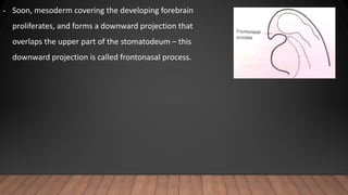
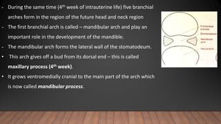
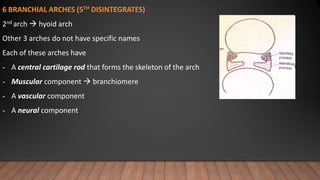

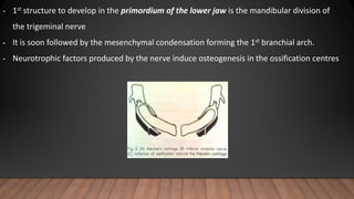


The document outlines the prenatal growth and development of the mandible, highlighting key stages from the 4th to 14th weeks of intrauterine life, including the roles of various branchial arches and the formation of Meckel's cartilage. It describes the processes of ossification and the morphological changes of the mandible during both prenatal and postnatal stages, detailing the influences of muscle growth and dental eruption on mandible development. Additionally, it discusses growth sites, the role of condylar cartilage, and the evolution of the mandible's shape and structure with age.