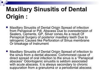
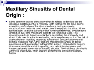
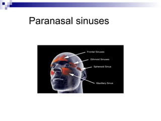
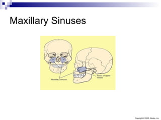
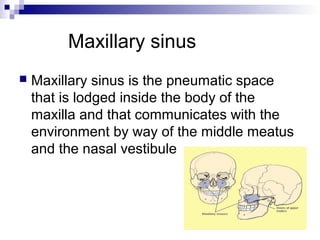
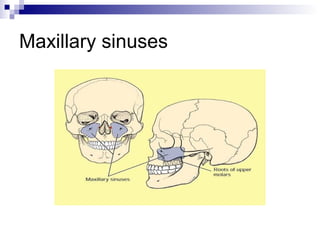
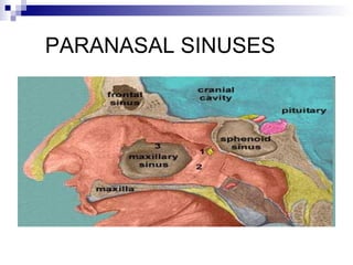
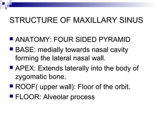
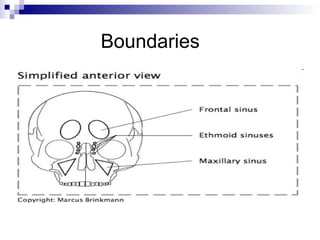
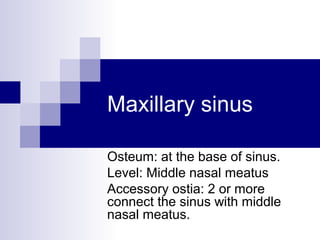
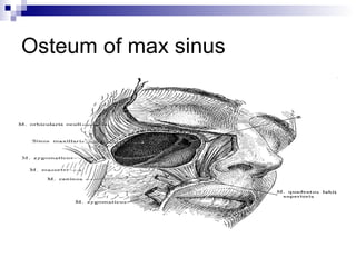
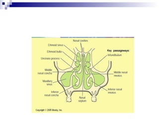
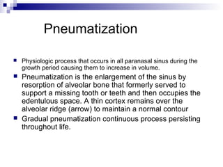
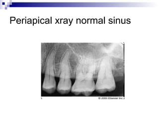
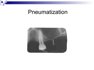
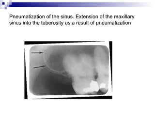
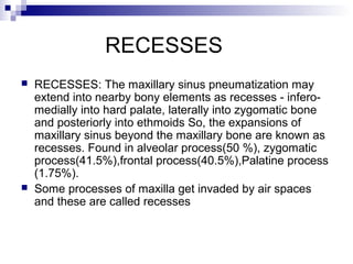
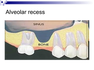
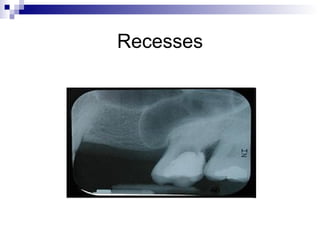
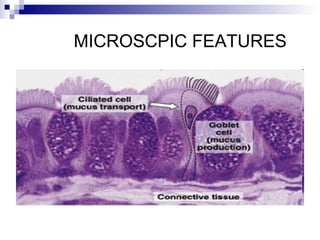
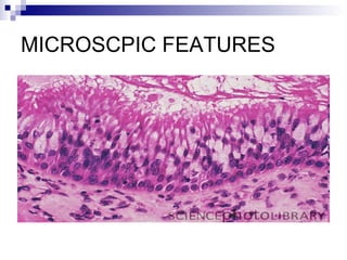
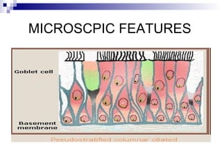
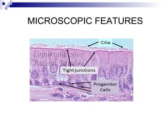
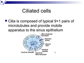
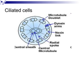
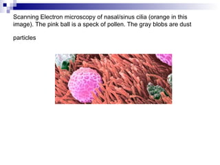
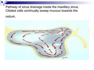
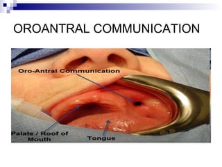
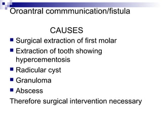
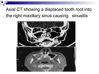
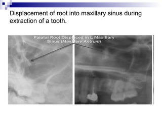
Paranasal sinuses
Paranasal sinuses
Paranasal sinuses
 Paranasal Sinuses (PNS) are air
containing bony spaces around the nasal
cavity. Usually lined by respiratory mucous
membrane of ciliated columnar epithelium
4 paired (bilateral) PNS are: Maxillary
Frontal, Sphenoidal ,Ethmoidal
Sinus
 LATIN word meaning a Fold or Pocket.
TYPES
named according to the bone in which they
lie.
1) The Maxillary sinuses: largest of the
paranasal sinuses ,are under the eyes in
the maxillary bones.
Maxillary Sinuses
Copyright © 2005, Mosby, Inc.
Types of paranasal sinuses
 2) The frontal sinuses: superior to eyes in
the frontal bone which forms the hard part
of the forehead.
FRONTAL SINUSES
Types of paranasal sinuses
 3) the ethmoid sinuses: which are formed
from several discrete air cells within the
ethmoid bone between the nose and eyes.
Ethmoid sinuses
Sphenoid sinus
 4) The sphenoid sinuses in the sphenoid
bone at the centre of skull base under the
pituitary gland.
Lateral Sinus Anatomy
Maxillary sinus
 Maxillary sinus is the pneumatic space
that is lodged inside the body of the
maxilla and that communicates with the
environment by way of the middle meatus
and the nasal vestibule
ANTRUM
 Maxillary sinus is also called “ maxillary
antrum” .ANTRUM IS A GREEK WORD
MEANING “CAVE”.
 ATTRIBUTED TO NATHIENEL
HIGHMOORE ENGLISH PHYSICIAN
1600.
Dr NATHANIEL HIGHMOORE
Maxillary sinuses
PARANASAL SINUSES
STRUCTURE OF MAXILLARY SINUS
 ANATOMY: FOUR SIDED PYRAMID
 BASE: medially towards nasal cavity
forming the lateral nasal wall.
 APEX: Extends laterally into the body of
zygomatic bone.
 ROOF( upper wall): Floor of the orbit.
 FLOOR: Alveolar process
PYRAMID
Boundaries
Walls of maxillary sinus
Occipitomental view
Size
 Adult maxillary sinus averages 34 mm in
anteroposterior direction
 Height= 33 mm
 Width= 23 mm.
 Volume of max sinus=15 to 20 ml.
Radiological view
Maxillary sinus
Osteum: at the base of sinus.
Level: Middle nasal meatus
Accessory ostia: 2 or more
connect the sinus with middle
nasal meatus.
Osteum of max sinus
Pneumatization
 Physiologic process that occurs in all paranasal sinus during the
growth period causing them to increase in volume.
 Pneumatization is the enlargement of the sinus by
resorption of alveolar bone that formerly served to
support a missing tooth or teeth and then occupies the
edentulous space. A thin cortex remains over the
alveolar ridge (arrow) to maintain a normal contour
 Gradual pneumatization continuous process persisting
throughout life.
PNEUMATIZATION
 EXPANSION OF MAXILLARY SINUS
GREATER BASALLY IMPINGING MORE
AND MORE ON ALVEOLAR PROCESS.
pneumatization???
 Pneumatization of alveolar process is
believed to result from disuse atrophy
initiated by tooth removal.
Periapical xray normal sinus
Pneumatization
Pneumatization of the sinus. Extension of the maxillary
sinus into the tuberosity as a result of pneumatization
RECESSES
 RECESSES: The maxillary sinus pneumatization may
extend into nearby bony elements as recesses - infero-
medially into hard palate, laterally into zygomatic bone
and posteriorly into ethmoids So, the expansions of
maxillary sinus beyond the maxillary bone are known as
recesses. Found in alveolar process(50 %), zygomatic
process(41.5%),frontal process(40.5%),Palatine process
(1.75%).
 Some processes of maxilla get invaded by air spaces
and these are called recesses
Alveolar recess
Recesses
 ZYGOMATIC RECESS= Superior alveolar nerves plus
vessels in proximity with sinus.
 Frontal Recess= invades and surrounds the contents of
infraorbital canal.
 Alveolopalatine recess; reduce the amount of bone
between dental apices and sinus space.most often
pneumatizes the floor of sinus adjacent to the roots of
the first molar.
 Fully developed alveolar recess= 3 depressions
separated by 2 incomplete bony septa.
 Anterior depression=premolar buds site
 Middle depression=molar buds, posterior depression=
third molar bud
Microscopic features
 3 layers surround the space of the max sinus :
 1) Epithelial Layer
 2) Basal lamina
 3) Subepithelial layer including the periosteum.
 Most numerous cells in max sinus are –
Columnar ciliated cells.
 Additional cells:Basal cells, columnar non
ciliated cells, mucus secreting goblet cells
Histology of max sinus
Epithelium
 Ciliated Pseudostratified, columnar
derived from olfactory epithelium of
middle meatus
 Most numerous cells in max sinus
epithelium Columnar ciliated cells.
 Additional cells:1)Basal cells 2)columnar
non ciliated cells 3)mucus secreting goblet
cells
MICROSCPIC FEATURES
MICROSCPIC FEATURES
MICROSCPIC FEATURES
MICROSCOPIC FEATURES
Ciliated cells
 Cilia is composed of typical 9+1 pairs of
microtubules and provide mobile
apparatus to the sinus epithelium
Ciliated cells
Scanning Electron microscopy of nasal/sinus cilia (orange in this
image). The pink ball is a speck of pollen. The gray blobs are dust
particles
Pathway of sinus drainage inside the maxillary sinus.
Ciliated cells continually sweep mucous towards the
ostium.
Ciliated cells
 The ciliated cells enclose the nucleus &
electron lucent cytoplasm with numerous
mitochondria & enzyme containing
organelles.
Ciliated cells
 The basal bodies serve as attachment of
ciliary microtubules.
 The cilia provide motile apparatus.
Ciliated cells
 By ciliary beating, the mucous blanket
lining the epithelial surface moves from
the interior of the sinus towards the nasal
cavity
Goblet cells
 Basal segment contains nucleus
 Goblet cells contain RER & SER along
with the Golgi apparatus all of which are
involved in the synthesis of secretory
substances
 This means that they contain all the
characteristics of secretory cells.
Goblet GLASS
GOBLET CELLS
Subepithelial layer
 Contain subepithelial glands and reach the sinus
lumen by way of excetory ducts.
 The glands contain both serous and mucous
acini i.e secrete serous as well as mucous
secretion.
 Myoepithelial cells surround the acini composed
of either both secretory cells
or a pure population of cells of either secretory
type.
Subepithelial layer
 The subepithelial layer also consists of
collagen bundles,fibroblasts,vessels and
nerves.
Composition
 serous secretion= water, neutral lipids,
proteins, carbohydrates.
 mucous secretion=compound
glycoprotein's and mucopolysaccharides
Subepithelial glands
 AUTONOMIC NERVOUS SYSTEM(ANS)
Control secretions from these glands.
Supplied to max sinus from max nerve
complex.
Functions Of Maxillary Sinus
 Warming/Humidification of air.
 Contribution to immune response i.e bactericidal
lysozyme .
 Lightening the skull
 Resonance to voice
 Assistance in regulation of intracranial pressure
 Enhance Facio-cranial resistance to shock
Nerve Supply
 Nerve Supply:
MAXILLARY DIVISION OF V
NERVE i.e V2
 1) Anterior, middle
and posterior superior
alveolar nerves,
 2) Infra orbital nerves
 3) greater palatine
nerve
Maxillary nerve
GREATER PALATINE NERVE
INNERVATION
 The innervation of the sinus is important from a
diagnostic standpoint. Post wall of the sinus
receives its supply from Posterior and Middle
Superior Alveolar nerves while anterior wall is by
Anterior Superior Alveolar Nerve. These nerves
travel enclosed in the wall of the sinus
innervating the related teeth (Wallace 1996).
Thus it is commonly seen that pain of the sinus
is mimicked as toothache and vice versa and is
difficult to distinguish
Arterial supply
 Major blood supply from branches of maxillary
artery
1) infraorbital artery
2) posterior superior dental artery
3) anterior superior dental artery
4) greater palatine
5) sphenopalatine
Smaller contribution from facial artery
both branches of external carotid artery.
EXTERNAL CAROTID
FACIAL ARTERY
SPHENOPALATINE ARTERY
Venous Drainage
 Venous Drainage: Via the Facial vein,
Sphenopalatine vein anteriorly and the
Pterygoid venous plexus posteriorly
Anterior, middle and superior dental veins
drain into the infra-orbital vein Pterygoid
plexus communicates with the cavernous
sinus by emissary veins
Venous Drainage
VENOUS DRAINAGE
CLINICAL IMPORTANCE
 The significance of the vascular drainage
of the sinus lies in the fact that apart from
the joining typical pathways in the maxilla
to the jugular veins, it can also drain
upward into the ethmoidal and frontal
sinuses and eventually reach the
cavernous sinus in the floor of the brain.
Spread of infections via this route is a
serious complication of maxillary sinus
infections
Lymph Drainage
 PREAURICULAR NODES
 Submandibular lymph node
 PAROTID node
 Facial node
CLINICAL CONSIDERATIONS
 Developmental anomalies
 Agenesis( complete absence of max sinus)
 Aplasia( altered development)
 Hypoplasia(underdevelopment)
 Supernumerary sinus(occurrence of 2
completely separated sinuses on the same side)
CLINICAL CONSIDERATIONS
 Pituitary gigantism: sinuses larger than normal
 Some congenital infections: sinuses smaller
than normal
e.g congenital syphilis
 Pathologically generated- functional and
systemic association
 Transfer of pathologic condition is through
Mechanical blood or lymphatic system.
Hypoplasia
Maxillary sinus hypoplasia presence of three of following four criteria:
 1. oval-shaped sinus
2. absence of pneumatization of the sinus
below the level of the nasal floor
 3. presence of medial wall of the sinus laterally
to a vertical line drawn tangentially to the
medial orbital border
 4. lateral extension of the sinus medial to a
vertical line drawn through the middle of the
orbit at the level of the infundibulum, in the
coronal plane
HYPOPLASIA
HYPOPLASIA OF MAXILLARY SINUS
MAXILLARY SINUS HYPOPLASIA
HYPOPLASIA OF MAXILLARY
SINUS
Clinical considerations
 Chronic infections of mucoperiosteal layer cause
neuralgia becoz of superior al n invovolvement.
 Neuralgia of maxillary nerve “tic doulorex”
may mimic sinus pain
 Non specific bacterial sinusistis
 Infections caused by
streptococci,staphylococci,pnemococci,virus of
common cold.
Clinical considerations/importance/implications
 1) Oroantral communication
accidental opening in the floor of the
antrum caused during extraction of
maxillary first molar which has a thin bone
separating the roots from the antrum.
a tract b/w oral cavity and max antrum not
lined by epithelium.
Fates of oro antral communication
 Either close spontaneouly
 Become epithelialized and persist as true
fistulae
OROANTRAL COMMUNICATION
Oroantral commmunication/fistula
CAUSES
 Surgical extraction of first molar
 Extraction of tooth showing
hypercementosis
 Radicular cyst
 Granuloma
 Abscess
Therefore surgical intervention necessary
EXTRACTION OF FIRST MOLAR
PERIAPICAL LESION OR
GRANULOMA CAUSING OAF
Pituitary gigantism
Tic doulourox( trigeminal neuralgia)
Malignant lesions
 Malignant lesions
adenocarcinoma
squamous cell carcinoma
osteosarcoma
fibrosarcoma
lymphosarcoma
Malignant lesions
 Primary manifestation in max teeth
1) pain
2) loosening
3) Supraeruption
4) bleeding in gingival tissue
MALIGNANT LESION
AXIAL VIEW CT SCAN
CORONAL VIEW CT SCAN
CT SCAN REPORT
CLINICAL
CONSIDERATIONS/IMPLICATIONS
 Infections introduced into the antrum
through periapical infections.
Infections introduced into the antrum
through periapical infections.
Maxillary sinusitis
Maxillary sinusitis
Signs symptoms
 When you begin to experience maxillary sinusitis you will
notice the following symptoms:
 • Nasal congestion
• Facial pain
• Differentiated facial sensations
• Night-time coughing
• Jawbone pain
• Teeth pain
• Runny nose
• Sinus pressure
• Chronic tooth aches
Axial CT showing a displaced tooth root into
the right maxillary sinus causing sinusitis
Maxillary Sinusitis of Dental
Origin :
 Maxillary Sinusitis of Dental Origin Spread of infection
from Periapical or Pdl. Abscess Due to overextension of
Sealers, Cements, GP, Silver cones As a result Of
Periapical Surgery of posterior maxillary teeth Due to
iatrogenic Causes like Perforation of Sinus membrane
Or breakage of Instrument
 Maxillary Sinusitis of Dental Origin Spread of infection to
the sinus from a dental abscess: Commonest cause of
direct spread of oral infection to the sinus is a “Periapical
abscess” Odontogenic sinusitis is seldom associated
with acute abscess. It is always secondary to chronic
suppuration from a granuloma or a periodontal abscess
Maxillary Sinusitis of Dental
Origin :
 Some common causes of maxillary sinusitis related to dentistry are the
iatrogenic displacement of a maxillary tooth root tip into the sinus during
extraction, perforation of the sinus membrane during exodontia,
andextrusion of materials used in root canal therapy into the sinus. When
teethadjacent to a lone-standing molar have been removed, alveolar bone
isresorbed over time mesial and distal to the remaining tooth. This
resorptionresults in thinner alveolar bone separating the oral cavity and
sinus. If ata later time the lone-standing molar requires extraction, the risk of
alveolarbone or maxillary tuberosity fracture with concomitant oroantral
commu-nication is high. Other oral and maxillofacial surgery or dental
procedures,such as maxillary orthognathic surgery, preprosthetic surgery,
sinusmembrane lifts and sinus grafting, and dental implant placement,
haveoccasionally been cited as causing sinusitis. The incidence of sinusitis
withthese procedures, when properly performed is almost nonexistent,
howev
Displacement of root into maxillary sinus during
extraction of a tooth.
Maxillary sinus for 2nd year BDS

Maxillary sinus for 2nd year BDS

  • 7.
  • 8.
  • 9.
    Paranasal sinuses  ParanasalSinuses (PNS) are air containing bony spaces around the nasal cavity. Usually lined by respiratory mucous membrane of ciliated columnar epithelium 4 paired (bilateral) PNS are: Maxillary Frontal, Sphenoidal ,Ethmoidal
  • 10.
    Sinus  LATIN wordmeaning a Fold or Pocket.
  • 11.
    TYPES named according tothe bone in which they lie. 1) The Maxillary sinuses: largest of the paranasal sinuses ,are under the eyes in the maxillary bones.
  • 12.
  • 13.
    Types of paranasalsinuses  2) The frontal sinuses: superior to eyes in the frontal bone which forms the hard part of the forehead.
  • 14.
  • 15.
    Types of paranasalsinuses  3) the ethmoid sinuses: which are formed from several discrete air cells within the ethmoid bone between the nose and eyes.
  • 16.
  • 17.
    Sphenoid sinus  4)The sphenoid sinuses in the sphenoid bone at the centre of skull base under the pituitary gland.
  • 18.
  • 19.
    Maxillary sinus  Maxillarysinus is the pneumatic space that is lodged inside the body of the maxilla and that communicates with the environment by way of the middle meatus and the nasal vestibule
  • 20.
    ANTRUM  Maxillary sinusis also called “ maxillary antrum” .ANTRUM IS A GREEK WORD MEANING “CAVE”.  ATTRIBUTED TO NATHIENEL HIGHMOORE ENGLISH PHYSICIAN 1600.
  • 21.
  • 22.
  • 23.
  • 24.
    STRUCTURE OF MAXILLARYSINUS  ANATOMY: FOUR SIDED PYRAMID  BASE: medially towards nasal cavity forming the lateral nasal wall.  APEX: Extends laterally into the body of zygomatic bone.  ROOF( upper wall): Floor of the orbit.  FLOOR: Alveolar process
  • 25.
  • 26.
  • 27.
  • 28.
  • 29.
    Size  Adult maxillarysinus averages 34 mm in anteroposterior direction  Height= 33 mm  Width= 23 mm.  Volume of max sinus=15 to 20 ml.
  • 30.
  • 31.
    Maxillary sinus Osteum: atthe base of sinus. Level: Middle nasal meatus Accessory ostia: 2 or more connect the sinus with middle nasal meatus.
  • 32.
  • 34.
    Pneumatization  Physiologic processthat occurs in all paranasal sinus during the growth period causing them to increase in volume.  Pneumatization is the enlargement of the sinus by resorption of alveolar bone that formerly served to support a missing tooth or teeth and then occupies the edentulous space. A thin cortex remains over the alveolar ridge (arrow) to maintain a normal contour  Gradual pneumatization continuous process persisting throughout life.
  • 35.
    PNEUMATIZATION  EXPANSION OFMAXILLARY SINUS GREATER BASALLY IMPINGING MORE AND MORE ON ALVEOLAR PROCESS.
  • 36.
    pneumatization???  Pneumatization ofalveolar process is believed to result from disuse atrophy initiated by tooth removal.
  • 37.
  • 38.
  • 39.
    Pneumatization of thesinus. Extension of the maxillary sinus into the tuberosity as a result of pneumatization
  • 40.
    RECESSES  RECESSES: Themaxillary sinus pneumatization may extend into nearby bony elements as recesses - infero- medially into hard palate, laterally into zygomatic bone and posteriorly into ethmoids So, the expansions of maxillary sinus beyond the maxillary bone are known as recesses. Found in alveolar process(50 %), zygomatic process(41.5%),frontal process(40.5%),Palatine process (1.75%).  Some processes of maxilla get invaded by air spaces and these are called recesses
  • 41.
  • 42.
  • 43.
     ZYGOMATIC RECESS=Superior alveolar nerves plus vessels in proximity with sinus.  Frontal Recess= invades and surrounds the contents of infraorbital canal.  Alveolopalatine recess; reduce the amount of bone between dental apices and sinus space.most often pneumatizes the floor of sinus adjacent to the roots of the first molar.  Fully developed alveolar recess= 3 depressions separated by 2 incomplete bony septa.  Anterior depression=premolar buds site  Middle depression=molar buds, posterior depression= third molar bud
  • 45.
    Microscopic features  3layers surround the space of the max sinus :  1) Epithelial Layer  2) Basal lamina  3) Subepithelial layer including the periosteum.  Most numerous cells in max sinus are – Columnar ciliated cells.  Additional cells:Basal cells, columnar non ciliated cells, mucus secreting goblet cells
  • 46.
  • 47.
    Epithelium  Ciliated Pseudostratified,columnar derived from olfactory epithelium of middle meatus  Most numerous cells in max sinus epithelium Columnar ciliated cells.  Additional cells:1)Basal cells 2)columnar non ciliated cells 3)mucus secreting goblet cells
  • 48.
  • 49.
  • 50.
  • 51.
  • 52.
    Ciliated cells  Ciliais composed of typical 9+1 pairs of microtubules and provide mobile apparatus to the sinus epithelium
  • 53.
  • 54.
    Scanning Electron microscopyof nasal/sinus cilia (orange in this image). The pink ball is a speck of pollen. The gray blobs are dust particles
  • 55.
    Pathway of sinusdrainage inside the maxillary sinus. Ciliated cells continually sweep mucous towards the ostium.
  • 56.
    Ciliated cells  Theciliated cells enclose the nucleus & electron lucent cytoplasm with numerous mitochondria & enzyme containing organelles.
  • 57.
    Ciliated cells  Thebasal bodies serve as attachment of ciliary microtubules.  The cilia provide motile apparatus.
  • 58.
    Ciliated cells  Byciliary beating, the mucous blanket lining the epithelial surface moves from the interior of the sinus towards the nasal cavity
  • 59.
    Goblet cells  Basalsegment contains nucleus  Goblet cells contain RER & SER along with the Golgi apparatus all of which are involved in the synthesis of secretory substances  This means that they contain all the characteristics of secretory cells.
  • 60.
  • 61.
  • 62.
    Subepithelial layer  Containsubepithelial glands and reach the sinus lumen by way of excetory ducts.  The glands contain both serous and mucous acini i.e secrete serous as well as mucous secretion.  Myoepithelial cells surround the acini composed of either both secretory cells or a pure population of cells of either secretory type.
  • 63.
    Subepithelial layer  Thesubepithelial layer also consists of collagen bundles,fibroblasts,vessels and nerves.
  • 64.
    Composition  serous secretion=water, neutral lipids, proteins, carbohydrates.  mucous secretion=compound glycoprotein's and mucopolysaccharides
  • 65.
    Subepithelial glands  AUTONOMICNERVOUS SYSTEM(ANS) Control secretions from these glands. Supplied to max sinus from max nerve complex.
  • 67.
    Functions Of MaxillarySinus  Warming/Humidification of air.  Contribution to immune response i.e bactericidal lysozyme .  Lightening the skull  Resonance to voice  Assistance in regulation of intracranial pressure  Enhance Facio-cranial resistance to shock
  • 68.
    Nerve Supply  NerveSupply: MAXILLARY DIVISION OF V NERVE i.e V2  1) Anterior, middle and posterior superior alveolar nerves,  2) Infra orbital nerves  3) greater palatine nerve
  • 69.
  • 70.
  • 71.
    INNERVATION  The innervationof the sinus is important from a diagnostic standpoint. Post wall of the sinus receives its supply from Posterior and Middle Superior Alveolar nerves while anterior wall is by Anterior Superior Alveolar Nerve. These nerves travel enclosed in the wall of the sinus innervating the related teeth (Wallace 1996). Thus it is commonly seen that pain of the sinus is mimicked as toothache and vice versa and is difficult to distinguish
  • 72.
    Arterial supply  Majorblood supply from branches of maxillary artery 1) infraorbital artery 2) posterior superior dental artery 3) anterior superior dental artery 4) greater palatine 5) sphenopalatine Smaller contribution from facial artery both branches of external carotid artery.
  • 73.
  • 74.
  • 75.
  • 76.
    Venous Drainage  VenousDrainage: Via the Facial vein, Sphenopalatine vein anteriorly and the Pterygoid venous plexus posteriorly Anterior, middle and superior dental veins drain into the infra-orbital vein Pterygoid plexus communicates with the cavernous sinus by emissary veins
  • 77.
  • 78.
  • 79.
    CLINICAL IMPORTANCE  Thesignificance of the vascular drainage of the sinus lies in the fact that apart from the joining typical pathways in the maxilla to the jugular veins, it can also drain upward into the ethmoidal and frontal sinuses and eventually reach the cavernous sinus in the floor of the brain. Spread of infections via this route is a serious complication of maxillary sinus infections
  • 80.
    Lymph Drainage  PREAURICULARNODES  Submandibular lymph node  PAROTID node  Facial node
  • 82.
  • 83.
    CLINICAL CONSIDERATIONS  Developmentalanomalies  Agenesis( complete absence of max sinus)  Aplasia( altered development)  Hypoplasia(underdevelopment)  Supernumerary sinus(occurrence of 2 completely separated sinuses on the same side)
  • 84.
    CLINICAL CONSIDERATIONS  Pituitarygigantism: sinuses larger than normal  Some congenital infections: sinuses smaller than normal e.g congenital syphilis  Pathologically generated- functional and systemic association  Transfer of pathologic condition is through Mechanical blood or lymphatic system.
  • 85.
    Hypoplasia Maxillary sinus hypoplasiapresence of three of following four criteria:  1. oval-shaped sinus 2. absence of pneumatization of the sinus below the level of the nasal floor  3. presence of medial wall of the sinus laterally to a vertical line drawn tangentially to the medial orbital border  4. lateral extension of the sinus medial to a vertical line drawn through the middle of the orbit at the level of the infundibulum, in the coronal plane
  • 86.
  • 87.
  • 88.
  • 89.
  • 90.
    Clinical considerations  Chronicinfections of mucoperiosteal layer cause neuralgia becoz of superior al n invovolvement.  Neuralgia of maxillary nerve “tic doulorex” may mimic sinus pain  Non specific bacterial sinusistis  Infections caused by streptococci,staphylococci,pnemococci,virus of common cold.
  • 91.
    Clinical considerations/importance/implications  1)Oroantral communication accidental opening in the floor of the antrum caused during extraction of maxillary first molar which has a thin bone separating the roots from the antrum. a tract b/w oral cavity and max antrum not lined by epithelium.
  • 92.
    Fates of oroantral communication  Either close spontaneouly  Become epithelialized and persist as true fistulae
  • 93.
  • 94.
    Oroantral commmunication/fistula CAUSES  Surgicalextraction of first molar  Extraction of tooth showing hypercementosis  Radicular cyst  Granuloma  Abscess Therefore surgical intervention necessary
  • 95.
  • 97.
  • 98.
  • 99.
  • 100.
    Malignant lesions  Malignantlesions adenocarcinoma squamous cell carcinoma osteosarcoma fibrosarcoma lymphosarcoma
  • 101.
    Malignant lesions  Primarymanifestation in max teeth 1) pain 2) loosening 3) Supraeruption 4) bleeding in gingival tissue
  • 102.
  • 103.
  • 104.
  • 105.
  • 106.
    CLINICAL CONSIDERATIONS/IMPLICATIONS  Infections introducedinto the antrum through periapical infections.
  • 107.
    Infections introduced intothe antrum through periapical infections.
  • 108.
  • 109.
  • 110.
    Signs symptoms  Whenyou begin to experience maxillary sinusitis you will notice the following symptoms:  • Nasal congestion • Facial pain • Differentiated facial sensations • Night-time coughing • Jawbone pain • Teeth pain • Runny nose • Sinus pressure • Chronic tooth aches
  • 111.
    Axial CT showinga displaced tooth root into the right maxillary sinus causing sinusitis
  • 112.
    Maxillary Sinusitis ofDental Origin :  Maxillary Sinusitis of Dental Origin Spread of infection from Periapical or Pdl. Abscess Due to overextension of Sealers, Cements, GP, Silver cones As a result Of Periapical Surgery of posterior maxillary teeth Due to iatrogenic Causes like Perforation of Sinus membrane Or breakage of Instrument  Maxillary Sinusitis of Dental Origin Spread of infection to the sinus from a dental abscess: Commonest cause of direct spread of oral infection to the sinus is a “Periapical abscess” Odontogenic sinusitis is seldom associated with acute abscess. It is always secondary to chronic suppuration from a granuloma or a periodontal abscess
  • 113.
    Maxillary Sinusitis ofDental Origin :  Some common causes of maxillary sinusitis related to dentistry are the iatrogenic displacement of a maxillary tooth root tip into the sinus during extraction, perforation of the sinus membrane during exodontia, andextrusion of materials used in root canal therapy into the sinus. When teethadjacent to a lone-standing molar have been removed, alveolar bone isresorbed over time mesial and distal to the remaining tooth. This resorptionresults in thinner alveolar bone separating the oral cavity and sinus. If ata later time the lone-standing molar requires extraction, the risk of alveolarbone or maxillary tuberosity fracture with concomitant oroantral commu-nication is high. Other oral and maxillofacial surgery or dental procedures,such as maxillary orthognathic surgery, preprosthetic surgery, sinusmembrane lifts and sinus grafting, and dental implant placement, haveoccasionally been cited as causing sinusitis. The incidence of sinusitis withthese procedures, when properly performed is almost nonexistent, howev
  • 114.
    Displacement of rootinto maxillary sinus during extraction of a tooth.

Editor's Notes

  • #13 The maxillary sinuses appear rectangular in the lateral image but they are really pyramidal in shape and have 3 walls. The apices project inferiorly and laterally There are several conic elevations at the floor of the maxillary sinuses for roots of the first and second molar teeth. Occasionally these roots can allow infections originating int eh teeth to travel to the sinuses.
  • #50 Maxillary sinus membrane. Light micrograph of a section through a mucous membrane from one of the two paired maxillary sinuses. The sinuses are the spaces in the facial bones of the skull, and the maxillary sinuses are found in the upper jaw bones (maxillae). The mucous membrane (mucosa) comprises columnar epithelial (surface) cells (across top) that include many goblet cells that secrete mucus. This outer layer is supported by a richly vascular (containing many blood vessels) layer called the lamina propria (across bottom). The mucosa serves to trap particles in inspired air, and also helps to humidify and heat inspired air
  • #108 Odontogenic periapical pathology originating from the premolar tooth has invaded the floor of the maxillarysinus (arrow). There is associated reactive sinus mucosal thickening