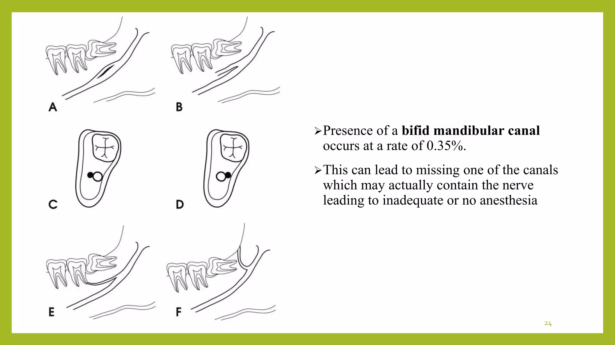
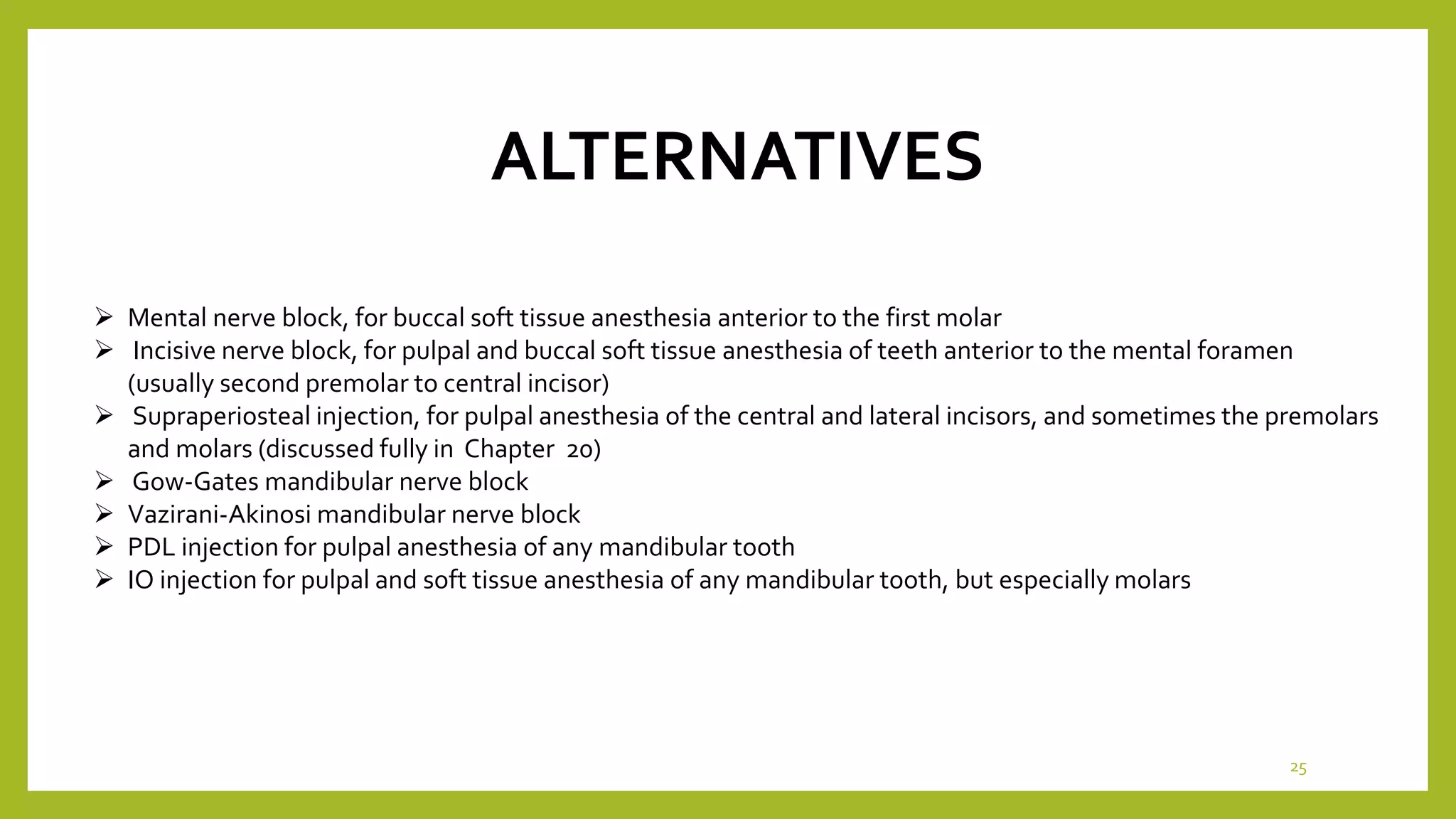
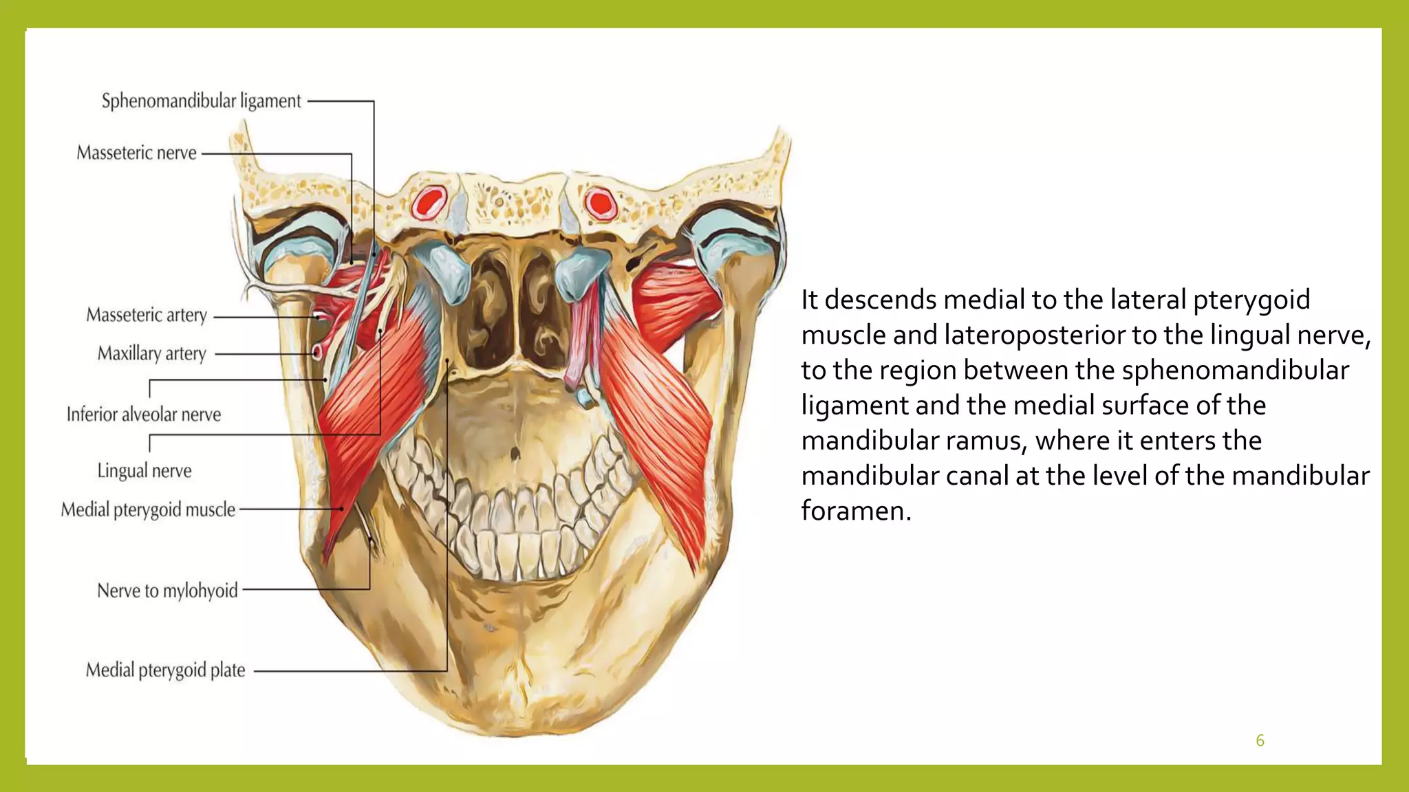
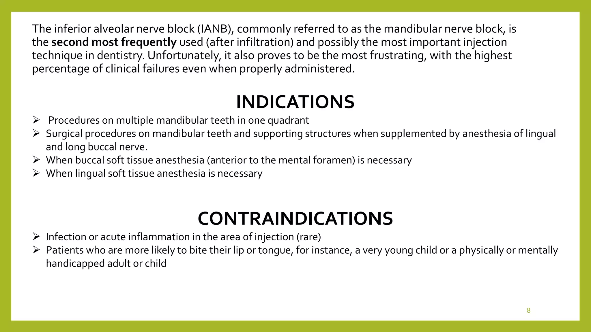
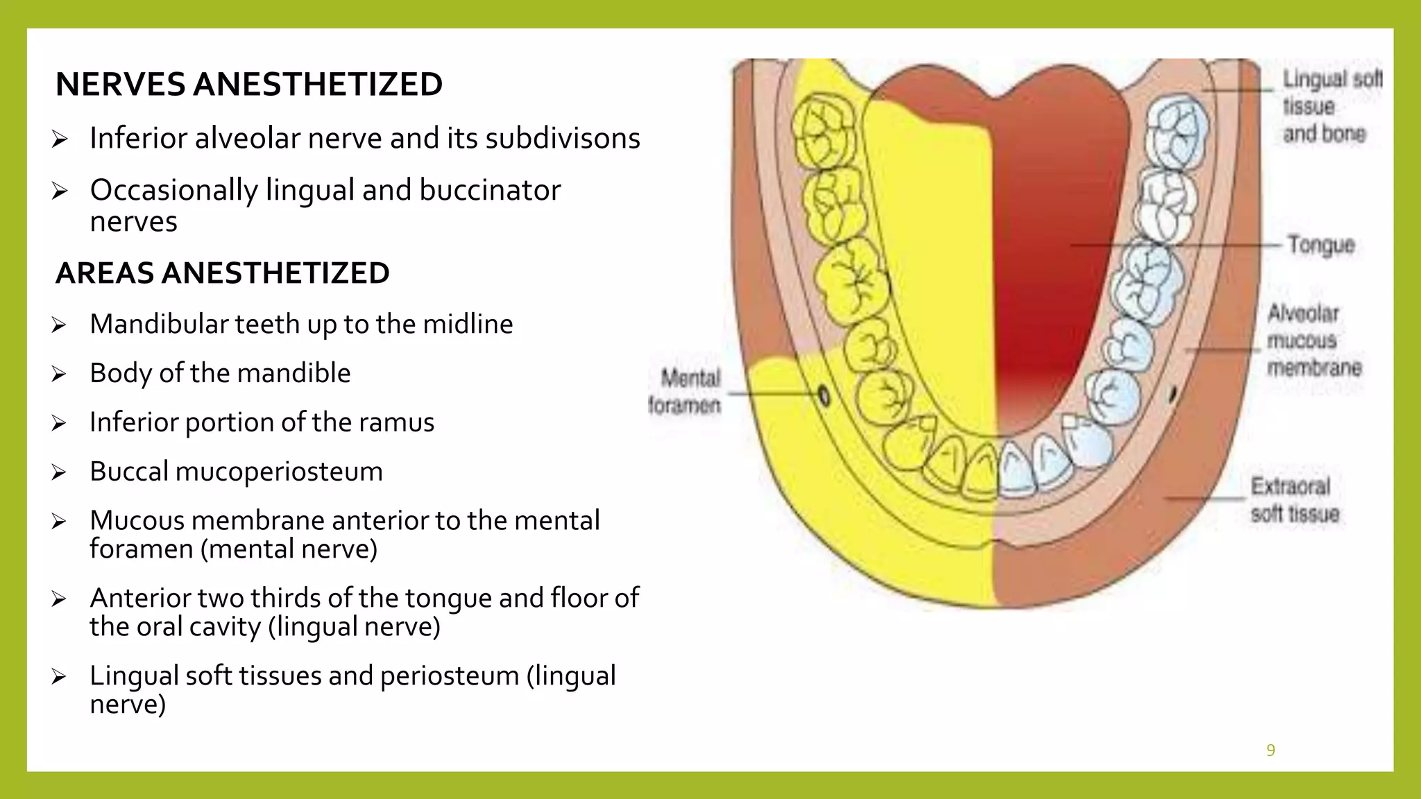
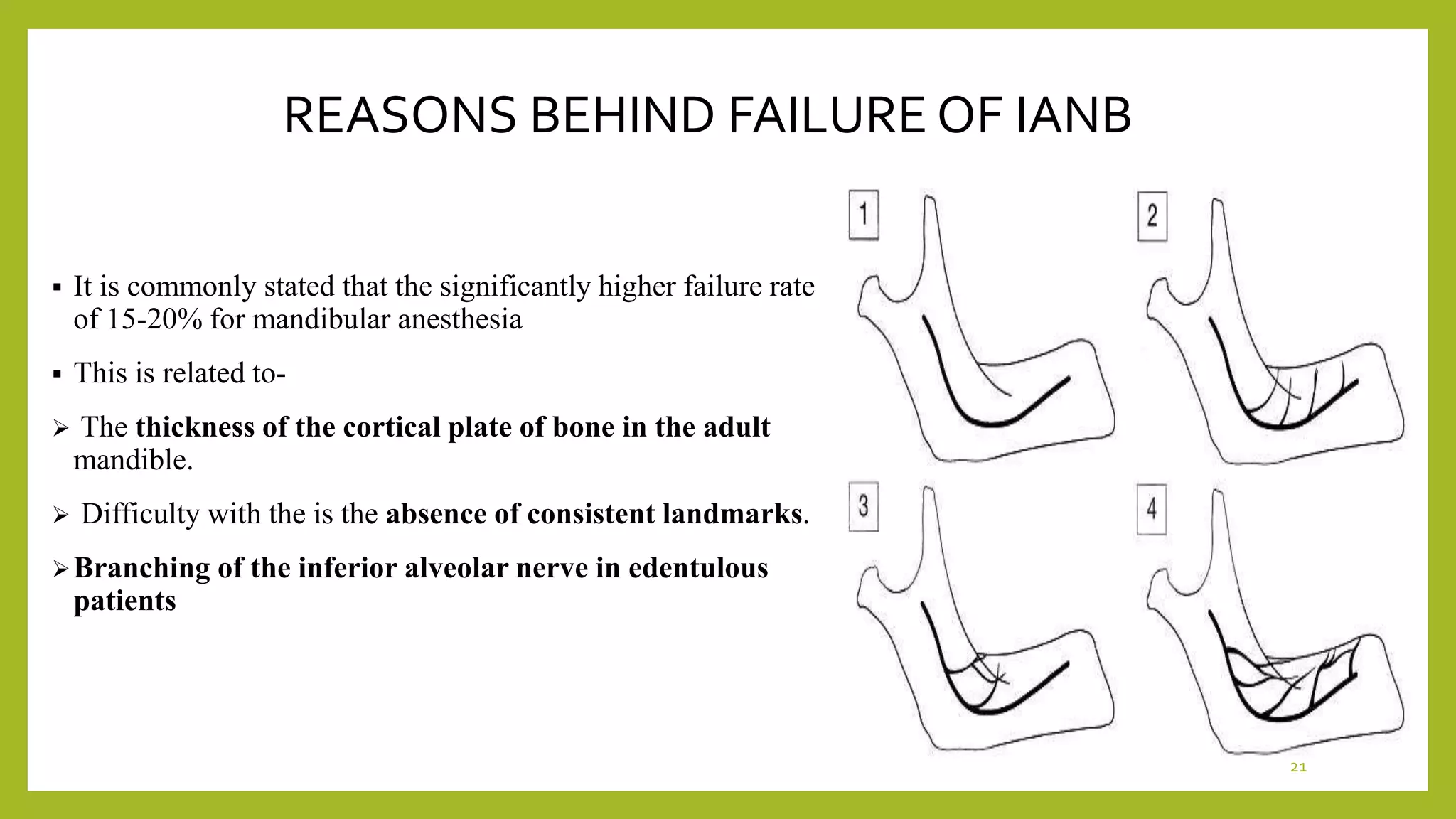
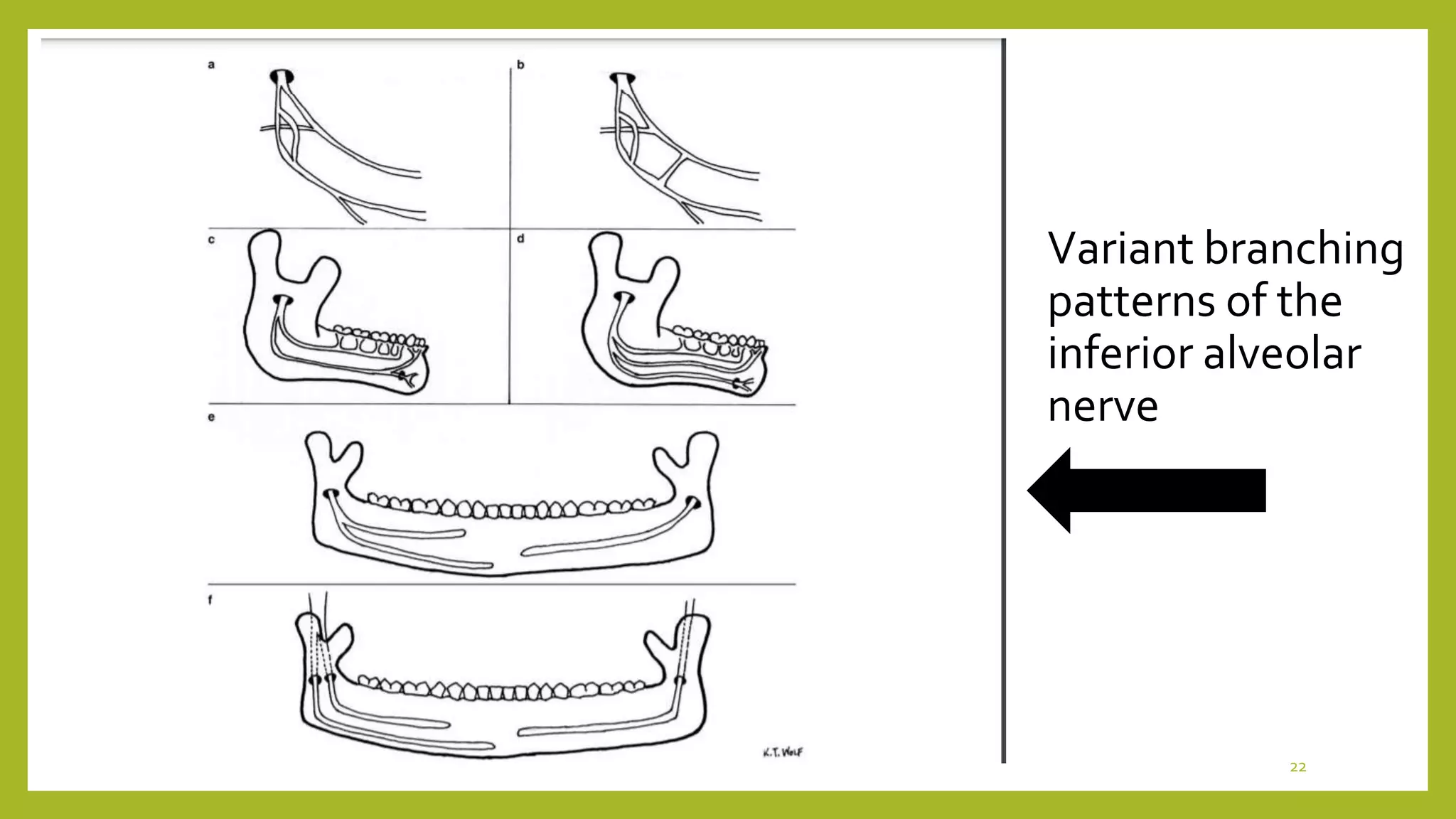
2. CONTENTS
Introduction
Indications and contraindications
Nerves and regions anesthesize
Landmarks
Techniques
Procedure
Signs and symptoms
Complications
Reasons of failure
2
3. Three large branches arise from trigeminal nerve V1-
Ophthalmic nerve, V2- Maxillary nerve, V3- Mandibular nerve
The third division V3- Mandibular nerve of the trigeminal nerve
gives off branches in three areas from the -
BRANCHES FROM UNDIVIDED NERVE BRANCHES FROM DIVIDED NERVE
• NERVUS SPINOSUS
• NERVE TO INTERNAL PTERYGOID MUSCLE
• ANTERIOR DIVISION
• POSTERIOR DIVISION
3
TRIGEMINAL NERVE
4. The posterior division of
trigeminal nerve is primarily
sensory with a small motor
component.
It descends for a short
distance downward and
medial to the lateral pterygoid
muscle, at which point it
branches into the
• Auriculotemporal Nerve
• Lingual Nerve
• Inferior alveolar nerves.
4
6. It descends medial to the lateral pterygoid
muscle and lateroposterior to the lingual nerve,
to the region between the sphenomandibular
ligament and the medial surface of the
mandibular ramus, where it enters the
mandibular canal at the level of the mandibular
foramen.
6
7. The nerve, artery, and vein travel anteriorly in the
mandibular canal as far forward as the mental
foramen, where the nerve divides into its
terminal branches near the apex of second
premolar into-
the Incisive nerve
the Mental nerve.
7
8. The inferior alveolar nerve block (IANB), commonly referred to as the mandibular nerve block, is
the second most frequently used (after infiltration) and possibly the most important injection
technique in dentistry. Unfortunately, it also proves to be the most frustrating, with the highest
percentage of clinical failures even when properly administered.
INDICATIONS
Procedures on multiple mandibular teeth in one quadrant
Surgical procedures on mandibular teeth and supporting structures when supplemented by anesthesia of lingual
and long buccal nerve.
When buccal soft tissue anesthesia (anterior to the mental foramen) is necessary
When lingual soft tissue anesthesia is necessary
CONTRAINDICATIONS
Infection or acute inflammation in the area of injection (rare)
Patients who are more likely to bite their lip or tongue, for instance, a very young child or a physically or mentally
handicapped adult or child
8
9. NERVES ANESTHETIZED
Inferior alveolar nerve and its subdivisons
Occasionally lingual and buccinator
nerves
AREAS ANESTHETIZED
Mandibular teeth up to the midline
Body of the mandible
Inferior portion of the ramus
Buccal mucoperiosteum
Mucous membrane anterior to the mental
foramen (mental nerve)
Anterior two thirds of the tongue and floor of
the oral cavity (lingual nerve)
Lingual soft tissues and periosteum (lingual
nerve)
9
10. LANDMARKS
Muccobuccal fold
Anterior border of ramus of mandible
External oblique ridge
Retromolar triangle
Internal oblique ridge
Pterygomandibular ligament
Buccal sucking pad
Pterygomandibular space
AREA OF INSERTION
Mucous membrane on the medial (lingual) side of the mandibular ramus, at the
intersection of two lines
One horizontal- representing the height of needle insertion,
Other vertical- representing the anteroposterior plane of injection
10
12. PROCEDURE
A long dental needle is recommended for the adult patient. A 25-gauge or a 27 gauge long needle is preferred.
a Assume the correct position.
(1)For a right IANB, a right-handed administrator should sit at the 8 o'clock position facing the patient
(2)For a left IANB, a right-handed administrator should sit at the 10 o'clock position facing in the same direction as the
patient
b Position the patient supine (recommended) or semisupine (if necessary).
The mouth should be opened wide to allow greater visibility of, and access to, the injection site.
c Locate the needle penetration (injection) site.
The posterior border of the mandibular ramus can be approximated intraorally by using the pterygomandibular raphe
as it turns superiorly toward the maxilla.
12
14. Three parameters must be considered during administration of IANB
The height of the injection
The anteroposterior placement of the needle (which helps to locate a precise needle entry point)
The depth of penetration (which determines the location of the inferior alveolar nerve)
14
15. HEIGHT OF INJECTION
Place the index finger or the thumb of your left hand in the coronoid notch
(a) An imaginary line extends posteriorly from the fingertip in the coronoid notch to the deepest part of the pterygomandibular
raphe (as it turns vertically upward toward the maxilla), determining the height of injection.
(b) The finger on the coronoid notch is used to pull the tissues laterally, stretching them over the injection site,
(c) The needle insertion point lies three fourths of the anteroposterior distance from the coronoid notch back to the deepest part
of the pterygomandibular raphe .
t
15
16. Site of needle insertion Verification of length of needle entry from the
anterior border of the ramus
Barrel of syringe brought to contralateral side and the needle
closeness to the bone is verified and solution deposited
16
17. TECHNIQUE IN PEDIATRIC PATIENT
The mandibular foramen is situated at a level lower to
the occulusal plane of primary teeth in pediatric
patient.
Hence the injection is made slightly lower and more
posteriorly than adult patients.
17
19. SIGNSAND SYMPTOMS
SUBJECTIVE SYMPTOMS
• Tingling and numbness of lower lip
• After the lingual nerve is affected numbness is felt on the tip of the tongue
OBJECTIVE SYMPTOMS
• Instrumentation necessary to demonstrate absence of pain sensation
19
20. DISADVANTAGES
One injection provides a wide area
of anesthesia - useful for quadrant
dentistry.
Wide area of anesthesia (not indicated for localized
procedures)
Rate of inadequate anesthesia (31% to 81%)
Intraoral landmarks not consistently reliable
Positive aspiration (10% to 15%, highest of all intraoral
injection techniques)
Lingual and lower lip anesthesia, discomfiting to many
patients and possibly dangerous (self-inflicted soft
tissue trauma) for certain individuals
Partial anesthesia possible where a bifid inferior
alveolar nerve and bifid mandibular canals are present;
cross-innervation in lower anterior region
ADVANTAGES
20
21. REASONS BEHIND FAILURE OF IANB
It is commonly stated that the significantly higher failure rate
of 15-20% for mandibular anesthesia
This is related to-
The thickness of the cortical plate of bone in the adult
mandible.
Difficulty with the is the absence of consistent landmarks.
Branching of the inferior alveolar nerve in edentulous
patients
21
23. Studies have shown the patients who are more anxious prior to the procedure; experience a
higher amount of pain.
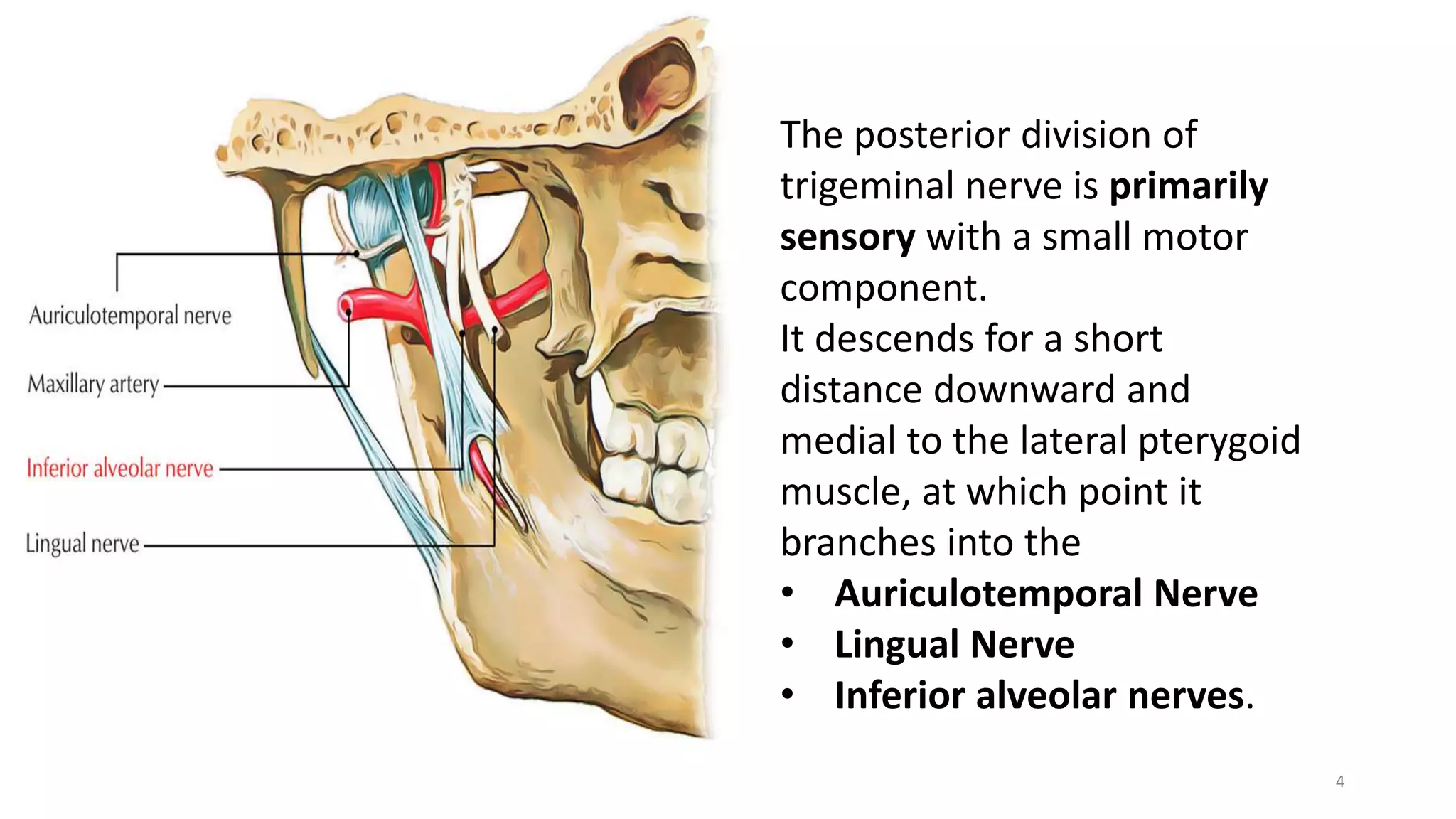
The mandibular hard tissue and soft tissue is supplied by a plexus of nerves. This plexus,
may allow sensation even if primary inferior alveolar nerve is blocked. In 1020% of the
cases, Mylohyoid nerve provides accessory innervation to mandibular molars
TYPE OF ANESTHETIC SOLUTION USED- Cohen et al. showed that 3% mepivacaine
is as effective as 2% lidocaine with 1:100 000 epinepthrine in achieving pulpal anaesthesia
with IANB.
Pulpal inflammation and abscess is a major problem when introducing anesthesia.[11]
Studies have shown changes in the impulse generation of nerve fibres in presence of
inflammation and also suggest changes in peripheral sensory fibers in presence of
inflammation.
23
24. Presence of a bifid mandibular canal
occurs at a rate of 0.35%.
This can lead to missing one of the canals
which may actually contain the nerve
leading to inadequate or no anesthesia
24
25. ALTERNATIVES
Mental nerve block, for buccal soft tissue anesthesia anterior to the first molar
Incisive nerve block, for pulpal and buccal soft tissue anesthesia of teeth anterior to the mental foramen
(usually second premolar to central incisor)
Supraperiosteal injection, for pulpal anesthesia of the central and lateral incisors, and sometimes the premolars
and molars (discussed fully in Chapter 20)
Gow-Gates mandibular nerve block
Vazirani-Akinosi mandibular nerve block
PDL injection for pulpal anesthesia of any mandibular tooth
IO injection for pulpal and soft tissue anesthesia of any mandibular tooth, but especially molars
25
26. Failure of inferior alveolar nerve block Exploring the alternatives- GAUTAM A. MADAN, M.D.S.; SONAL G. MADAN, M.D.S.; ARJUN D. MADAN, M.D.S. 26
27. REFERENCES
Handbook of Local Anesthesia 6th ed. Stanley F. Malamed, DDS
Monheims – local anesthesia and pain control in dental practice- C RICHARD BENNET
Sicher and dubrul- Oral Anatomy
Articles-
A basic review on the inferior alveolar nerve block techniques Hesham Khalil- Department of
Maxillofacial Surgery, College of Dentistry, King Saud University, Riyadh, Saudi Arabia
FAILURE OF INFERIOR ALVEOLAR NERVE BLOCK (IANB) AND TECHNIQUES TO AVOID
IT. - Dr. Harsh Rajvanshi, 2Dr. Sandra Ernest, 3Dr. Hafsa Effendi, 4Dr. Sarah Afridi, 5Dr. Madhur
Chhabra, 6Dr. Navneet Kaur
Inferior alveolar nerve block: Alternative technique- K. Thangavelu, R. Kannan, and N. Senthil Kumar
Failure of inferior alveolar nerve block Exploring the alternatives- GAUTAM A. MADAN, M.D.S.;
SONAL G. MADAN, M.D.S.; ARJUN D. MADAN, M.D.S.
Variant Inferior Alveolar Nerves and Implications for Local Anesthesia Kevin T. Wolf, BS,* Everett J.
Brokaw, BA,* Andrea Bell, DMD, MS,† and Anita Joy, BDS, PhD‡
27
The inferior alveolar nerve is the The incisive nerve remains within the mandibular canal and forms a nerve plexus that innervates the pulpal tissues of the mandibular first premolar, canine, and incisors via the dental branches.
A- This imaginary line should be parallel to the occlusal plane of the mandibular molar teeth.
In most patients, this line lies 6 to 10 mm above the occlusal plane
B- making them taut, and enabling needle insertion to be less traumatic, while providing better visibility.
C- The line should begin at the midpoint of the notch and terminate at the deepest (most posterior) portion of the pterygomandibular raphe as the raphe bends vertically upward toward the palate.
The position of the mandibular foramen changes during growth. However, the mandibular foramen is below the occlusive plane in children. The foramen is always situated on the line, where the ramus is narrowest, two‐thirds of the way back from the anterior concavity.
Type 1: Presence of one single trunk with no branching.
Type 2: Presence of a series of separate nerve branches (most common type).
Type 3: Presence of a molar plexus.
Type 4: Presence of proximal and distal plexuses.
Indeed it is generally acknowledged that mandibular infiltration is successful in cases where the patient has a full primary dentition. Once a mixed dentition develops, it is a general rule of teaching that the mandibular cortical plate of bone has thickened to the degree that infiltration might not be effective, leading to the recommendation that “mandibular block” techniques should now be employed.
It has been reported failure rates for the IANB are commonly high, ranging from 31% and 41% in mandibular second and first molars to 42%, 38%, and 46% in second and first premolars and canines, respectively, 9 and 81% in lateral incisors.
Summary diagram indicating variant branching patterns of the inferior alveolar nerve. (a) The inferior alveolar nerve can split high in the infratemporal fossa and then reunite to enter the mandibular canal as a single nerve. (b) During its course in the infratemporal fossa, the inferior alveolar nerve may be connected via small nerve branches to the lingual nerve. (c) A single inferior alveolar nerve entering the mandibular canal may course through the mandible as 2 distinct branches. These branches may be located within a single mandibular canal, or within 2 independent mandibular canals. (d) A single inferior alveolar nerve entering the mandibular canal may course through the mandible as 3 distinct branches. (e) Variations of the inferior alveolar nerve within the mandible may be unilateral. (f) A unilateral presentation of the inferior alveolar nerve may include a bifid nerve entering the mandibular canal as 2 distinct nerves, and continuing to course through the mandible as independent nerves.
6 The incidence of variations associated with the anatomy of the mandibular nerve and its branches necessitate that clinicians adopt alternate techniques of local anesthesia in order to achieve expected results. Alternate techniques to achieve anesthesia of the inferior alveolar nerve may include delivery of the anesthetic solution at a higher anatomical level in order to sufficiently anesthetize all branches of the mandibular nerve, including any additional inferior alveolar branches that may be present.57,58 Specific techniques that target the lingual nerve could also be used in situations where collateral anatomical connections between the inferior alveolar and lingual nerves are suspected. Preoperative imaging evaluation can provide evidence of such variations in the dental office, and the growing use of cone beam CT imaging for routine diagnostic purposes can increase the clinician’s ability to identify and manage such variations in the population
Diagram of configuration of the bifid mandibular canal is classified into four types as follows by Naitoh et al
1) The forward canal included with confluence (A)
or without confluence (B).
2) The bucco-lingual canal from the buccal or lingual wall (C and D).
3) The dental canal reached to the root apex (E).
4) The retromolar canal branched to the retromolar foramen (F).