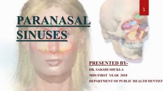
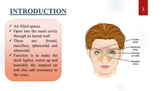
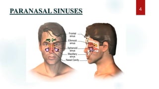
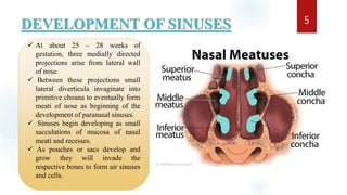
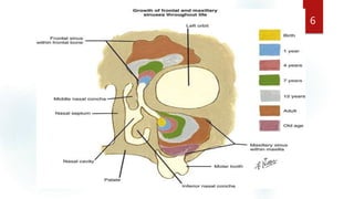
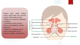
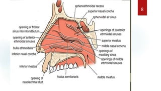
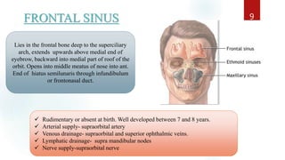
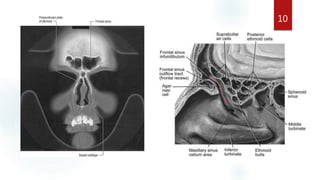
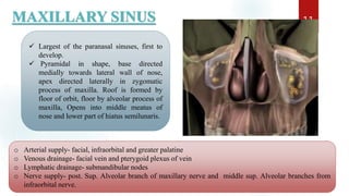
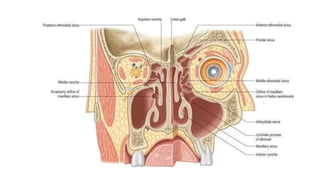
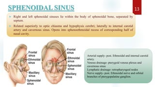
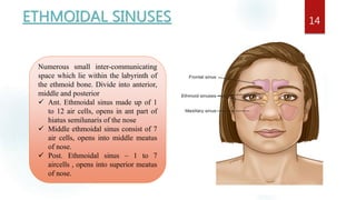
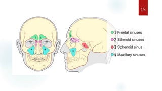
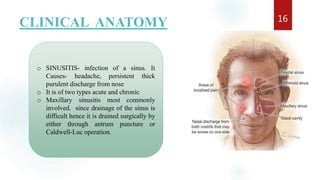
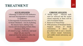
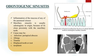
PARANASAL
SINUSES
PRESENTED BY-
DR. SAKSHI SHUKLA
MDS FIRST YEAR 2018
DEPARTMENT OF PUBLIC HEALTH DENTISTR
1
CONTENTS
 INTRODUCTION
 DEVELOPMENT
 PARANASAL SINUSES
o FRONTAL SINUS
o MAXILLARY SINUS
o ETHMOIDAL SINUS
o SPHENOIDAL SINUS
 CLINICAL ANATOMY
 CONCLUSION
 REFERENCES
2
INTRODUCTION 3
 Air filled spaces
 Open into the nasal cavity
through its lateral wall
 These are- frontal,
maxillary, sphenoidal and
ethmoidal
 Function is to make the
skull lighter, warm up and
humidify the inspired air
and also add resonance to
the voice.
PARANASAL SINUSES 4
DEVELOPMENT OF SINUSES 5
 At about 25 – 28 weeks of
gestation, three medially directed
projections arise from lateral wall
of nose.
 Between these projections small
lateral diverticula invaginate into
primitive choana to eventually form
meati of nose as beginning of the
development of paranasal sinuses.
 Sinuses begin developing as small
sacculations of mucosa of nasal
meati and recesses.
 As pouches or sacs develop and
grow they will invade the
respective bones to form air sinuses
and cells.
6
` 7
Sinuses have small orifices
(ostia) which open into recesses
(meati) of the nasal cavities.
• Meati are covered by turbinates
(conchae).
• Turbinates consist of bony
shelves surrounded by erectile
soft tissue.
• There are 3 turbinates and 3
meati in each nasal cavity
(superior, middle, and inferior).
8
FRONTAL SINUS 9
Lies in the frontal bone deep to the superciliary
arch, extends upwards above medial end of
eyebrow, backward into medial part of roof of the
orbit. Opens into middle meatus of nose into ant.
End of hiatus semilunaris through infundibulum
or frontonasal duct.
 Rudimentary or absent at birth. Well developed between 7 and 8 years.
 Arterial supply- supraorbital artery
 Venous drainage- supraorbital and superior ophthalmic veins.
 Lymphatic drainage- supra mandibular nodes
 Nerve supply-supraorbital nerve
10
MAXILLARY SINUS 11
 Largest of the paranasal sinuses, first to
develop.
 Pyramidal in shape, base directed
medially towards lateral wall of nose,
apex directed laterally in zygomatic
process of maxilla. Roof is formed by
floor of orbit, floor by alveolar process of
maxilla, Opens into middle meatus of
nose and lower part of hiatus semilunaris.
o Arterial supply- facial, infraorbital and greater palatine
o Venous drainage- facial vein and pterygoid plexus of vein
o Lymphatic drainage- submandibular nodes
o Nerve supply- post. Sup. Alveolar branch of maxillary nerve and middle sup. Alveolar branches from
infraorbital nerve.
12
SPHENOIDAL SINUS
 Right and left sphenoidal sinuses lie within the body of sphenoidal bone, separated by
septum.
 Related superiorly to optic chiasma and hypophysis cerebri; laterally to internal carotid
artery and cavernous sinus. Opens into sphenoethmoidal recess of corresponding half of
nasal cavity.
13
Arterial supply- post. Ethmoidal and internal carotid
artery.
Venous drainage- pterygoid venous plexus and
cavernous sinus
Lymphatic drainage- retropharyngeal nodes
Nerve supply- post. Ethmoidal nerve and orbital
branches of pterygopalatine ganglion.
ETHMOIDAL SINUSES 14
Numerous small inter-communicating
space which lie within the labyrinth of
the ethmoid bone. Divide into anterior,
middle and posterior
 Ant. Ethmoidal sinus made up of 1
to 12 air cells, opens in ant part of
hiatus semilunaris of the nose
 Middle ethmoidal sinus consist of 7
air cells, opens into middle meatus
of nose.
 Post. Ethmoidal sinus – 1 to 7
aircells , opens into superior meatus
of nose.
15
CLINICAL ANATOMY 16
o SINUSITIS- infection of a sinus. It
Causes- headache, persistent thick
purulent discharge from nose
o It is of two types acute and chronic
o Maxillary sinusitis most commonly
involved. since drainage of the sinus is
difficult hence it is drained surgically by
either through antrum puncture or
Caldwell-Luc operation.
ACUTE SINUSITIS
It is the acute inflammation of the sinus mucosa with severe
pain in sinus area.
17
SUBACUTE MAXILLARY SINUSITIS 18
 It is the intermediate stage between
acute and chronic sinusitis. There is
pain only in the form of the local
discomfort.
 Patient has persistent discharge.
 The voice is nasal, throat is sore with
constant irritating cough. Patient can
not sleep well.
 The disease may take a long course
over week or months.
CHRONIC SINUSITIS
The term Chronic sinusitis is poorly defined but is best considered as
persistent incompletely resolved acute sinusitis.
19
MMT: Mucous Membrane
Thickening
Ofs: Opacified Frontal Sinus
Oes: Opacified Ethmoid Sinus
Oms: Opacified Maxillary Sinus
M: Mucocoele
TREATMENT 20
ACUTE SINUSITIS
1)Drainage is achieved with antibiotics
and nasal decongestants or extraction.
2) Antibiotics
3) Decongestant:Xylometazoline (0.1%)
4) Steam Inhalation : Acts by hydrating
the mucous layer, making it less
viscous and encouraging normal ciliary
clearance of the sinus.
(5) Antral Lavage : Antibiotics and nasal
drops fail, pus must be
removed from the antrum to allow the
sinus mucosa to
recover
CHRONIC SINUSITIS
 Dental origin : Affected teeth
must be removed and the socket
closed surgically as there will be
risk of oroantral fistula.
 Removal of Nasal Polyp
 With presence of Oroantral fistula:
Surgical closure of fistula
Antrostomy: If above all
procedures fails to cure c sinusitis
the inferior meatal Antrostomy
and middle meatal antrostomy can
be done.
ODONTOGENIC SINUSITIS 21
 Inflammation of the mucosa of any of
the paranasal sinuses
 Maxillary sinusitis is usually
odontogenic in origin, because of its
close proximity with the maxillary
teeth.
 Cause may be-
o infection- periapical abscess
o Allergy
o Trauma
o Displaced tooth or root
o neoplasm
OROANTRAL COMMUNICATION AND FISTULA 22
 An oroantral perforation is an unnatural communication between the oral cavity
and maxillary sinus
 An oroantral fistula is an epithelialized, pathological, unnatural communication
between these two cavities.
can be mainly due to-
 extraction
 Periapical lesions
 Trauma
 Chronic infections of maxillary sinus
 During surgery
 Infected maxillary implant denture teratomatous
destruction of maxilla

23
24Treatment - Cases where oroantral
communication is recent and formation of
fistula is not established.
o Immediate surgery repair to achieve primary
closure
o Simultaneous antibiotic prophylaxis to
prevent sinus infection
Cases seen more than 24 hours after accident:- It is
preferable to defer the surgical closure until the gingival
edges of the fistula have healed soundly (approx 3
weeks)
- Supportive measures should be given
- Maxillary sinus should be gently irrigated with warm
normal saline, if there is purulent discharge or signs of
acute or chronic sinusitis is seen.
• Cases of long duration(more than a
month):
- Surgical closure is required
ANTROLITHS IN MAXILLARY SINUS 25
Antroliths are calcified
masses found in the maxillary
sinus.
• There is calcification of
masses of stagnant mucus in
site of previous
inflammation, root fragments
or bone chips.
• Asymptomatic but if continue
to grow patient complain of blood
stain nasal discharge or facial
pain.
• Removal if it is symptomatic
ODONTOGENIC CYST LESIONS 26
Odontogenic cysts
are the most common
group of extrinsic
lesions that encroach
on the maxillary
sinuses.
• The cyst enlarges ,the
sinus decrease in size
• The result is radioopaque
line between
the cyst and the air
space of the sinus.
27
CARCINOMA 28
Carcinoma of maxillary sinus
arises from mucosal lining ,
symptoms depend on direction of
growth-
o Proptosis or diplopia
o Bulging or ulceration of palate
o Swelling of the face
o Pain
o Nasal obstruction, epixtasis,
epiphora
BENIGN TUMOR 29
Benign tumors in the sinus may arise from the
lining as polyps and
papillomas, from bone as osteoma or from
maxillary teeth as
odontomes.
MALIGNANT TUMOR 30
Squamous cell carcinoma is
the most common malignant
tumor of the paranasal
sinuses.
DEVELOPMENTALANOMALIES 31
• Crouzon syndrome : Early synostosis (fusion) of
sutures produces hypoplasia of the maxilla and
therefore the maxillary sinus together with the high
arched palate.
• Treacher Collins syndrome : Associated with grossly and
symmetrically underdeveloped maxillary sinuses
and Malar bones.
• Binder syndrome : Hypoplasia of middle third of the face
with smaller maxillary length and maxillary
sinus hypoplasia.
CONCLUSION 32
Paranasal Sinuses (PNS) are air containing bony spaces around
the nasal cavity. There are 4 pairs of paranasal
sinuses(bilaterally) but maxillary sinus is considered most
important to dentists due to close proximity of maxillary sinus
to orbit, alveolar ridge, diseases involving these structures may
produce confusing symptoms. Hence a precise information
about the surgical anatomy is essential to dental practitioners.
the close anatomical relationship of the maxillary sinus and the
roots of maxillary molars, premolars and in some instances
canines, can also lead to several endodontic complications.
Clinicians must be particularly cautious when performing
dental procedures involving the maxillary posterior teeth.
REFERENCES
 Malik N. A. Textbook Of Oral And Maxillofacial Surgery,Jaypee Brothers Medical
Publishers Ltd ;( 4th Edition)2015
 Chaurasia B. D. Human Anatomy, CBS publishers and distributers(7th edition)
vol.3,2015
33
34
THANK YOU

Paranasal sinuses

  • 1.
    PARANASAL SINUSES PRESENTED BY- DR. SAKSHISHUKLA MDS FIRST YEAR 2018 DEPARTMENT OF PUBLIC HEALTH DENTISTR 1
  • 2.
    CONTENTS  INTRODUCTION  DEVELOPMENT PARANASAL SINUSES o FRONTAL SINUS o MAXILLARY SINUS o ETHMOIDAL SINUS o SPHENOIDAL SINUS  CLINICAL ANATOMY  CONCLUSION  REFERENCES 2
  • 3.
    INTRODUCTION 3  Airfilled spaces  Open into the nasal cavity through its lateral wall  These are- frontal, maxillary, sphenoidal and ethmoidal  Function is to make the skull lighter, warm up and humidify the inspired air and also add resonance to the voice.
  • 4.
  • 5.
    DEVELOPMENT OF SINUSES5  At about 25 – 28 weeks of gestation, three medially directed projections arise from lateral wall of nose.  Between these projections small lateral diverticula invaginate into primitive choana to eventually form meati of nose as beginning of the development of paranasal sinuses.  Sinuses begin developing as small sacculations of mucosa of nasal meati and recesses.  As pouches or sacs develop and grow they will invade the respective bones to form air sinuses and cells.
  • 6.
  • 7.
    ` 7 Sinuses havesmall orifices (ostia) which open into recesses (meati) of the nasal cavities. • Meati are covered by turbinates (conchae). • Turbinates consist of bony shelves surrounded by erectile soft tissue. • There are 3 turbinates and 3 meati in each nasal cavity (superior, middle, and inferior).
  • 8.
  • 9.
    FRONTAL SINUS 9 Liesin the frontal bone deep to the superciliary arch, extends upwards above medial end of eyebrow, backward into medial part of roof of the orbit. Opens into middle meatus of nose into ant. End of hiatus semilunaris through infundibulum or frontonasal duct.  Rudimentary or absent at birth. Well developed between 7 and 8 years.  Arterial supply- supraorbital artery  Venous drainage- supraorbital and superior ophthalmic veins.  Lymphatic drainage- supra mandibular nodes  Nerve supply-supraorbital nerve
  • 10.
  • 11.
    MAXILLARY SINUS 11 Largest of the paranasal sinuses, first to develop.  Pyramidal in shape, base directed medially towards lateral wall of nose, apex directed laterally in zygomatic process of maxilla. Roof is formed by floor of orbit, floor by alveolar process of maxilla, Opens into middle meatus of nose and lower part of hiatus semilunaris. o Arterial supply- facial, infraorbital and greater palatine o Venous drainage- facial vein and pterygoid plexus of vein o Lymphatic drainage- submandibular nodes o Nerve supply- post. Sup. Alveolar branch of maxillary nerve and middle sup. Alveolar branches from infraorbital nerve.
  • 12.
  • 13.
    SPHENOIDAL SINUS  Rightand left sphenoidal sinuses lie within the body of sphenoidal bone, separated by septum.  Related superiorly to optic chiasma and hypophysis cerebri; laterally to internal carotid artery and cavernous sinus. Opens into sphenoethmoidal recess of corresponding half of nasal cavity. 13 Arterial supply- post. Ethmoidal and internal carotid artery. Venous drainage- pterygoid venous plexus and cavernous sinus Lymphatic drainage- retropharyngeal nodes Nerve supply- post. Ethmoidal nerve and orbital branches of pterygopalatine ganglion.
  • 14.
    ETHMOIDAL SINUSES 14 Numeroussmall inter-communicating space which lie within the labyrinth of the ethmoid bone. Divide into anterior, middle and posterior  Ant. Ethmoidal sinus made up of 1 to 12 air cells, opens in ant part of hiatus semilunaris of the nose  Middle ethmoidal sinus consist of 7 air cells, opens into middle meatus of nose.  Post. Ethmoidal sinus – 1 to 7 aircells , opens into superior meatus of nose.
  • 15.
  • 16.
    CLINICAL ANATOMY 16 oSINUSITIS- infection of a sinus. It Causes- headache, persistent thick purulent discharge from nose o It is of two types acute and chronic o Maxillary sinusitis most commonly involved. since drainage of the sinus is difficult hence it is drained surgically by either through antrum puncture or Caldwell-Luc operation.
  • 17.
    ACUTE SINUSITIS It isthe acute inflammation of the sinus mucosa with severe pain in sinus area. 17
  • 18.
    SUBACUTE MAXILLARY SINUSITIS18  It is the intermediate stage between acute and chronic sinusitis. There is pain only in the form of the local discomfort.  Patient has persistent discharge.  The voice is nasal, throat is sore with constant irritating cough. Patient can not sleep well.  The disease may take a long course over week or months.
  • 19.
    CHRONIC SINUSITIS The termChronic sinusitis is poorly defined but is best considered as persistent incompletely resolved acute sinusitis. 19 MMT: Mucous Membrane Thickening Ofs: Opacified Frontal Sinus Oes: Opacified Ethmoid Sinus Oms: Opacified Maxillary Sinus M: Mucocoele
  • 20.
    TREATMENT 20 ACUTE SINUSITIS 1)Drainageis achieved with antibiotics and nasal decongestants or extraction. 2) Antibiotics 3) Decongestant:Xylometazoline (0.1%) 4) Steam Inhalation : Acts by hydrating the mucous layer, making it less viscous and encouraging normal ciliary clearance of the sinus. (5) Antral Lavage : Antibiotics and nasal drops fail, pus must be removed from the antrum to allow the sinus mucosa to recover CHRONIC SINUSITIS  Dental origin : Affected teeth must be removed and the socket closed surgically as there will be risk of oroantral fistula.  Removal of Nasal Polyp  With presence of Oroantral fistula: Surgical closure of fistula Antrostomy: If above all procedures fails to cure c sinusitis the inferior meatal Antrostomy and middle meatal antrostomy can be done.
  • 21.
    ODONTOGENIC SINUSITIS 21 Inflammation of the mucosa of any of the paranasal sinuses  Maxillary sinusitis is usually odontogenic in origin, because of its close proximity with the maxillary teeth.  Cause may be- o infection- periapical abscess o Allergy o Trauma o Displaced tooth or root o neoplasm
  • 22.
    OROANTRAL COMMUNICATION ANDFISTULA 22  An oroantral perforation is an unnatural communication between the oral cavity and maxillary sinus  An oroantral fistula is an epithelialized, pathological, unnatural communication between these two cavities. can be mainly due to-  extraction  Periapical lesions  Trauma  Chronic infections of maxillary sinus  During surgery  Infected maxillary implant denture teratomatous destruction of maxilla 
  • 23.
  • 24.
    24Treatment - Caseswhere oroantral communication is recent and formation of fistula is not established. o Immediate surgery repair to achieve primary closure o Simultaneous antibiotic prophylaxis to prevent sinus infection Cases seen more than 24 hours after accident:- It is preferable to defer the surgical closure until the gingival edges of the fistula have healed soundly (approx 3 weeks) - Supportive measures should be given - Maxillary sinus should be gently irrigated with warm normal saline, if there is purulent discharge or signs of acute or chronic sinusitis is seen. • Cases of long duration(more than a month): - Surgical closure is required
  • 25.
    ANTROLITHS IN MAXILLARYSINUS 25 Antroliths are calcified masses found in the maxillary sinus. • There is calcification of masses of stagnant mucus in site of previous inflammation, root fragments or bone chips. • Asymptomatic but if continue to grow patient complain of blood stain nasal discharge or facial pain. • Removal if it is symptomatic
  • 26.
    ODONTOGENIC CYST LESIONS26 Odontogenic cysts are the most common group of extrinsic lesions that encroach on the maxillary sinuses. • The cyst enlarges ,the sinus decrease in size • The result is radioopaque line between the cyst and the air space of the sinus.
  • 27.
  • 28.
    CARCINOMA 28 Carcinoma ofmaxillary sinus arises from mucosal lining , symptoms depend on direction of growth- o Proptosis or diplopia o Bulging or ulceration of palate o Swelling of the face o Pain o Nasal obstruction, epixtasis, epiphora
  • 29.
    BENIGN TUMOR 29 Benigntumors in the sinus may arise from the lining as polyps and papillomas, from bone as osteoma or from maxillary teeth as odontomes.
  • 30.
    MALIGNANT TUMOR 30 Squamouscell carcinoma is the most common malignant tumor of the paranasal sinuses.
  • 31.
    DEVELOPMENTALANOMALIES 31 • Crouzonsyndrome : Early synostosis (fusion) of sutures produces hypoplasia of the maxilla and therefore the maxillary sinus together with the high arched palate. • Treacher Collins syndrome : Associated with grossly and symmetrically underdeveloped maxillary sinuses and Malar bones. • Binder syndrome : Hypoplasia of middle third of the face with smaller maxillary length and maxillary sinus hypoplasia.
  • 32.
    CONCLUSION 32 Paranasal Sinuses(PNS) are air containing bony spaces around the nasal cavity. There are 4 pairs of paranasal sinuses(bilaterally) but maxillary sinus is considered most important to dentists due to close proximity of maxillary sinus to orbit, alveolar ridge, diseases involving these structures may produce confusing symptoms. Hence a precise information about the surgical anatomy is essential to dental practitioners. the close anatomical relationship of the maxillary sinus and the roots of maxillary molars, premolars and in some instances canines, can also lead to several endodontic complications. Clinicians must be particularly cautious when performing dental procedures involving the maxillary posterior teeth.
  • 33.
    REFERENCES  Malik N.A. Textbook Of Oral And Maxillofacial Surgery,Jaypee Brothers Medical Publishers Ltd ;( 4th Edition)2015  Chaurasia B. D. Human Anatomy, CBS publishers and distributers(7th edition) vol.3,2015 33
  • 34.

Editor's Notes

  • #9 Hiatus – eccentric groove.
  • #14 Hypophysic- pituitary body
  • #24 Painful purulent, passage of fluid
  • #28 Endoscopic surgery
  • #30 surgical
  • #31 Chemotherapy or radiation therapy