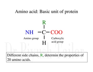
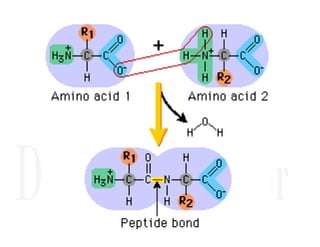
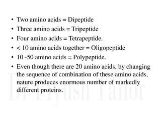
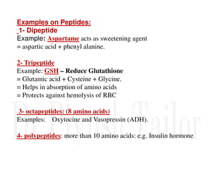
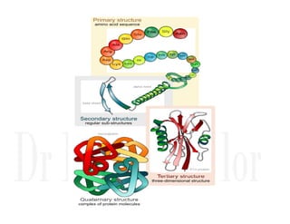
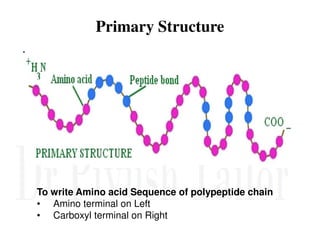
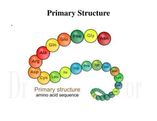
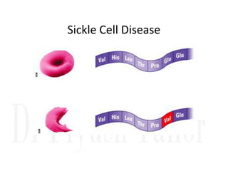
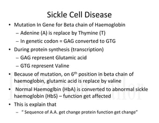
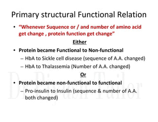
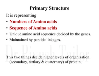
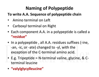
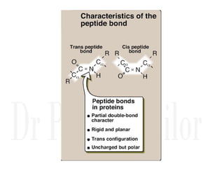
1. Proteins are polymers of amino acids linked by peptide bonds. Changes to the primary structure, including the sequence or number of amino acids, can alter a protein's function.
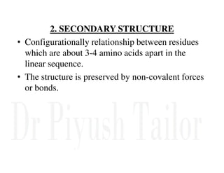
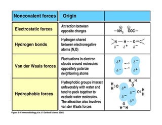
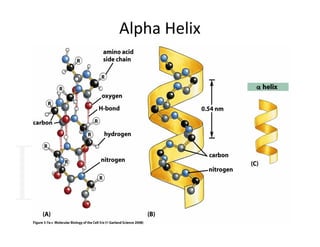
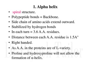
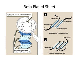
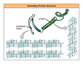
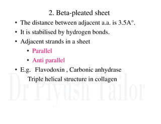
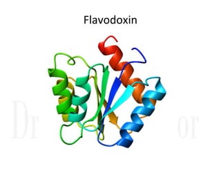
2. Secondary structure forms due to interactions between residues that are near each other in the primary structure. Common secondary structures include alpha helices and beta sheets.
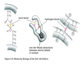
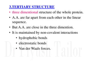
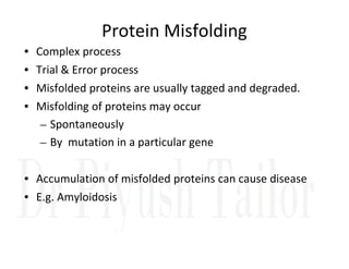
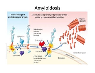
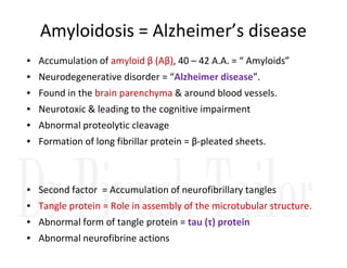
3. Tertiary structure describes the three-dimensional structure of the whole protein, influenced by interactions between residues far apart in the primary structure. Misfolded proteins can cause diseases like amyloidosis.
Protein structures determine their functions. There are four levels of protein structure:
1) Primary structure is the amino acid sequence
2) Secondary structure involves local patterns like alpha helices and beta sheets
3) Tertiary structure describes the overall 3D shape formed by secondary structures
4) Quaternary structure refers to the arrangement of multiple polypeptide chains
The most common secondary structures are alpha helices, stabilized by hydrogen bonds between amino acids i and i+4, and beta sheets formed by hydrogen bonding between strands. Protein structure enables functions like catalysis, transport, and information transfer.
The document discusses various topics related to protein structure and function. It defines different types of bonds in proteins including peptide bonds, disulfide bonds, and hydrogen bonds. It describes the 20 common amino acids that make up proteins and different secondary structures such as alpha helices and beta sheets. It discusses the four levels of protein structure - primary, secondary, tertiary, and quaternary structure. It also covers protein folding driven by hydrophobic interactions and hydrogen bonding, as well as denaturation of proteins.
Proteins are linear polymers of amino acids linked by peptide bonds. There are 20 different amino acids that make up proteins. Proteins have four levels of structure - primary, secondary, tertiary, and quaternary. The primary structure is the linear sequence of amino acids, secondary structure involves folding into shapes like alpha helices and beta sheets, tertiary structure is the overall 3D shape of the protein, and quaternary structure involves the joining of multiple protein subunits. Proteins play many important roles in the body such as enzymes, transport, structure, and regulation.
Amino acids are the building blocks of proteins. They contain carbon, hydrogen, oxygen, nitrogen and a distinctive side chain. There are 20 common amino acids that serve as monomers for protein synthesis. Amino acids differ in their side chains, which influence solubility. The general structure includes an amino group, carboxyl group, hydrogen and R group. Amino acids form proteins through peptide bonds between their amino and carboxyl groups. Proteins have primary, secondary, tertiary and quaternary levels of structure determined by amino acid sequence and bonding.
Amino acids are the building blocks of proteins. There are 20 common amino acids that make up proteins through peptide bonds. Amino acids have four groups attached to the alpha carbon including an amino group, carboxyl group, hydrogen, and a variable side chain. Amino acids can be classified based on their structure and properties. Proteins have four levels of structure - primary, secondary, tertiary, and quaternary - which determine their shape and function.
This document discusses principles of protein structure, including primary, secondary, and supersecondary structure. It covers the following key points:
- Primary structure refers to the amino acid sequence of a protein. There are 20 common amino acids that make up protein sequences.
- Secondary structure includes common elements like alpha helices and beta sheets. Alpha helices are right-handed coils stabilized by hydrogen bonds between amino acids four positions apart in the sequence. Beta sheets consist of beta strands connected laterally or anti-parallel by hydrogen bonds.
- Supersecondary structure refers to recurrent structural motifs formed by combinations of secondary structure elements, like beta-alpha-beta motifs or helix-loop-helix motifs. Larger domains
Amino acids, Structure of Protein and Amino acid metabolism Pramod Pandey
This document provides an overview of amino acids and protein structure and metabolism. It begins with definitions of amino acid structure and classifications based on properties. It then discusses protein structure at the primary, secondary, tertiary and quaternary levels. Key metabolic pathways of amino acids are covered, including transamination, deamination, the urea cycle, and the metabolism of specific amino acids like glycine, phenylalanine, tyrosine, and tryptophan.
Amino acids are the building blocks of proteins and peptides. They contain non-polar, polar, and charged R groups. Peptides are formed through condensation reactions between amino acids, linking them through peptide bonds. There are four levels of protein structure - primary, secondary, tertiary, and quaternary. The secondary structure involves folding into alpha helices or beta sheets. The alpha helix is stabilized by hydrogen bonds between residues four places apart in the sequence. Tertiary structure describes the three-dimensional folding of the polypeptide chain. Quaternary structure involves the interaction of multiple polypeptide chains in a protein.
The document summarizes key aspects of amino acids and protein structure in 3 paragraphs or less:
Amino acids are the building blocks of proteins. They contain common structural features and exist in L- and D-forms. In proteins, amino acids are exclusively in the L-conformation. Amino acids are classified based on the properties of their side chains into nonpolar, aromatic, polar, positively charged, and negatively charged categories.
Protein structure is hierarchical, progressing from primary to secondary, tertiary, and quaternary levels. The primary structure is the amino acid sequence. Secondary structures include alpha helices, beta sheets, and turns formed by hydrogen bonding. Tertiary structure refers to the overall 3
Proteins are made up of amino acid chains that fold into complex three-dimensional shapes determined by interactions between their side chains. This folding allows proteins to perform a vast array of functions in the body, including building muscles and tissues, catalyzing biochemical reactions, transporting molecules, and regulating processes. The structure of a protein is hierarchical, progressing from its linear amino acid sequence (primary structure) through localized folding patterns (secondary structure) to its overall three-dimensional conformation (tertiary structure), which enables it to carry out its specific role.
This document provides information on enzymes. It begins by defining enzymes as soluble, colloidal organic catalysts formed by living cells that are specific, protein in nature, and inactive at 0°C. It notes that all enzymes are proteins and that activity is lost through denaturation or dissociation. The document discusses enzymes as proteins that often require cofactors. It provides details on different types of enzymes based on their structure, location, and method of secretion. The rest of the document covers enzyme nomenclature, classification, specificity, and mechanisms of action. It discusses factors that affect enzyme activity such as temperature, pH, product concentration, and more.
Principle of protein structure and functionAsheesh Pandey
The document discusses principles of protein structure, including primary, secondary, and tertiary structure. It covers amino acids and their properties, peptide bonds, and common structural elements like the alpha helix. Specifically, it defines primary structure as the amino acid sequence, discusses the 20 common amino acids and their characteristics like chirality. It also covers dihedral angles, Ramachandran plots, common secondary structures like the alpha helix and their properties, including hydrogen bonding patterns and characteristic phi and psi angles.
The document discusses amino acids and their properties. It introduces the 20 standard amino acids that make up mammalian proteins, as well as 2 additional amino acids coded by DNA in a non-standard manner. Amino acids have different structures depending on their variable R groups, and can be classified based on properties like polarity, acidity, and charge. Peptides are formed from amino acids joined by peptide bonds. The peptide bond gives peptides some rigidity and planar structure. Important peptides include glutathione, bradykinin, and peptide hormones. The primary structure of a protein refers to the specific sequence of amino acid residues that make it up.
Proteins are polymers made up of amino acid monomers linked by peptide bonds. They are classified based on their number of amino acids, with proteins containing more than 50 amino acids. Proteins have four levels of structure - primary, secondary, tertiary, and quaternary. The primary structure is the linear sequence of amino acids. Secondary structures include alpha helices and beta sheets formed by hydrogen bonding between amino acids. Tertiary structure involves folding of the polypeptide into a compact 3D structure. Quaternary structure involves the assembly of multiple polypeptide subunits.
Peptide and polypeptide, protein structure.pptxRASHMI M G
peptide and polypeptide-meaning and definition, peptide bond, protein structures- primary, secondary, tertiary, supersecondary, quaternary structures denaturation and solubilities of proteins.
Describes the structural organisation of proteins with example and its determination, interrelationship b/w structure and function of proteins, also biologically important peptides is covered.
by Dr. N. Sivaranjani, MD
Proteins are polymers of amino acids that are involved in most body functions and life processes. They have a primary structure defined by their amino acid sequence and levels of organization including secondary structures like alpha helices and beta sheets, tertiary structure describing the overall 3D shape, and potentially quaternary structure for multi-chain proteins. Proteins can be classified based on their function, solubility, composition and structure. Protein folding and structures are stabilized by various bonds and interactions.
Proteins are macromolecules composed of amino acid subunits linked by peptide bonds. There are 20 common amino acids that make up proteins. Amino acids are classified based on properties like charge, polarity, and essentiality. Proteins form through peptide bond formation between amino acid residues, resulting in primary, secondary, tertiary, and quaternary structure levels that determine a protein's shape and function.
Enzymes are protein catalysts that accelerate biochemical reactions without being consumed. They achieve this by lowering the activation energy of reactions, allowing substrates to be converted to products more easily. Enzymes are specific in their action and only catalyze certain reactions. They work by binding substrates in their active sites and facilitating the biochemical transformations. The structure and conditions of the active site, as well as factors like temperature and pH, influence an enzyme's activity level and specificity.
This document discusses amino acids, peptides, and proteins. It begins by defining them as monomers (amino acids), polymers of a few monomers (peptides), and polymers of many monomers (proteins). It then covers the structures and properties of amino acids, including the 20 that are found in proteins. Peptide bond formation is explained as linking amino acids together. Various proteins are classified and examples given, including simple proteins like albumins and globulins, and structural proteins like keratins, collagens, and elastins. The roles and importance of proteins in the body are also summarized.
Here are the key points from the protein separation methods reading:
- There are several methods used to separate proteins including precipitation, electrophoresis, chromatography, and ultracentrifugation.
- Precipitation separates proteins based on differences in solubility at various pH, temperatures, or in the presence of salts, organic solvents, or other reagents. It is a crude separation method.
- Electrophoresis separates proteins based on their charge and size by applying an electric current through a gel or liquid. Common types are PAGE and SDS-PAGE.
- Chromatography separates proteins based on differences in how they interact with a stationary phase compared to a mobile phase as they flow through a column. Key types are ion-exchange
- Amino acids are the building blocks of proteins and contain an amino group, carboxyl group, hydrogen atom, and side chain bonded to an alpha carbon.
- There are 20 standard amino acids that vary in properties based on their side chains. Phenylalanine, methionine, and tryptophan are hydrophobic while cysteine forms disulfide bridges.
- Proteins are made of chains of amino acids that can form different structures like fibrous or globular proteins depending on their shape and solubility.
The document discusses the primary structure of proteins, which is the specific sequence of amino acids. It explains that genetic diseases can result from abnormal amino acid sequences that cause improper folding and loss of function. Determining the normal and mutated protein sequences can help diagnose or study the disease. It then goes on to describe various methods used to determine a protein's primary structure, including identifying amino acid composition through chromatography, N-terminal sequencing using Edman reagent, cleaving large proteins into fragments, and DNA sequencing to translate the nucleotide sequence into the amino acid sequence.
Proteins are composed of amino acids and have four levels of structure - primary, secondary, tertiary, and quaternary. The primary structure is the amino acid sequence. Common secondary structures include alpha helices and beta pleated sheets formed by hydrogen bonding. Tertiary structure describes the overall 3D shape formed by interactions between amino acid side chains. Globular proteins fold such that hydrophobic residues are buried inside and hydrophilic residues face outward, while fibrous proteins form insoluble fibers.
General structure of amino acid
Specific learning objective (SLO): Amino acid as Ampholytes (acid and base), Zwitter ions.
Classification of amino acid on the basis of side chain, chemical composition, Nutritional Requirement and metabolic fate.
Derived amino acids.
Optical properties of amino acids.
Acid-Base properties and Buffer characteristic.
Biological Important Peptides
Proteins based on nutritional value
This document discusses amino acids and proteins. It begins by defining proteins and polypeptides as chains of amino acids, with proteins being longer chains. It then defines amino acids as molecules containing both a carboxyl and amino group. There are 20 naturally occurring amino acids that are used to build proteins. The document discusses the structures and properties of amino acids and how they depend on pH. It also covers peptide bonds and the four levels of protein structure: primary, secondary, tertiary, and quaternary. Examples of protein structures like collagen, hemoglobin and myoglobin are provided. The roles of proteins in the human body and daily protein requirements are summarized at the end.
Sexuality - Issues, Attitude and Behaviour - Applied Social Psychology - Psyc...PsychoTech Services
A proprietary approach developed by bringing together the best of learning theories from Psychology, design principles from the world of visualization, and pedagogical methods from over a decade of training experience, that enables you to: Learn better, faster!
Authoring a personal GPT for your research and practice: How we created the Q...Leonel Morgado
Thematic analysis in qualitative research is a time-consuming and systematic task, typically done using teams. Team members must ground their activities on common understandings of the major concepts underlying the thematic analysis, and define criteria for its development. However, conceptual misunderstandings, equivocations, and lack of adherence to criteria are challenges to the quality and speed of this process. Given the distributed and uncertain nature of this process, we wondered if the tasks in thematic analysis could be supported by readily available artificial intelligence chatbots. Our early efforts point to potential benefits: not just saving time in the coding process but better adherence to criteria and grounding, by increasing triangulation between humans and artificial intelligence. This tutorial will provide a description and demonstration of the process we followed, as two academic researchers, to develop a custom ChatGPT to assist with qualitative coding in the thematic data analysis process of immersive learning accounts in a survey of the academic literature: QUAL-E Immersive Learning Thematic Analysis Helper. In the hands-on time, participants will try out QUAL-E and develop their ideas for their own qualitative coding ChatGPT. Participants that have the paid ChatGPT Plus subscription can create a draft of their assistants. The organizers will provide course materials and slide deck that participants will be able to utilize to continue development of their custom GPT. The paid subscription to ChatGPT Plus is not required to participate in this workshop, just for trying out personal GPTs during it.
ESR spectroscopy in liquid food and beverages.pptxPRIYANKA PATEL
With increasing population, people need to rely on packaged food stuffs. Packaging of food materials requires the preservation of food. There are various methods for the treatment of food to preserve them and irradiation treatment of food is one of them. It is the most common and the most harmless method for the food preservation as it does not alter the necessary micronutrients of food materials. Although irradiated food doesn’t cause any harm to the human health but still the quality assessment of food is required to provide consumers with necessary information about the food. ESR spectroscopy is the most sophisticated way to investigate the quality of the food and the free radicals induced during the processing of the food. ESR spin trapping technique is useful for the detection of highly unstable radicals in the food. The antioxidant capability of liquid food and beverages in mainly performed by spin trapping technique.
ESA/ACT Science Coffee: Diego Blas - Gravitational wave detection with orbita...Advanced-Concepts-Team
Presentation in the Science Coffee of the Advanced Concepts Team of the European Space Agency on the 07.06.2024.
Speaker: Diego Blas (IFAE/ICREA)
Title: Gravitational wave detection with orbital motion of Moon and artificial
Abstract:
In this talk I will describe some recent ideas to find gravitational waves from supermassive black holes or of primordial origin by studying their secular effect on the orbital motion of the Moon or satellites that are laser ranged.
Immersive Learning That Works: Research Grounding and Paths ForwardLeonel Morgado
We will metaverse into the essence of immersive learning, into its three dimensions and conceptual models. This approach encompasses elements from teaching methodologies to social involvement, through organizational concerns and technologies. Challenging the perception of learning as knowledge transfer, we introduce a 'Uses, Practices & Strategies' model operationalized by the 'Immersive Learning Brain' and ‘Immersion Cube’ frameworks. This approach offers a comprehensive guide through the intricacies of immersive educational experiences and spotlighting research frontiers, along the immersion dimensions of system, narrative, and agency. Our discourse extends to stakeholders beyond the academic sphere, addressing the interests of technologists, instructional designers, and policymakers. We span various contexts, from formal education to organizational transformation to the new horizon of an AI-pervasive society. This keynote aims to unite the iLRN community in a collaborative journey towards a future where immersive learning research and practice coalesce, paving the way for innovative educational research and practice landscapes.
The debris of the ‘last major merger’ is dynamically youngSérgio Sacani
The Milky Way’s (MW) inner stellar halo contains an [Fe/H]-rich component with highly eccentric orbits, often referred to as the
‘last major merger.’ Hypotheses for the origin of this component include Gaia-Sausage/Enceladus (GSE), where the progenitor
collided with the MW proto-disc 8–11 Gyr ago, and the Virgo Radial Merger (VRM), where the progenitor collided with the
MW disc within the last 3 Gyr. These two scenarios make different predictions about observable structure in local phase space,
because the morphology of debris depends on how long it has had to phase mix. The recently identified phase-space folds in Gaia
DR3 have positive caustic velocities, making them fundamentally different than the phase-mixed chevrons found in simulations
at late times. Roughly 20 per cent of the stars in the prograde local stellar halo are associated with the observed caustics. Based
on a simple phase-mixing model, the observed number of caustics are consistent with a merger that occurred 1–2 Gyr ago.
We also compare the observed phase-space distribution to FIRE-2 Latte simulations of GSE-like mergers, using a quantitative
measurement of phase mixing (2D causticality). The observed local phase-space distribution best matches the simulated data
1–2 Gyr after collision, and certainly not later than 3 Gyr. This is further evidence that the progenitor of the ‘last major merger’
did not collide with the MW proto-disc at early times, as is thought for the GSE, but instead collided with the MW disc within
the last few Gyr, consistent with the body of work surrounding the VRM.
Mending Clothing to Support Sustainable Fashion_CIMaR 2024.pdfSelcen Ozturkcan
Ozturkcan, S., Berndt, A., & Angelakis, A. (2024). Mending clothing to support sustainable fashion. Presented at the 31st Annual Conference by the Consortium for International Marketing Research (CIMaR), 10-13 Jun 2024, University of Gävle, Sweden.
When I was asked to give a companion lecture in support of ‘The Philosophy of Science’ (https://shorturl.at/4pUXz) I decided not to walk through the detail of the many methodologies in order of use. Instead, I chose to employ a long standing, and ongoing, scientific development as an exemplar. And so, I chose the ever evolving story of Thermodynamics as a scientific investigation at its best.
Conducted over a period of >200 years, Thermodynamics R&D, and application, benefitted from the highest levels of professionalism, collaboration, and technical thoroughness. New layers of application, methodology, and practice were made possible by the progressive advance of technology. In turn, this has seen measurement and modelling accuracy continually improved at a micro and macro level.
Perhaps most importantly, Thermodynamics rapidly became a primary tool in the advance of applied science/engineering/technology, spanning micro-tech, to aerospace and cosmology. I can think of no better a story to illustrate the breadth of scientific methodologies and applications at their best.
Travis Hills of MN is Making Clean Water Accessible to All Through High Flux ...Travis Hills MN
By harnessing the power of High Flux Vacuum Membrane Distillation, Travis Hills from MN envisions a future where clean and safe drinking water is accessible to all, regardless of geographical location or economic status.
2. INTRODUCTION
• Abnormality in protein structure will lead to
major diseases with profound alterations in
metabolic functions.
• Polymers of amino acids
• Linked by peptid bond
3. Amino acid: Basic unit of protein
Amino acid: Basic unit of protein
COO-
NH3+ C
R
H
Different side chains, R, determin the properties of
20 amino acids.
Amino group Carboxylic
acid group
4.
5. • Two amino acids = Dipeptide
• Three amino acids = Tripeptide
• Four amino acids = Tetrapeptide.
• < 10 amino acids together = Oligopeptide
• 10 -50 amino acids = Polypeptide.
• Even though there are 20 amino acids, by changing
the sequence of combination of these amino acids,
nature produces enormous number of markedly
different proteins.
6. Examples on Peptides:
1- Dipeptide
Example: Aspartame acts as sweetening agent
= aspartic acid + phenyl alanine.
2- Tripeptide
Example: GSH – Reduce Glutathione
= Glutamic acid + Cysteine + Glycine.
= Helps in absorption of amino acids
= Protects against hemolysis of RBC
3- octapeptides: (8 amino acids)
Examples: Oxytocine and Vasopressin (ADH).
4- polypeptides: more than 10 amino acids: e.g. Insulin hormone
7. Type of Protein Structure
1. Primary
2. Secondary
3. Tertiary
4. Quaternary
4. Quaternary
8.
9. Primary Structure
.
To write Amino acid Sequence of polypeptide chain
• Amino terminal on Left
• Carboxyl terminal on Right
14. Sickle Cell Disease
• Mutation In Gene for Beta chain of Haemoglobin
– Adenine (A) is replace by Thymine (T)
– In genetic codon = GAG converted to GTG
• During protein synthesis (transcription)
– GAG represent Glutamic acid
– GTG represent Valine
• Because of mutation, on 6th position in beta chain of
haemoglobin, glutamic acid is replace by valine
• Normal Haemoglbin (HbA) is converted to abnormal sickle
haemoglobin (HbS) – function get affected
• This is explain that
– “ Sequence of A.A. get change protein function get change”
16. Pre-pro-insulin to Pro-insulin to Insulin
• Pre-pro-insulin & pro-Insulin ,both are inactive
• Proinsulin
– single chain with 84 amino acids
– Inactive form
• Insulin
• Insulin
– double chain (A & B chain) with 51 amino acids
– Active form
• When Proinsulin is converted to insulin
– Sequence & number of amino acid, both get change
– So Function of protein get change
– Protein (insulin) became non-functional to functional
17. Thalassemia
• Normal HbA = Alpha (141 A.A.) & Beta (146 A.A.) Chain
• Mutation
– Non sense type of mutation (one of the etiology)
– Premature termination of haemoglobin chain synthesis
– Alpha or Beta either absent or short
– Alpha or Beta thalassemia occur
• Number of amino acid get changed
• Functional protein (HbA) become non-functional
18. Primary structural Functional Relation
• “Whenever Suquence or / and number of amino acid
get change , protein function get change”
Either
• Protein became Functional to Non-functional
– HbA to Sickle cell disease (sequence of A.A. changed)
– HbA to Thalassemia (Number of A.A. changed)
Or
• Protein became non-functional to functional
– Pro-insulin to Insulin (sequence & number of A.A.
both changed)
24. Extra 2 Arginine….Added in Human Insulin
• Is it make any change in charge?
– More positive
– More Negative
– No any change
• What shell be added more to make it neutral
(Net Charge Zero = Zwitter Ion )?
– Positive ions (H+)
– Negative ions (HCO3-)
• Can it change pI of insulin ?
– Decrease
– Increase
– Unchange
25. Extra 2 Arginine….Added in Human Insulin
• Human insulin has isoelectric pH of 5.4. what
can be pI of this newly for recombinant insulin?
– More than 5.4
– Less than 5.4
• In human body, this new recomb-insulin is going
to expose pH of _______ .
• Recombinant insulin pI (6.7) is nearer to
physiological pH (7.4) than human insulin pI
(5.4). So what will be effect on it’s solubility?
– More soluble
– Less soluble (precipitation)
27. Primary Structure
It is representing
• Numbers of Amino acids
• Sequence of Amino acids
• Unique amino acid sequence decided by the genes.
• Maintained by peptide linkages.
This two things decide higher levels of organization
(secondary, tertiary & quaternary) of protein.
28. Naming of Polypeptide
To write A.A. Sequence of polypeptide chain
• Amino terminal on Left
• Carboxyl terminal on Right
• Each component A.A. in a polypeptide is called a
“residue”
“residue”
• In a polypeptide , all A.A. residues suffixes (-ine,
-an, -ic, or -ate) changed to -yl, with the
exception of the C-terminal amino acid.
• E.g. Tripeptide = N-terminal valine, glycine, & C-
terminal leucine
• “valylglycylleucine”
29. Charecteristics of a peptide bond
• It’s a partial double bond.
• ‘trans’ in nature
• Freedom of rotation with limitation to “R” group
• Having rotation of 180 degree
• single bond = 360 degree
• double bond = 0 degree
• Distance is 1.32 A°
• single bond(1.42A°)
• double bond(1.27A°).
• Angles of rotation known as Ramchandran angles
34. Sequencing of the peptide from its N-terminal end
• Edman reagent = Phenylisothiocyanate
• Label N-terminal A.A. residue under mildly alkaline
conditions.
• Phenylthiohydantoin (PTH) create instability in the N-
terminal peptide bond
• It can be selectively hydrolyzed without cleaving the
other peptide bonds
37. Gene to Peptide sequence
• Has limitations
– Not able to predict positions of disulfide bonds
– Not able to identify any posttranslational
modification.
• Therefore, direct protein sequencing is an
extremely important tool.
38. Branched and Circular proteins
• In linear polypeptide chains, there is
• Interchain Disulphide bridges.
• In different polypeptide chains
• Interchain = In same protein.
• Intrachain = Same polypeptide chain.
43. 2. SECONDARY STRUCTURE
• Configurationally relationship between residues
which are about 3-4 amino acids apart in the
linear sequence.
• The structure is preserved by non-covalent forces
or bonds.
or bonds.
44.
45. Disulfide bonds:
=Covalent linkage between
sulfhydryl group (–SH)
=two cysteines may be far
aways in the primary sequence
or may even be different
polypeptide chains;
=contributes stability
=Prevents denatured
= E.g. Immunoglobulins
46.
47.
48.
49. Hydrophobic interactions
• Non polar side chains = Remain in the interior
• Polar side chains = Remain on the surface
Hydrogen bonds
• Oxygen- or Nitrogen-bound hydrogen of side chains
• Alcohol groups of serine and threonine
• Can form hydrogen bonds with Oxygen of a carboxyl group of a
peptide bond (electron-rich atoms)
Ionic interactions
• Negatively charged groups, carboxyl group (–COO-) in the side
chain of aspartate or glutamate.
• Positively charged groups, such as the amino group (–NH3
+) in the
side chain of lysine .
52. 1. Alpha helix
• spiral structure.
• Polypeptide bonds = Backbone.
• Side chain of amino acids extend outward.
• Stabilized by hydrogen bonds
• In each turn = 3.6 A.A. residues.
• Distance between each A.A. residue is 1.5A°
• Right handed.
• As A.A. in the proteins are of L-variety.
• Proline and hydroxyproline will not allow the
formation of α-helix.
53.
54. Example of Alpha Helix
1. Keratins = fibrous proteins in Hair & Nail
• It’s rigidity is determined by the number of
disulfide bonds
2. Myoglobin = globular & flexible molecule.
(Contrast to Keratin)
55. It can disturb Alpha helix
• Proline
– Secondary amino group is not geometrically
compatible
– it inserts a kink in the chain
• Large numbers of charged amino acids
– As it forms more ionic bonds or by electrostatically
repelling each other.
– E.g. glutamate, aspartate, histidine, lysine, or
arginine
• Amino acids with bulky side chains or branched
– Tryptophane, Valine, Isoleucine
58. 2. Beta-pleated sheet
• The distance between adjacent a.a. is 3.5A°.
• It is stabilised by hydrogen bonds.
• Adjacent strands in a sheet
• Parallel
• Anti parallel
• Anti parallel
• E.g. Flavodoxin , Carbonic anhydrase
Triple helical structure in collagen
60. Beta Bands (beta turn)
• Reverse the direction of a polypeptide chain
• Helping it form a compact, globular shape.
• Found on the surface of protein molecules
61. Supersecondary Structure (Motif)
Combining of α-helices and β-sheets
Form primarily the core region—that is, the
interior of the molecule.
Connected by loop β-bends
Supersecondary structures are usually produced
by packing side chains from adjacent secondary
structural elements close to each other.
67. 3.TERTIARY STRUCTURE
• three dimentional structure of the whole protein.
• A.A. are far apart from each other in the linear
sequence.
• But A.A. are close in the three dimention.
• It is maintained by non-covalent interactions
• hydrophobic bonds
• electrostatic bonds
• Van der Waals forces.
68.
69. Domain
• Compact Globular Functional Unit of a protein.
• Independent region of the protein
• May be multiple = if protein has > 200 amino acids.
• Core of a domain is built from combinations of motifs.
• Folding of domain peptide chain is independently of
folding in other domains.
70. Protein Folding
• Three-dimensional shape of the functional protein.
• Due to Interactions between A.A. side chains
– Charges of a.a. side chains = attraction and repulsion
– Hydrogen bonds
– Hydrophobic interactions
– Disulfide bonds
• Process of trial and error
• Tests many configuration
• This results in a correctly folded protein with a low-
energy state
71. Denaturation of Protein
Secondary, Tertiary and Quaternary structures of
protein molecules broken down.
Primary structure is not altered
Unfolding & Disorganization of Protein
Decreases the solubility
So protein precipitated
Loss of biological activity.
Denatured proteins are re-natured when the
physical agent is removed. (less possible)
72. Factor which can does “Denaturation”
Heating = Cooking, Heat coagulation test
Urea = Renal Failure
X-ray
Ultra - violet rays
High pressure = Cooking
Organic solvent = Alcohol as antiseptic
Metals = Mercury poisoning
Acid & Alkali = Digestion
73. Chaperones = In protein folding
• Specialized group of proteins for the proper folding.
• Chaperones = “Heat Shock” proteins
• Information for Correct folding is in primary structure of
protein.
• Interact with the polypeptide at various stages
• Some chaperones keeps protein unfolded until its
synthesis is finished.
• Some Chaperones tends to fold so that their vulnerable,
exposed regions do not become tangled(mixed).
• Some denatured protein can not be re-folded properly,
– Because folding starts with stages its synthesis
– Folding does not wait for synthesis be totally completed.
74.
75. 4.QUATERNARY STRUCTURE
• Certain polypeptides aggregate
• form one functional protein
• known as quaternary structure.
• The protein will lose its function when the
subunits are dissociated.
• Stabilised by
• hydrogen bonds
• electrostatic bonds
• hydrophobic bonds
• van der Waals forces.
76. • Depending on the number of the monomers
• Dimer(2)
• Tetramer(4).
• Each polypeptide chain is termed as Subunit or
Monomer.
• For example,
– Hemoglobin = 2 alpha and 2 beta chains
– Immunoglobulin G = 2 heavy & 2 light chains
– Creatine kinase is dimer
– Lactate dehydrogenase is a tetramer.
77. Protein Misfolding
• Complex process
• Trial & Error process
• Misfolded proteins are usually tagged and degraded.
• Misfolding of proteins may occur
– Spontaneously
By mutation in a particular gene
– By mutation in a particular gene
• Accumulation of misfolded proteins can cause disease
• E.g. Amyloidosis
79. Amyloidosis = Alzheimer’s disease
• Accumulation of amyloid β (Aβ), 40 – 42 A.A. = “ Amyloids”
• Neurodegenerative disorder = “Alzheimer disease”.
• Found in the brain parenchyma & around blood vessels.
• Neurotoxic & leading to the cognitive impairment
• Abnormal proteolytic cleavage
• Formation of long fibrillar protein = β-pleated sheets.
• Second factor = Accumulation of neurofibrillary tangles
• Tangle protein = Role in assembly of the microtubular structure.
• Abnormal form of tangle protein = tau (τ) protein
• Abnormal neurofibrine actions
80. Prion Protein(PrP) & Prion disease
• PrP is a host protein.
• Present on the surface of neurons and Glial cells.
• Infective Prion Protein
– No any change in amino acid and gene sequences
– No change in primary structure differences
– No any alternate post-translational modifications
– changes in the three-dimensional conformation
– number of α-helices noninfectious PrP (PrPc) are
replaced by β-sheets in the infectious form(PrPsc).
– highly resistant to proteolytic degradation
– Accumulation of insoluble aggregates of fibrils
84. Prion Protein(PrP) & Prion disease
• The Prion Protein (PrP) as the causative agent of
– Transmissible Spongiform Encephalopathies (TSEs)
– Creutzfeldt Jakob disease in humans
– Scrapie in sheep
– Bovine spongiform encephalopathy in cattle
• Infective agent is thus an altered version of a normal
protein
• Which acts as a “template” for converting the normal
protein to the pathogenic conformation.
• TSEs are invariably fatal
• No treatment is currently available
85. Can
dsRNA (double stranded RNA)
&
RISC (RNA Induce Silencer Complex)
be useful to treat
Prion Disease
?
86.
87.
88. CLASSIFICATION OF PROTEINS
BASED ON FUNCTIONS
a) Catalytic proteins = Enzymes
b) Structural proteins = Collagen, Elastin
c) Contractile proteins = Actin, Myosin
d) Transport proteins = Hemoglobin, Myoglobin,
Albumin, Transferrin.
e) Regulatory proteins = ACTH, Insulin,
Growth hormone.
f) Genetic proteins = Histones
g) Protective = Immunoglobing,
Interferon, Clotting factors
89. Type of Protein based on Composition & Solubility
Simple proteins (Only Polypeptide Chain)
1. Albumins
2. Globulins
3. Protamines More of arginine and lysine = Strongly basic
E.g. protamine zinc insulin
4. Prolamines:
Proline but lack in lysine.
E.g. zein from corn, gliadin of wheat
5. Lectins:
High affinity to sugar groups.
human blood group A1 RBCs.
6. Scleroproteins:
Form supporting tissues.
E.g. Collagen = Cartilage & Tendon, keratin of hair,nail
90. Conjugated proteins (Protein + Non Protein group)
1. Glycoproteins: = Blood group antigens
2. Lipoproteins: = HDL, LDL,IDL,VLDL
3. Nucleoproteins: = Histones.
4. Chromoproteins = Hemoglobin , Flavoprotein
5. Phosphoproteins: = Casein of Milk , Vitellin of Egg yolk.
6. Metalloproteins: = Hemoglobin(iron), Cytochrome(iron),
Tyrosinase(copper) , Carbonic anhydrase(zinc)
Derived proteins
• Degradation products of the native proteins.
• Progressive hydrolysis of protein results in smaller & smaller chains
• protein→ peptones→ pepSdes→ amino acids
91. Type of Protein Based on Nutritional Value
Nutritionally rich proteins:
• Complete proteins or First class proteins.
• Contains all the essential amino acids in required proportion.
• E.g. Casein of milk.
Incomplete proteins:
• Lack one essential amino acid.
• Cannot promote body growth but able to sustain growth in adults.
• Proteins of Pulses = Deficient in Methionine
Proteins of Cereals = Deficient in Lysine.
• If both are combined in the diet , adequate growth may be obtained.
Poor proteins:
• Lack in many essential amino acids
• Zein of Corn lacks Tryptophan and Lysine.