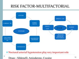
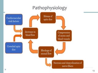
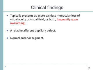
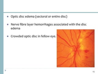
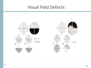
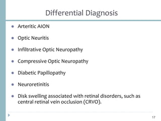
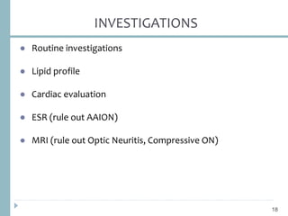
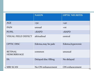
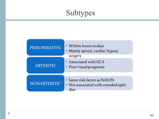
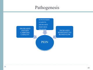
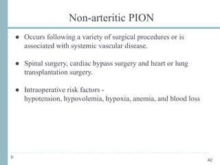
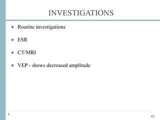
Ischemic Optic Neuropathy
Dr. Sriniwas Atal
MD Resident
Ophthalmology
BPKLCOS, IOM
LAYOUT
2
 INTRODUCTION
 ANTERIOR ISCHEMIC OPTIC NEUROPATHY
 BLOOD SUPPLY OF OPTIC NERVE HEAD
 NA-AION
 A-AION
 PION
 Case Report
 REFERENCES
Introduction
● Acute, painless optic neuropathy due to circulatory
insufficiency.
● > 50 years of age
● Most frequently at the optic nerve head (intraocular
insufficiency), Anterior Ischemic Optic Neuropathy (AION)
● Less frequently behind the optic neve head (intraorbital
insufficiency), Posterior Ischemic Optic Neuropathy (PION)
3
Anterior Ischemic Optic Neuropathy
● Most common cause of acute optic neuropathy in older age
groups.
● Rapid onset of painless, unilateral visual loss.
● Decreased visual acuity, visual field, or both.
4
1. Non-Arteritic Anterior Ischemic Optic Neuropathy
(NAAION)
2. Arteritic Anterior Ischemic Optic Neuropathy (AAION)
5
Non-arteritic Anterior Ischemic Neuropathy
(NAAION)
● 90-95% of AION
● Mean age - 60 yrs
● Less severe visual loss compared to Arteritic AION.
● Arcuate and altitudinal visual field defects.
6
RISK FACTORS
● Crowded optic disc
● “Disc at risk”
● Small optic disc plus small or absent cup
● Axonal crowding and compression
7
Blood supply
8
9
Blood flow
The blood flow in the ONH is calculated by using the following
formula (Hayreh et al):
Perfusion pressure = Mean BP – Intraocular pressure (IOP)
(Mean BP = Diastolic BP + 1/3 Pulse pressure)
From this formula, it is evident that the blood flow depends upon
(a) Resistance to blood flow, (b) BP and (c) IOP.
10
Autoregulation
● Helps compensate for any decrease in the blood flow.
● Operates only over a critical range of perfusion pressure.
● Rise or fall of perfusion pressure beyond the critical range
leads to autoregulation failure.
11
RISK FACTOR-MULTIFACTORIAL
Noctural arterial hypotension playvery importantrole
H’RGIC SHOCK
V
ASOSPASTIC
DISORDER
COLLAGEN
V
ASULAR DS
ABSENT CUP
RAISED
IOP
PROLONG
PRESSURE
ONEYEBALL
SMALL
CUP
SYSTEMIC
OCULAR
HTN/DM
H’RGIC
SHOCK
V
ASOSPASTIC
DISORDER
THYROID DS
COLLAGEN
V
ASCULAR
DS
CARDIAC DS
12
Drugs - Sildenafil, Amiodarone, Cocaine
Pathophysiology
13
Clinical findings
● Typically presents as acute painless monocular loss of
visual acuity or visual field, or both, frequently upon
awakening.
● A relative afferent pupillary defect.
● Normal anterior segment.
14
● Optic disc edema (sectoral or entire disc)
● Nerve fibre layer hemorrhages associated with the disc
edema
● Crowded optic disc in fellow eye.
15
Visual Field Defects
16
Differential Diagnosis
● Arteritic AION
● Optic Neuritis
● Infiltrative Optic Neuropathy
● Compressive Optic Neuropathy
● Diabetic Papillopathy
● Neuroretinitis
● Disk swelling associated with retinal disorders, such as
central retinal vein occlusion (CRVO).
17
INVESTIGATIONS
● Routine investigations
● Lipid profile
● Cardiac evaluation
● ESR (rule out AAION)
● MRI (rule out Optic Neuritis, Compressive ON)
18
19
Treatment
● None proven
● Aspirin
● Levodopa or carbidopa
● Hyperbaric O2
● Topical - Brimonidine
20
Surgical treatment
● Ischemic Optic Nerve Decompression Trial (IONDT)
● Optic nerve sheath fenestration, with drainage of perineural
subarachnoid cerebrospinal fluid.
● No effect of surgery on visual outcome.
21
Prognosis
● Improvement may take up to 6 months
● ~15% risk for fellow eye involvement in 2 years
● < 5 % risk for recurrent AION (the same eye)
● A significant visual field defect persists
22
Goal of Management
● Eliminating other potential treatable optic neuropathies such
as optic neuritis or a compressive optic neuropathy.
● Identifying any potential modifiable vascular risk factors.
23
Arteritic Anterior Ischemic Optic Neuropathy
24
● 5-10% of AION
● Short posterior ciliary artery vasculitis Infarction of optic
nerve head
● Commonly seen in elderly, mean age 70 yrs
● Severe visual loss in majority (<6/60)
● >50% caused by Giant Cell Arteritis (GCA)
● Other etiologies include:
Systemic Lupus Erythematosus
Wegener’s Granulomatosis
Behçet's disease
Churg Strauss Syndrome
Polyarteritis Nodosa
25
Giant Cell Arteritis
● Granulomatous necrotizing arteritis
● Mainly involves large and medium-size arteries :
Major aortic branches
Superficial temporal artery(STA)
Ophthalmic artery
Posterior ciliary arteries
Proximal vertebral arteries.
26
● Headache (most common)
● Jaw claudication (pathognomic)
● Temporal artery or scalp tenderness
● Malaise, Anorexia, Weight loss, Fever
● Superficial temporal arteritis is characterized by pulseless,
thickened, tender, inflamed and nodular arteries.
27
● 20% of GCA patients experience severe visual loss
● AION is the most common ophthalmic manifestation of GCA
● 30% have preceding transient visual loss (amaurosis fugax)
● 54% have visual acuity of counting fingers - No perception of
light
● >50% second eye involved within hours - weeks
● When temporal arteritis is suspected as the cause of visual
loss, treatment should not be delayed for results of laboratory
investigations or biopsy to prevent blindness in fellow eye.
28
● Optic disc pallor (pallid, chalky optic disc edema)
● Choroidal ischemia may be seen - peripapillary pallor and
edema deep to retina
● Disc of fellow eye is normal
● Cotton wool spots
29
Fundus Examination
● CRAO
● Anterior Ischaemic syndrome
● Ophthalmic artery occlusion
● Delay in choroidal perfusion or non-perfusion
● PION can occur
30
 Fluorescein fundusangiography
In earlystageshows filling defect in opticdisc, peripapillary choroid or
choroidal watershedarea 31
INVESTIGATIONS
● Westergren sedimentation rate
● C-reactive protein (reliable indicator than ESR)
● Complete blood count
● Fluorescein angiography
● Temporal artery biopsy - ‘gold standard’
2-3 cm specimen taken to avoid skip lesion
32
Treatment
● Steroids
● Methotrexate
● Aspirin
● Rheumatology consultation & follow-up
33
Regimen
● IV Methylprednisolone 1 g/day for 3 days.
● Followed by oral Prednisolone 1-2mg/kg/day for 3 days.
● Then 50-60mg/day (not less than 0.75mg/kg/day) for 4 weeks
until symptom resolution and ESR/CRP normalisation.
● Then tapering by 10mg/day every 2 weeks until 20mg/kg.
● Tapering then titrated against ESR/CRP and symptoms.
34
35
Posterior Ischemic Optic Neuropathy(PION)
● Uncommon
● No disc edema
● The mechanism of ischemia is presumed to be insufficiency
of pial arterial vascular supply to the optic nerve distal to the
optic nerve head.
36
Diagnosis
● Rule out other types of retrobulbar optic neuropathies,
including traumatic, infiltrative, inflammatory, toxic,
radiation, and compressive types.
37
CLINICAL FEATURES
● Sudden, painless unilateral or bilateral vision loss.
● Relative afferent pupillary defect in unilateral cases as well as
in bilateral cases when the degree of vision loss is unequal
between the two eyes.
● Normal disc at presentation.
● Optic disc atrophy ensues in 4-6 weeks.
38
Subtypes (Sadda et al)
1. Arteritic PION (associated with temporal arteritis)
2. Non-arteritic PION
39
Subtypes
• Within hours todays
• Mainly spinal, cardiac bypass
surgery
PERIOPERATIVE
• Associated with GCA
• Poor visualprognosis
ARTERITIC
• Same risk factor asNAION
• Not associated with crowdedoptic
disc
NONARTERITIC
40
Pathogenesis
PION
DECREASED
OXYGEN
CARRYING
CAPACITY
HYPOTENSION
LEADS TO
DECREASED
PERFUSION
PRESSURE
INCREASED
RESISTANCETO
BLOOD FLOW
41
Non-arteritic PION
● Occurs following a variety of surgical procedures or is
associated with systemic vascular disease.
● Spinal surgery, cardiac bypass surgery and heart or lung
transplantation surgery.
● Intraoperative risk factors -
hypotension, hypovolemia, hypoxia, anemia, and blood loss
42
INVESTIGATIONS
● Routine investigations
● ESR
● CT/MRI
● VEP - shows decreased amplitude
43
TREATMENT
● No effective treatment
● Steroid
● Aspirin
● IOP lowering agents
● Hemodynamic correction
44
Case of A-AION
45
 A 77-year-old Chinese man complained of bilateral,
simultaneous onset vision loss for 5 days, accompanied by
severe headache on right side and jaw pain. The visual acuities
were no light perception in both eyes.The bilateral superficial
temporal arteries were palpable and tenderness.
46
Fundus photographs at presentation of the patient showing severe
bilateral optic disc swollen, with “chalky white” pallid appearance;
there are splinter hemorrhage and cotton wool spots on the retina in
the right eye. The choroid showing diffused atrophy around the optic
disc
47
The orbital fat-suppression T1-weighted magnetic resonance imaging with
contract showing the enhancement of the optic nerve sheath
in the right eye, (a):axial and (b): sagittal. The white arrows indicate the optic
nerve sheath; the MRA of the cerebral vascular is unremarkable (c)
48
 The right temporal artery biopsy was performed and revealed
the occlusion of the luminal owing to the intimal proliferation
and infiltration
 Methylprednisolone 1 g/d was intravenousfor 3 days followed
by prednisone 1 mg/kg/d.
 The vision acuity maintained NLP bilateral after treatment,
whereas the headache and jaw pain disappeared. The oral
prednisone was weaned and methotrexate was added as the
immunosuppressive agent for long treatment.
Take Home Message
● ION is an ophthalmic emergency.
● Patients with GCA+ION are in danger of catastrophic,
irreversible, bilateral blindness that may be prevented by
prompt treatment with corticosteroids.
● Thus, any patient > 50 presenting with ION an immediate
workup to rule out GCA.
49
● There is no effective treatment for ION.
● Limited efficacy for prophylaxis.
● Aspirin 100mg daily.
● Control of cardiovascular risk factors.
● Suspect GCA !!!
● Avoid prolonged surgical time and dramatic shifts in body
perfusion during surgrey.
50
References
 Walsh and Hoyt's Clinical Neuro-ophthalmology, 6th edition.
Volume 3.
 Myron Yanoff and Jay S. Duker. Ophthalmology, 5th edition
2019. Elsevier.
 Albert and Jakobiec’s, Miller, Azar, Bloo Principle and Practice
of Ophthalmology. Volume III. Elsevier.
 AAO. Basic and clinical science course. Neuro-ophthalmology
.Volume 5, 2019-20
 Tian et al. BMC Ophthalmology (2018) 18:282
https://doi.org/10.1186/s12886-018-0953-5
51
Ischemic Optic Neuropathy 1.pptx

Ischemic Optic Neuropathy 1.pptx

  • 1.
    Ischemic Optic Neuropathy Dr.Sriniwas Atal MD Resident Ophthalmology BPKLCOS, IOM
  • 2.
    LAYOUT 2  INTRODUCTION  ANTERIORISCHEMIC OPTIC NEUROPATHY  BLOOD SUPPLY OF OPTIC NERVE HEAD  NA-AION  A-AION  PION  Case Report  REFERENCES
  • 3.
    Introduction ● Acute, painlessoptic neuropathy due to circulatory insufficiency. ● > 50 years of age ● Most frequently at the optic nerve head (intraocular insufficiency), Anterior Ischemic Optic Neuropathy (AION) ● Less frequently behind the optic neve head (intraorbital insufficiency), Posterior Ischemic Optic Neuropathy (PION) 3
  • 4.
    Anterior Ischemic OpticNeuropathy ● Most common cause of acute optic neuropathy in older age groups. ● Rapid onset of painless, unilateral visual loss. ● Decreased visual acuity, visual field, or both. 4
  • 5.
    1. Non-Arteritic AnteriorIschemic Optic Neuropathy (NAAION) 2. Arteritic Anterior Ischemic Optic Neuropathy (AAION) 5
  • 6.
    Non-arteritic Anterior IschemicNeuropathy (NAAION) ● 90-95% of AION ● Mean age - 60 yrs ● Less severe visual loss compared to Arteritic AION. ● Arcuate and altitudinal visual field defects. 6
  • 7.
    RISK FACTORS ● Crowdedoptic disc ● “Disc at risk” ● Small optic disc plus small or absent cup ● Axonal crowding and compression 7
  • 8.
  • 9.
  • 10.
    Blood flow The bloodflow in the ONH is calculated by using the following formula (Hayreh et al): Perfusion pressure = Mean BP – Intraocular pressure (IOP) (Mean BP = Diastolic BP + 1/3 Pulse pressure) From this formula, it is evident that the blood flow depends upon (a) Resistance to blood flow, (b) BP and (c) IOP. 10
  • 11.
    Autoregulation ● Helps compensatefor any decrease in the blood flow. ● Operates only over a critical range of perfusion pressure. ● Rise or fall of perfusion pressure beyond the critical range leads to autoregulation failure. 11
  • 12.
    RISK FACTOR-MULTIFACTORIAL Noctural arterialhypotension playvery importantrole H’RGIC SHOCK V ASOSPASTIC DISORDER COLLAGEN V ASULAR DS ABSENT CUP RAISED IOP PROLONG PRESSURE ONEYEBALL SMALL CUP SYSTEMIC OCULAR HTN/DM H’RGIC SHOCK V ASOSPASTIC DISORDER THYROID DS COLLAGEN V ASCULAR DS CARDIAC DS 12 Drugs - Sildenafil, Amiodarone, Cocaine
  • 13.
  • 14.
    Clinical findings ● Typicallypresents as acute painless monocular loss of visual acuity or visual field, or both, frequently upon awakening. ● A relative afferent pupillary defect. ● Normal anterior segment. 14
  • 15.
    ● Optic discedema (sectoral or entire disc) ● Nerve fibre layer hemorrhages associated with the disc edema ● Crowded optic disc in fellow eye. 15
  • 16.
  • 17.
    Differential Diagnosis ● ArteriticAION ● Optic Neuritis ● Infiltrative Optic Neuropathy ● Compressive Optic Neuropathy ● Diabetic Papillopathy ● Neuroretinitis ● Disk swelling associated with retinal disorders, such as central retinal vein occlusion (CRVO). 17
  • 18.
    INVESTIGATIONS ● Routine investigations ●Lipid profile ● Cardiac evaluation ● ESR (rule out AAION) ● MRI (rule out Optic Neuritis, Compressive ON) 18
  • 19.
  • 20.
    Treatment ● None proven ●Aspirin ● Levodopa or carbidopa ● Hyperbaric O2 ● Topical - Brimonidine 20
  • 21.
    Surgical treatment ● IschemicOptic Nerve Decompression Trial (IONDT) ● Optic nerve sheath fenestration, with drainage of perineural subarachnoid cerebrospinal fluid. ● No effect of surgery on visual outcome. 21
  • 22.
    Prognosis ● Improvement maytake up to 6 months ● ~15% risk for fellow eye involvement in 2 years ● < 5 % risk for recurrent AION (the same eye) ● A significant visual field defect persists 22
  • 23.
    Goal of Management ●Eliminating other potential treatable optic neuropathies such as optic neuritis or a compressive optic neuropathy. ● Identifying any potential modifiable vascular risk factors. 23
  • 24.
    Arteritic Anterior IschemicOptic Neuropathy 24 ● 5-10% of AION ● Short posterior ciliary artery vasculitis Infarction of optic nerve head ● Commonly seen in elderly, mean age 70 yrs ● Severe visual loss in majority (<6/60)
  • 25.
    ● >50% causedby Giant Cell Arteritis (GCA) ● Other etiologies include: Systemic Lupus Erythematosus Wegener’s Granulomatosis Behçet's disease Churg Strauss Syndrome Polyarteritis Nodosa 25
  • 26.
    Giant Cell Arteritis ●Granulomatous necrotizing arteritis ● Mainly involves large and medium-size arteries : Major aortic branches Superficial temporal artery(STA) Ophthalmic artery Posterior ciliary arteries Proximal vertebral arteries. 26
  • 27.
    ● Headache (mostcommon) ● Jaw claudication (pathognomic) ● Temporal artery or scalp tenderness ● Malaise, Anorexia, Weight loss, Fever ● Superficial temporal arteritis is characterized by pulseless, thickened, tender, inflamed and nodular arteries. 27
  • 28.
    ● 20% ofGCA patients experience severe visual loss ● AION is the most common ophthalmic manifestation of GCA ● 30% have preceding transient visual loss (amaurosis fugax) ● 54% have visual acuity of counting fingers - No perception of light ● >50% second eye involved within hours - weeks ● When temporal arteritis is suspected as the cause of visual loss, treatment should not be delayed for results of laboratory investigations or biopsy to prevent blindness in fellow eye. 28
  • 29.
    ● Optic discpallor (pallid, chalky optic disc edema) ● Choroidal ischemia may be seen - peripapillary pallor and edema deep to retina ● Disc of fellow eye is normal ● Cotton wool spots 29 Fundus Examination
  • 30.
    ● CRAO ● AnteriorIschaemic syndrome ● Ophthalmic artery occlusion ● Delay in choroidal perfusion or non-perfusion ● PION can occur 30
  • 31.
     Fluorescein fundusangiography Inearlystageshows filling defect in opticdisc, peripapillary choroid or choroidal watershedarea 31
  • 32.
    INVESTIGATIONS ● Westergren sedimentationrate ● C-reactive protein (reliable indicator than ESR) ● Complete blood count ● Fluorescein angiography ● Temporal artery biopsy - ‘gold standard’ 2-3 cm specimen taken to avoid skip lesion 32
  • 33.
    Treatment ● Steroids ● Methotrexate ●Aspirin ● Rheumatology consultation & follow-up 33
  • 34.
    Regimen ● IV Methylprednisolone1 g/day for 3 days. ● Followed by oral Prednisolone 1-2mg/kg/day for 3 days. ● Then 50-60mg/day (not less than 0.75mg/kg/day) for 4 weeks until symptom resolution and ESR/CRP normalisation. ● Then tapering by 10mg/day every 2 weeks until 20mg/kg. ● Tapering then titrated against ESR/CRP and symptoms. 34
  • 35.
  • 36.
    Posterior Ischemic OpticNeuropathy(PION) ● Uncommon ● No disc edema ● The mechanism of ischemia is presumed to be insufficiency of pial arterial vascular supply to the optic nerve distal to the optic nerve head. 36
  • 37.
    Diagnosis ● Rule outother types of retrobulbar optic neuropathies, including traumatic, infiltrative, inflammatory, toxic, radiation, and compressive types. 37
  • 38.
    CLINICAL FEATURES ● Sudden,painless unilateral or bilateral vision loss. ● Relative afferent pupillary defect in unilateral cases as well as in bilateral cases when the degree of vision loss is unequal between the two eyes. ● Normal disc at presentation. ● Optic disc atrophy ensues in 4-6 weeks. 38
  • 39.
    Subtypes (Sadda etal) 1. Arteritic PION (associated with temporal arteritis) 2. Non-arteritic PION 39
  • 40.
    Subtypes • Within hourstodays • Mainly spinal, cardiac bypass surgery PERIOPERATIVE • Associated with GCA • Poor visualprognosis ARTERITIC • Same risk factor asNAION • Not associated with crowdedoptic disc NONARTERITIC 40
  • 41.
  • 42.
    Non-arteritic PION ● Occursfollowing a variety of surgical procedures or is associated with systemic vascular disease. ● Spinal surgery, cardiac bypass surgery and heart or lung transplantation surgery. ● Intraoperative risk factors - hypotension, hypovolemia, hypoxia, anemia, and blood loss 42
  • 43.
    INVESTIGATIONS ● Routine investigations ●ESR ● CT/MRI ● VEP - shows decreased amplitude 43
  • 44.
    TREATMENT ● No effectivetreatment ● Steroid ● Aspirin ● IOP lowering agents ● Hemodynamic correction 44
  • 45.
    Case of A-AION 45 A 77-year-old Chinese man complained of bilateral, simultaneous onset vision loss for 5 days, accompanied by severe headache on right side and jaw pain. The visual acuities were no light perception in both eyes.The bilateral superficial temporal arteries were palpable and tenderness.
  • 46.
    46 Fundus photographs atpresentation of the patient showing severe bilateral optic disc swollen, with “chalky white” pallid appearance; there are splinter hemorrhage and cotton wool spots on the retina in the right eye. The choroid showing diffused atrophy around the optic disc
  • 47.
    47 The orbital fat-suppressionT1-weighted magnetic resonance imaging with contract showing the enhancement of the optic nerve sheath in the right eye, (a):axial and (b): sagittal. The white arrows indicate the optic nerve sheath; the MRA of the cerebral vascular is unremarkable (c)
  • 48.
    48  The righttemporal artery biopsy was performed and revealed the occlusion of the luminal owing to the intimal proliferation and infiltration  Methylprednisolone 1 g/d was intravenousfor 3 days followed by prednisone 1 mg/kg/d.  The vision acuity maintained NLP bilateral after treatment, whereas the headache and jaw pain disappeared. The oral prednisone was weaned and methotrexate was added as the immunosuppressive agent for long treatment.
  • 49.
    Take Home Message ●ION is an ophthalmic emergency. ● Patients with GCA+ION are in danger of catastrophic, irreversible, bilateral blindness that may be prevented by prompt treatment with corticosteroids. ● Thus, any patient > 50 presenting with ION an immediate workup to rule out GCA. 49
  • 50.
    ● There isno effective treatment for ION. ● Limited efficacy for prophylaxis. ● Aspirin 100mg daily. ● Control of cardiovascular risk factors. ● Suspect GCA !!! ● Avoid prolonged surgical time and dramatic shifts in body perfusion during surgrey. 50
  • 51.
    References  Walsh andHoyt's Clinical Neuro-ophthalmology, 6th edition. Volume 3.  Myron Yanoff and Jay S. Duker. Ophthalmology, 5th edition 2019. Elsevier.  Albert and Jakobiec’s, Miller, Azar, Bloo Principle and Practice of Ophthalmology. Volume III. Elsevier.  AAO. Basic and clinical science course. Neuro-ophthalmology .Volume 5, 2019-20  Tian et al. BMC Ophthalmology (2018) 18:282 https://doi.org/10.1186/s12886-018-0953-5 51