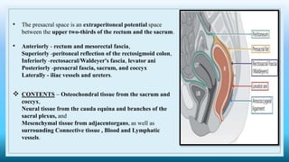
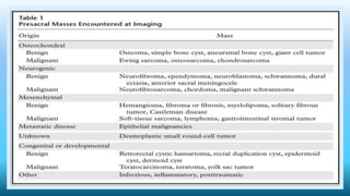
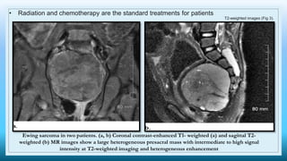
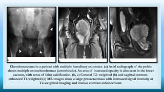
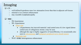
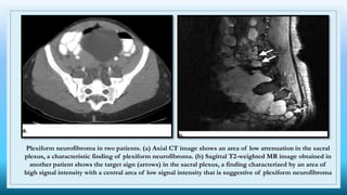
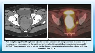
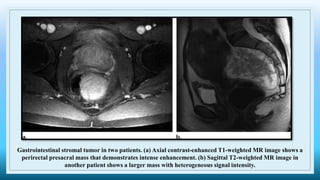
2. • The presacral space is an extraperitoneal potential space
between the upper two-thirds of the rectum and the sacrum.
• Anteriorly - rectum and mesorectal fascia,
Superiorly -peritoneal reflection of the rectosigmoid colon,
Inferiorly -rectosacral/Waldeyer's fascia, levator ani
Posteriorly -presacral fascia, sacrum, and coccyx
Laterally - iliac vessels and ureters.
CONTENTS – Osteochondral tissue from the sacrum and
coccyx,
Neural tissue from the cauda equina and branches of the
sacral plexus, and
Mesenchymal tissue from adjacentorgans, as well as
surrounding Connective tissue , Blood and Lymphatic
vessels.
5. GIANT CELL TUMOR
• most common sacral tumors after chordoma
• 2nd–4th decades, F>M
• locally aggressive, lytic, destructive, expansile lesion, often in an eccentric
location
• commonly involve both sides of the midline and may extend across the
sacroiliac joints
6. CT - osteolytic or radiolucent lesion , soft-tissue mass within the bone; usually no
surrounding sclerosis (80-85%) , the mass may have a thin sclerotic rim
MR –
• T1
○ low to intermediate signal (solid component)
○ low signal rim
• T2: heterogeneous high signal with areas of low signal intensity due to hemosiderin
or fibrosis
• T1 C+ (Gd): solid components will enhance
7. Giant cell tumor. (a) Lateral radiograph shows a lytic lesion involving S1 and S2. (b) Sagittal T2-weighted MR image
shows that the lesion has heterogeneous signal intensity. (c) Axial contrast-enhanced T1-weighted MR image shows
that the lesion demonstrates intense enhancement. (d) Axial CT image obtained with bone window settings clearly
shows lytic bone destruction.
8. EWING SARCOMA
• Three to ten percent of all primary Ewing sarcomas occur in the spine, but metastatic involvement
is more common
• Ages Group - 5 and 30
9. • Aggressive, demonstrating permeative bone lysis, osseous expansion, or sclerosis
• lamellated (onion skin) periosteal reaction
• Associated soft-tissue masses are common
• MRI - T1: low to intermediate signal
T1 C+ (Gd): heterogeneous but prominent enhancement
T2: heterogeneously high signal, may see hair on end low signal striations
IMAGING
10. T2-weighted images (Fig 3).
• Radiation and chemotherapy are the standard treatments for patients
Ewing sarcoma in two patients. (a, b) Coronal contrast-enhanced T1- weighted (a) and sagittal T2-
weighted (b) MR images show a large heterogeneous presacral mass with intermediate to high signal
intensity at T2-weighted imaging and heterogeneous enhancement
12. Plain radiograph –
o lytic (50%)
o intralesional calcifications: ~70% (rings and arcs calcification or popcorn
calcification), characteristic chondroid matrix
o endosteal scalloping: affecting more than two-thirds of the cortical thickness
o moth-eaten appearance or permeative appearance in higher grade tumors
CT –
o attenuation of the non-mineralized portion is often lower than that of muscle due to the
relatively high water content of hyaline cartilage
IMAGING
13. MRI –
o T1: low to intermediate signal.
o T2: very high intensity in non-mineralized/calcified portions - the cartilage is a
hydrophilic tissue with high water content
o gradient echo/SWI: blooming of mineralized/calcified portions.
o T1 C+ (Gd) :
■ enhancement can be septal and peripheral rim-like corresponding to fibrovascular
septation between lobules of hyaline cartilage - rings and arcs enhancement
pattern
■ most demonstrate heterogeneous moderate to intense contrast enhancement.
14. Chondrosarcoma in a patient with multiple hereditary exostoses. (a) Axial radiograph of the pelvis
shows multiple osteochondromas (arrowheads). An area of increased opacity is also seen in the lower
sacrum, with areas of faint calcification. (b, c) Coronal T2- weighted (b) and sagittal contrast-
enhanced T1-weighted (c) MR images show a large presacral mass with increased signal intensity at
T2-weighted imaging and intense contrast enhancement.
16. NEUROFIBROMAS
• benign neural tumors
• Neurofibromas most often occur in isolation, although multiple tumors may occur in
patients with neurofibromatosis type 1 (NF1)
17. Imaging
CT –
o well-defined hypodense mass low attenuation lower than that in adjacent soft tissues
o minimal or no contrast enhancement
o may resemble lymphadenopathy
MRI –
o T1: hypointense
o T2 : hyperintense
■ target sign
● a hyperintense rim (myxoid material ) and central area of a low signal (dense
central area of collagenous stroma ) may be seen
● although this sign is highly suggestive of neurofibroma, it is occasionally also
seen in schwannomas and malignant peripheral nerve sheath tumors
■ fascicular sign
o T1 C+ (Gd): heterogeneous enhancement
18. Plexiform neurofibroma in two patients. (a) Axial CT image shows an area of low attenuation in the sacral
plexus, a characteristic finding of plexiform neurofibroma. (b) Sagittal T2-weighted MR image obtained in
another patient shows the target sign (arrows) in the sacral plexus, a finding characterized by an area of
high signal intensity with a central area of low signal intensity that is suggestive of plexiform neurofibroma
19. SCHWANNOMAS
• benign nerve sheath tumors
• peak incidence in the 5th to 6th decades
• When they occur in patients with neurofibromatosis type 2 (NF2), schwannomas
usually present by the 3rd decade
• Multiple schwannomas are characteristic of neurofibromatosis type 2
• large mass, and they may remodel or erode through the sacral bone
20. • well-circumscribed masses which displace adjacent structures without direct invasion
• cystic and fatty degeneration is common
• calcification is rare
CT –
o heterogeneous presacral soft-tissue mass, with or without calcifications.
o intense contrast enhancement
o adjacent bone remodeling with smooth corticated edges
MR –
o T1: isointense or hypointense
o T1 C+ (Gd): intense enhancement
o T2: heterogeneously hyperintense, cystic degenerative areas may be present, especially in larger
tumors
o T2*: larger tumors often have areas of hemosiderin
Imaging
21. Schwannoma in two patients. (a) Axial T2-weighted MR image shows a high-signal-intensity presacral mass
with scattered cystic areas. (b) Sagittal contrast-enhanced T1-weighted MR image shows that the mass
demonstrates heterogeneous enhancement. (c) Axial contrast-enhanced CT image in another patient shows a
heterogeneously enhancing presacral mass.
22. PERINEURAL CYSTS
• CSF-filled dilatations of the nerve root sheath at the dorsal root ganglion (posterior nerve root
sheath)
• most commonly occur at the level of the second and third sacral nerve roots
• cyst wall is continuous with the arachnoid and dura, and the cyst cavity occupies the space
between the perineurium and endoneurium
• may be congenital or traumatic
• Stenosis of the ostium of the nerve root sheath allows cerebrospinal fluid (CSF) to flow in with
arterial pulsation and restricts outflow
23. CT –
o cystic mass that is isoattenuating relative to CSF at the neural foramina
o Bone remodeling and enlargement of the neural foramina may be seen, a result of
increased CSF pressure
o Because communicate with CSF, perineural cysts fill with contrast material at CT
myelography
MRI –
o T1: low signal
o T2:
■ high signal
■ will show chemical shift at fat-fluid boundary with epidural fat
o T1 C+: no enhancement
o DWI/ADC: facilitated diffusion ~3000 x 10-6 mm2/s
Imaging
24. A peri-neural cyst at the level of S2 segment lateralizing to right with smooth
scalloping of the related bony skeleton and mild effacement of the related right
sided sacral nerve roots
25. DURAL ECTASIA
• ballooning or widening of the dural sac which can result in posterior vertebral scalloping and is
associated with herniation of nerve root sleeves.
• defective microfibrils weaken the dural sac, usually in the lumbosacral spine, where CSF
pressure is greatest, which leads to incompetence
• Dural ectasia is present in 56%–65% of patients with Marfan syndrome
26. • widening of the interpediculate distance and posterior vertebral body scalloping
• Widening of the dural sac, dilatation of the nerve root sleeves, and scalloping of the posterior
vertebral bodies may be seen in the lumbosacral spine
Dural ectasia in a patient with Marfan syndrome. Axial contrast-enhanced CT images obtained with
bone (a) and soft-tissue (b) window settings show dural ectasia with bone remodeling in the sacral
foramina.
27. ANTERIOR MYELOMENINGOCELES
• Anterior myelomeningoceles occur when the dural sac herniates through a defect in the anterior
surface of the sacrum
• Dural sac is composed of both the outer dura and the inner arachnoid membranes, and it
contains CSF and, occasionally, neural elements
• manifest in the 1st decade of life
• Currarino triad, which comprises an anorectal malformation, a sacrococcygeal osseous defect,
and a presacral mass. The associated presacral mass may be a teratoma, anterior sacral
meningocele, dermoid cyst, hamartoma, or enteric duplication cyst and is autosomal dominant
in more than 50% of patients.
• classic clinical presentation of headache during valsalva owing to increased cerebrospinal fluid
(CSF) pressure transmitted via the connection between the meningocele and subdural space
28. Imaging
pelvic radiographs - unilateral anterior sacral defect may be seen with a rounded, concave border
with scalloping/destruction of the surrounding bone, often referred to as the “scimitar sign”
CT - Osseous defects, such as vertebral body scalloping, hypoplasia, and aplasia, may accompany
an anterior sacral meningocele and are best assessed at CT
MRI - the signal intensity of the content within the meningocele should parallel the CSF signal on
all sequences
• Biopsy and aspiration should not be performed, given the risk of introduction of pathogens
directly into the spinal meninges, which can result in meningitis.
29. Well defined, rounded defect in the lower
sacrum with a huge fluid collection in the
pelvis communicating with the spinal thecal
sac - typical of a sacral meningocoele.
MRI confirms the CT findings indicating that the
huge fluid collection in the pelvis is connected to the
thecal sac and is not the bladder or other pathological
cyst.
30. A 17-year-old-male with anterior
sacral meningocele.
Sagittal T2 weighted image showing
a sacral defect (black arrow) and a
small anterior cystic mass with
demonstrable direct communication
with the dural sac (white arrow).
31. PARAGANGLIOMAS
• neuroendocrine neoplasms that arise from the paraganglia, accessory organs of the
peripheral nervous system
• Typically, spinal paragangliomas are intradural, extramedullary lesions located in the
region of the cauda equina and filum terminale
• more common in men,
32. • Paragangliomas are highly vascular tumors that demonstrate an intense, early blush at
angiography that persists into the late arterial and early venous phases
• well-circumscribed mass that is isointense on T1-weighted MR images and iso- to hyperintense
on T2-weighted
• Hemorrhage is common, and a low-signal-intensity rim (the cap sign) may be seen. Intense
enhancement is seen on contrast-enhanced images, and flow voids are common
33. Paraganglioma in a pregnant woman. Axial (a) and sagittal (b) T2-weighted MR images
show a large presacral mass that is iso- to hyperintense with a hypointense rim, a finding
known as the cap sign.
34. CHORDOMAS
• most common primary sacral tumor
• from notochordal rests and are limited to the clivus, spine, and sacrum on the basis of their cell
of origin
• typically located in the midline or paramedian region
• most common in the 4th–7th decades of life
35. • Chordomas are destructive lytic lesions that may extend across the sacroiliac joints
• Calcification is common in sacrococcygeal lesions
• large presacral soft-tissue component is present, with soft tissues extending into the sacral canal.
CT
o centrally located
o well-circumscribed
o destructive lytic lesion, sometimes with marginal sclerosis
o expansile soft-tissue mass soft-tissue mass is often disproportionately large relative to the
bony destruction
o irregular intratumoral calcifications (thought to represent sequestra of normal bone rather
than dystrophic calcifications)
o moderate enhancement
low to intermediate signal intensity on T1-weighted MR images and high signal intensity on T2-
weighted variable, often only moderate, contrast enhancement
36. Chordoma. (a) Axial contrastenhanced CT image shows a homogeneous mass anterior to the distal
sacrum and coccyx. (b) Sagittal T2-weighted MR image shows the hyperintense mass. (c) Axial
contrast-enhanced T1-weighted MR image shows that the mass demonstrates only mild enhancement.
38. HEMANGIOMAS
• most common soft-tissue abnormality
• most common tumors in infants and usually appear within the 1st week of life
• three times more common in girls
39. Imaging
• intermediate signal intensity on T1-weighted MR images and high signal intensity and flow
voids on T2-weighted images
• On T2-weighted images, clusters of high-signal-intensity lobules indicate cystic vascular spaces
that contain stagnant blood.
• Areas of increased signal intensity on T1- and T2- weighted images correlate with haemorrhage
and areas of fat deposition.
• Phleboliths are common and appear as low-signal-intensity foci on both T1- and T2-weighted
images.
40. Perirectal hemangioma in two patients. (a) Sagittal short inversion time inversion-recovery MR image
shows a perirectal mass with heterogeneous high signal intensity. (b) Axial contrast-enhanced T1-weighted
MR image shows that the mass demonstrates heterogeneous enhancement. (c) Axial contrast enhanced CT
image obtained in another patient shows phleboliths, a typical finding of perirectal hemangioma.
42. • CT - they typically appear as a hypovascular mass that contains macroscopic fat with areas of
soft-tissue attenuation interspersed
• T1-weighted MR imaging, the fatty areas have high signal intensity that drops out when fat is
suppressed.
Imaging
43. Myelolipoma.
(a) Axial contrast-enhanced CT
image shows a well-circumscribed
presacral mass with macroscopic
fat.
(b, c) Axial (b) and sagittal
(c) T1-weighted MR images show that
the areas of fat demonstrate increased
signal intensity.
(d) Sagittal T1-weighted fat-
suppressed MR image shows dropout
of signal intensity in the areas of fat.
44. SOLITARY FIBROUS TUMORS
• slow-growing mass in middle-aged adults
• Solitary fibrous tumors are mesenchymal tumors of fibroblastic or myofibroblastic origin
45. Imaging
CT - well-circumscribed mass with soft-tissue attenuation that typically demonstrates intense
contrast enhancement
Areas of hemorrhage, necrosis, and cystic change may be seen.
MRI - low to intermediate signal intensity on T1- and T2- weighted MR images a result of
fibrous tissue, but areas of myxoid or cystic degeneration may demonstrate high signal intensity
on T2-weighted images.
Hypervascularity with prominent enhancement and flow voids is common.
46. Solitary fibrous tumor.
(a) Axial contrast-enhanced CT image shows a homogeneous mass that is
isoattenuating relative to muscle.
(b) Sagittal T1-weighted MR image shows flow voids (arrows) in the low-signal-
intensity mass.
(c) (c, d) Axial
(c) and coronal
(d) contrast-enhanced T1-weighted MR images show that the mass demonstrates
intense enhancement. (e) Angiogram shows the tumor, which demonstrates
increased vascularity
47. LYMPHOMAS
• CT - lymphomas have soft-tissue attenuation with homogeneous enhancement
• MR imaging, they are characterized by an area of low signal intensity on T1-weighted images
and an area of high signal intensity on T2-weighted images
48. Non-Hodgkin–type B-cell lymphoma. (a) Axial contrast-enhanced CT image shows an abnormal area of
homogeneous enhancement in the rectal and perirectal soft tissues. (b) Positron emission tomographic
(PET)/CT image shows an area of intense uptake that corresponds to the abnormal rectal and perirectal
soft tissues seen at CT
49. GASTROINTESTINAL STROMAL TUMORS
• most commonly occur in older patients and have a slight male predominance
• Gastrointestinal stromal tumors usually involve the outer muscular layer and tend to be
exophytic. When located in the anorectal region, they tend to expand to the rectal wall, resulting
in a focal well-circumscribed mural mass
50. Imaging
• CT - soft-tissue attenuation with a central area of low attenuation resulting from hemorrhage,
necrosis, or cyst formation.
• Peripheral enhancement is common
• MRI - uniform intermediate signal intensity is seen on T1-weighted MR images and an area of
heterogeneous high signal intensity is seen on T2-weighted images, with heterogeneous
enhancement. Extension into the ischiorectal fossa, prostate, or vagina may be present.
51. Gastrointestinal stromal tumor in two patients. (a) Axial contrast-enhanced T1-weighted MR image shows a
perirectal presacral mass that demonstrates intense enhancement. (b) Sagittal T2-weighted MR image in
another patient shows a larger mass with heterogeneous signal intensity.
53. DEVELOPMENTAL CYSTS
• Developmental cysts are the most common congenital condition in the presacral space and
include epidermoid, dermoid, enteric, tailgut (also known as retrorectal cystic hamartoma), and
duplication cysts
• female predilection
• 13% of these cysts may undergo malignant transformation, and for this reason, they are
removed
• its typically manifest during childhood, and they are relatively rare in adults.
• Tailgut cysts are the most common asymptomatic presacral lesions that are incidentally found in
adults
54. • Tailgut cysts are often multiloculated cysts containing mucin and lack a muscular layer, a
differentiating feature from rectal duplication cysts, which can be confirmed on endorectal
ultrasound
• Rectal duplication cysts may be associated with other congenital abnormalities of the anorectal
region and bladder/urethra.
• Sacrococcygeal teratoma is the most common presacral mass in children containing all three
germ-cell lineages.
• Benign mature teratomas tend to be predominantly cystic containing fat, sebum, calcification and
soft tissue from dermoid plugs.
55. • Thin-walled and may be uni- or multilocular.
USG - Internal echoes related to mucoid material or inflammatory debris may be seen.
CT –
o low attenuation , with no associated enhancement, and associated thin calcifications are
rare
o If secondarily infected - thick-walled with surrounding inflammatory change, and they
may contain air if a fistula is present.
MRI –
• hypointense on T1 (although they may be hyperintense if they contain mucoid material),
hyperintense on T2.
• Focal irregular wall thickening with enhancement is suggestive of malignant degeneration
Imaging
56. Rectal duplication cyst. (a) Sagittal T2-weighted MR image shows a large hyperintense presacral cystic
mass. (b) Coronal contrast-enhanced T1-weighted MR image shows a thin area of peripheral
enhancement around the mass.
57. ON IMAGING - CONGENITAL DEVELOPMENTAL CYSTS,
● well defined, unilocular or multilocular, cystic masses ranging from simple to complex in their internal
contents
A 45-year-old-female with tailgut cyst. (a) Axial T2 weighted, (b) fat-saturated pre-contrast and (c) fat-saturated post-contrast
subtraction T1 weighted images (T1WI) through the pelvis showing a presacral multiloculated cystic mass posterior to the
rectum without post-contrast enhancement (arrows in a, c). High signal intensity on pre-contrast T1WI (arrow in b),
suggesting haemorrhage or proteinaceous debris. Surgical resection confirmed tailgut cyst.
58. GERM CELL TUMORS
• occur secondary to disorganization of totipotent primitive neural cells during embryogenesis
• degree of differentiation determines the tumor type, with teratomas, yolk sac tumors, and
embryonal cell carcinomas demonstrating lower degrees of differentiation
• Sacrococcygeal teratoma is the most common presacral germ cell tumor in children and the
most common solid tumor in neonates
59. • Benign teratomas contain only mature tissues, including fluid, fat, calcification, and a small
amount of soft tissue. Benign teratomas are predominantly cystic, with fluid attenuation at CT.
Areas of bone, fat, and calcification may be seen.
• T1-weighted MR imaging, areas that contain fat have high signal intensity, and those that
contain bone and calcifications demonstrate signal void
60. Sacrococcygeal teratoma in two patients. (a) Image from a barium enema study shows widening of the
presacral space. (b, c) Axial (b) and sagittal (c) contrast-enhanced CT images in another patient show a large
multiloculated solid and cystic mass that extends into the sacral canal and foramina. Associated bone
remodeling and destruction are seen
61. • Yolk sac tumors have a more aggressive imaging appearance than teratomas and are
characterized by more heterogeneous soft tissues and areas of hemorrhage and necrosis
• CT, areas of low attenuation with no enhancement are seen, and at T1-weightd MR imaging,
areas of hyperintensity are seen, a finding related to hemorrhage.
• Yolk sac tumors demonstrate heterogeneous contrast enhancement and often invade adjacent
organs.
62. Yolk sac tumor. Axial (a) and sagittal (b) contrast-enhanced CT images show a heterogeneously enhancing
presacral mass that extends into the sacral canal.