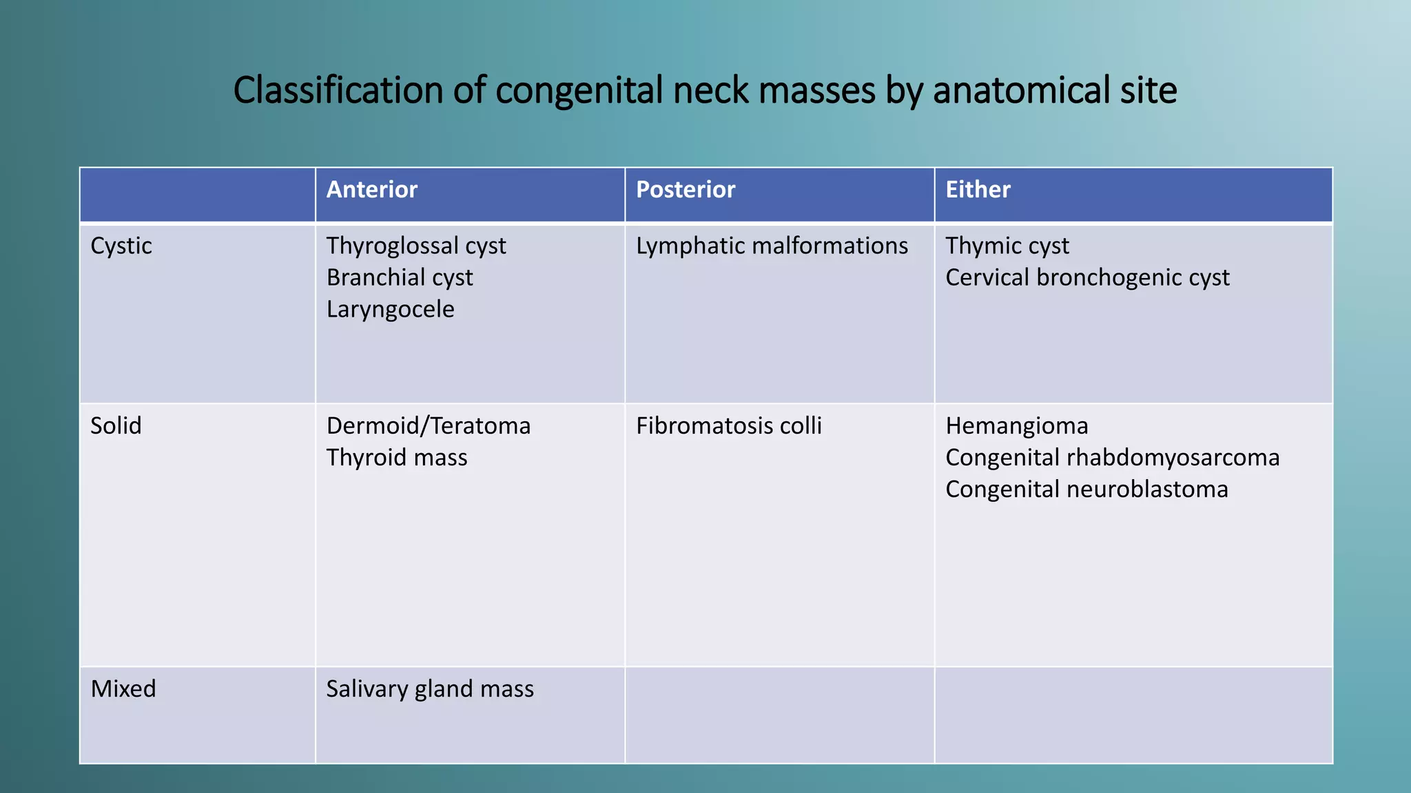
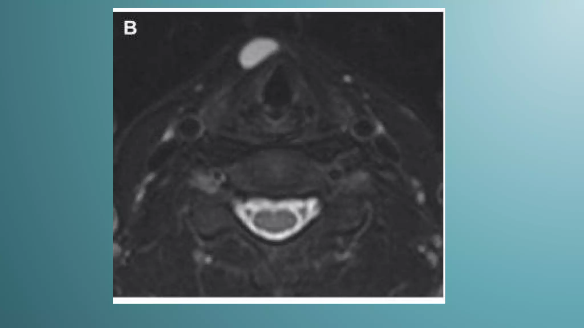
Congenital Neck Mass
Pradeep Kumar
INTRODUCTION
• Cervical congenital masses are
uncommon group of lesions,
usually diagnosed in infancy and
childhood.
• Most lesions occur due to
developmental defect.
• Most of them are cystic.
• They are usually slow-growing
masses and typically cause
symptoms only because of
enlargement or infection
Classification of congenital neck masses by anatomical site
Anterior Posterior Either
Cystic Thyroglossal cyst
Branchial cyst
Laryngocele
Lymphatic malformations Thymic cyst
Cervical bronchogenic cyst
Solid Dermoid/Teratoma
Thyroid mass
Fibromatosis colli Hemangioma
Congenital rhabdomyosarcoma
Congenital neuroblastoma
Mixed Salivary gland mass
Diagnosis
• Age: most developmental defects manifest in infancy or childhood
• Physical examination: cystic Vs solid, narrowing diagnosis,
determining suitable imaging modality.
• USG: the size and extent of the mass, relationship to surrounding
normal structures, cystic/solid nature of the lesion.
• CT: evaluation of soft-tissue planes adjacent to larger masses not
entirely visualized with USG, detecting calcification and vascularity.
• MRI: supplemental information for accurate preoperative
planning, especially in cases of extension into the mediastinum or
deep spaces of the neck.
Thyroglossal duct cyst
• Most common congenital neck mass- 70% of congenital neck
anomalies; second most common benign neck mass, after benign
lymphadenopathy
• 50% present before 20 years of age, rest in young adulthood
• Thyroid primordium originates at the level of the foramen cecum-
junction of the anterior two-thirds and posterior one-third of the
tongue.
• During its migration into final position in the inferior part of the neck
by the 7th week of gestation thyroid gland is connected to the tongue
by thyroglossal duct
• Duct normally involutes by the 8th–10th gestational week.
• Persistence of any portion of the thyroglossal duct may lead to cystic
lesions.
Thyroglossal duct cyst
• The size :0.5 to 6 cm (most
between 1.5 and 3 cm)
• Always within 2 cm of the
midline usually on the left
(if paramedian)
• 80% at or below the level of
the hyoid bone (20% above
the hyoid bone).
• Rarely, as a mass in the
floor of the mouth.
Thyroglossal duct cyst
• At USG: anechoic mass with a thin outer
wall(40%)
• Cysts may also manifest as hypoechoic
masses, often with increased through-
transmission.
• May be either homogeneous or
heterogeneous with variable degrees of fine
to coarse internal echoes.
Continue-
• CT: smooth, well-circumscribed mass along thyroglossal duct
path with thin wall and homogeneous attenuation (10–18
HU).
• Usually unilocular; septations may be seen occasionally.
• Peripheral rim enhancement in contrast-enhanced images.
• Hypo on T1 and is hyperintense on T2-weighted
images(variable signal in presence of proteinaceous debris)
Branchial Cleft Cysts
• 1st branchial cleft cyst
• Arises along the residual tract of the 1st branchial cleft
extending from the EAC through parotid gland to the
submandibular triangle
• 5%–8% of all branchial cleft defects
• Commonly seen in middle-aged women
• Symptom of recurrent abscesses or inflammation around
the ear or at angle of the mandible
Inflamed first branchial cleft cyst (c)
occupies the tail of the parotid gland.
Thick enhancing wall (arrowheads)
indicates ongoing inflammation.
Inflamed first branchial cleft cyst (c) occupies
the tail of the parotid gland
Inflamed parotid cyst in parotid tail cannot be distinguished from first branchial cleft cyst on
the CT images. Pathology showed the parotid duct to be inflamed and ectatic.
Branchial Cleft Cysts
• 2nd Branchial cleft cyst:
• Constitutes 75% of branchial cleft anomalies
• Age: 10 and 40 years of age (whereas fistulas or
sinuses present during the 1st decade of life)
• The “classic” location: anteromedial border of
the SCM muscle, lateral to the carotid space,
and at the posterior margin of the
submandibular gland causing displacement of
SCM posterolaterally, vessels of the carotid
space medially or posteromedially, and SMG
anteriorly
• USG: sharply marginated,
round to ovoid,
compressible centrally
anechoic mass with a thin
peripheral wall that
displaces the surrounding
soft tissues Occasionally,
fine, indistinct internal
echoes, of debris, may be
seen.
An infected 2nd BCC: thick-walled
round cystic mass just posteroinferior
to the mandibular angle
Classic dis- placement of submandibular gland
(SG) anteriorly, the SCM (SM) displaced
posterolaterally, and the jugular vein (arrow)
and carotid artery (arrowhead) displaced
posteromedially
Second Branchial Cleft Cyst. Axial non-contrast MRI images (above) and
sagittal contrast-enhanced MRI images below. There is a cystic mass filled
with a simple fluid surrounded by a homogeneously enhancing thin-wall in
the right neck anteriorly. The cyst is located anterior to the right
sternocleidomastoid muscle and inferoposterior to the right parotid gland
and is most consistent with a second branchial cleft cyst.
3rd branchial cleft cyst
4th branchial cleft cyst
Diagram for
Branchial
cleft cysts.
Lymphangioma
• Lymphangiomas arise from early
sequestration of embryonic
lymphatic channels, most
commonly developing along the
jugular chain.
• Four types of lymphangioma are
described based on microscopic size
of dilated lymphatic channels-
•Cystic hygroma
•cavernous lymphangioma
•Capillary lymphangioma and
•Vasculolymphatic
malformation.
Cystic hygromas are the most common
form of lymphangioma; 75% of these occur
in the neck, usually centered in the
posterior triangle or the sub-mandibular
space.
These lesions are characteristically
infiltrative in nature and do not respect
facial planes. The mediastinum and axilla
are common sites of their extension.
Sudden enlargement may occur due to
hemorrhage or infection.
.
• USG: Multi-locular cystic masses, with
septations of variable thickness.
Fluid-fluid levels may be present when
the lesions are complicated by
hemorrhage
CT shows these as poorly
circumscribed,
multi-loculated,
hypodense masses with
fluid attenuation . Higher
density may be observed
in infected or hemorrhagic
lesions.
MRI typically demonstrates low or
intermediate signal intensity on
T1-weighted images and
hyper-intensity on T2-weighted images.
T1 hyper-intensity may be seen with
hemorrhage or lipid content. Fluid level
may also be seen.
Thymic cyst
Thymic cysts are uncommon lesions that arise
from persistence of the thymopharyngeal
duct.
These occur adjacent to the carotid sheath
anywhere from the hyoid bone to the anterior
mediastinum.
The thymic cysts may have a similar
appearance to third and fourth BCCs, being
differentiated only by the presence of thymic
tissue within thymic cysts.
The cysts usually present as a unilocular cystic
mass extending inferiorly within the neck,
paralleling the sternocleidomastoid muscle,
or as a dumbbell-shaped left cervicothoracic
cystic mass.
Laryngocele
A laryngocele is considered to have a congenital
derivation, although it usually manifests in adults.
It is dilatation of the laryngeal saccule, a small pouch
arising from the roof of the ventricle.
Laryngoceles are of three types—internal, external, and
mixed.
Fifteen percent of laryngoceles are associated with
carcinoma, with tumor occluding the orifice of the
laryngeal ventricle.
A laryngocele may become infected and is then called a
laryngopyocele.
A sharply defined oval or round lucent area in the para-laryngeal soft tissues on a radiograph is diagnostic of
laryngocele.
On CT, a laryngocele may is seen as a well-defined fluid, with an air or soft tissue attenuation mass in the lateral
aspect of the superior para-laryngeal space .
Demonstration of a connection between the air sac and the airway confirms the diagnosis.
It may contain an air-fluid level. The presence of soft tissue within it suggests an underlying laryngeal neoplasm.
• Retention cysts of sublingual glands
• Simple: confined to the sublingual
space
• Plunging/ diving: extend posteriorly
into the sub-mandibular space or
through a mylohyoid defect.
RANULA
1. US: unilocular, well-defined, cystic
lesion in the sub-mental region deep to
the mylohyoid muscle.
2 May contain fine internal echoes,
usually due to presence of debris from
previous episodes of inflammation.
Teratoma
• Teratomas are usually histologically benign lesions that
manifest in children often in the newborn.
• They originate from two or more embryonic germ layers.
• Teratomas of the neck, nasopharynx, pharynx, and oral
cavity are usually large, bulky masses.
• Clearly evident and recognized at birth.
• Many of these are diagnosed at routine prenatal
ultrasound examinations.
Usually multiloculated,
heterogeneous masses
with calcification with
scattered lipid foci.
• MRI – T1WI: Hypo- to isointense cysts and solid tissue, +/-
hyperintense fat –
• T2WI: Cystic portions hyperintense, solid tissue variable – T1 C+:
Heterogeneous enhancement of solid tissue
Dermoid and epidermoid cysts
Dermoid cysts are lined by the epithelium and differ
from epidermoid cysts in that they contain skin
appendages such as sebaceous glands and hair
follicles within the cyst wall.
Complex dermoid cysts contain mesodermal
elements like cartilage, bone, and fat.
Dermoid cysts usually manifest during the second
and third decades of life as slow-growing mass in
the sub-mandibular or sublingual space.
Epidermoid cysts manifest earlier in life, with most
lesions evident during infancy.
Epidermoid cysts: usually show fluid-density
material. Post contrast, the lesion wall may be
imperceptible or may show subtle rim
enhancement.
Dermoid cyst: Heterogeneous appearance may
be seen due to the presence of echogenic fat,
osseous, or dental elements.
On non-contrast CT, dermoid cyst usually
appears as a low-density, unilocular,
well-circumscribed mass. Fat, mixed-density
.
Fibromatosis colli
Fibromatosis colli is a rare form of infantile
fibromatosis that occurs within the
sternocleidomastoid muscle .
The importance of fibromatosis colli, above all
else, is that it must be recognised as a the
benign lesion
Presentation is usually with torticollis, and is
most frequently related to birth trauma (e.g.
forceps delivery) or malposition in the womb. It
is unilateral in most cases.
• Ultrasound is the imaging modality of choice.
• The sternocleidomastoid is diffusely enlarged in a fusiform manner, with
resultant shortening and therefore head turned away from the affected side
(mastoid process drawn down towards the ipsilateral head of clavicle)
• Echogenicity may vary. Colour Doppler interrogation may reveal a high resistance
waveform. The enlarged area often moves synchronously with the rest of the
SCM muscle on real time sonography
Congenital Goiter
• The result of iodine excess or deficiency,
intrauterine exposure to antithyroid drugs or
congenital metabolic disorders of thyroid
synthesis.
• Ultrasound diagnosis is based on the
demonstration of a solid, anteriorly located
symmetric mass, which may result in
hyperextension of the fetal head.
• Polyhydramnios is common due to mechanical
obstruction of the esophagus.
Ectopic thyroid
USG:
• Absence of thyroid tissue in normal position
• Echogenic mass with vascularity.
• Color Doppler provides information about its activity.
CT/ MRI:
• Provides better anatomical relation, extension.
• Hyperdense mass.
USG: circumscribed hyperechoic lesion
in base of tongue
Axial and sag CECT: circumscribed hyper
enhancing mass in base of tongue with
asset thyroid gland n normal position
Ectopic thymus
• Ectopic thymic tissue may
occur anywhere along the
path of descent through
the thymopharyngeal
duct.
• When it presents as a
neck mass, it can be
mistaken for a tumor.
• The ectopic part of the
thymus has the same echo
characteristics as the
normal thymus.
Vascular Lesions
• Infantile hemangiomas rarely present at birth.
• Normal rate of endothelial turnover, lesion grows proportionate to child,
progressive dilation of vessels.
• Shows rapid endothelial proliferation followed by slow involution.
• Hemangiomas may arise in or around critical structures, such as the airway,
and rapid growth can result in progressive stridor and airway compromise.
•USG: homogeneously
hyperechoic with high
diastolic & low RI-
hemangioma & variable
flow in vascular
malformations.
Vascular lesions
Venous malformations frequently affect the oral
cavity, extracranial head, and neck and cause
airway compromise due to their size.
About 14% of venous malformations involving
skeletal muscle occur in the head and neck
region. The masseter and pterygoid muscles are
most frequently involved, followed by the trapezius
and sternocleidomastoid muscles
•CECT: intense clustering of enhancing vessels
•MRI: Hyperintense lobulated lesions in T2 weighted images
•TREATMENT:
–Steroids, interferon, laser, embolisation and excision
Jugular ectasia
Infections/inflammatory lesions
• Adenitis
• Abscesses
• Acute suppurative thyroiditis
• Cellulitis
• Lymphadenitis
Cervical Neuroblastoma
• Neuroblastoma of the head and neck frequently represents
metastasis from neural crest sympathetic precursor cells in the
adrenal gland.
• The tumor arises from the sympathetic ganglia along the spinal
column in a paramedian position from the base of the skull to the
pelvis in addition to the adrenal medulla, the organ of Zuckerkandl
• Extension through the intervertebral foramina with dumbbell masses
is of particular importance in perivertebral space locations due to
possible spinal cord compression.
• Rhabdomyosarcoma is a malignant tumor that arises
from skeletal muscle cells.
• It is the most common pediatric soft-tissue sarcoma and
represents the second most common head and neck
malignancy..
Rhabdomyosarcoma:
• Inhomogeneous solid mass
+/- intratumoral hemorrhage.
Bone destruction is common.
• The tumor displays strong
enhancement with occasional
necrotic areas.
Thank You

Congenital neck mass radiology pk final

  • 1.
  • 2.
    INTRODUCTION • Cervical congenitalmasses are uncommon group of lesions, usually diagnosed in infancy and childhood. • Most lesions occur due to developmental defect. • Most of them are cystic. • They are usually slow-growing masses and typically cause symptoms only because of enlargement or infection
  • 5.
    Classification of congenitalneck masses by anatomical site Anterior Posterior Either Cystic Thyroglossal cyst Branchial cyst Laryngocele Lymphatic malformations Thymic cyst Cervical bronchogenic cyst Solid Dermoid/Teratoma Thyroid mass Fibromatosis colli Hemangioma Congenital rhabdomyosarcoma Congenital neuroblastoma Mixed Salivary gland mass
  • 6.
    Diagnosis • Age: mostdevelopmental defects manifest in infancy or childhood • Physical examination: cystic Vs solid, narrowing diagnosis, determining suitable imaging modality. • USG: the size and extent of the mass, relationship to surrounding normal structures, cystic/solid nature of the lesion. • CT: evaluation of soft-tissue planes adjacent to larger masses not entirely visualized with USG, detecting calcification and vascularity. • MRI: supplemental information for accurate preoperative planning, especially in cases of extension into the mediastinum or deep spaces of the neck.
  • 7.
    Thyroglossal duct cyst •Most common congenital neck mass- 70% of congenital neck anomalies; second most common benign neck mass, after benign lymphadenopathy • 50% present before 20 years of age, rest in young adulthood • Thyroid primordium originates at the level of the foramen cecum- junction of the anterior two-thirds and posterior one-third of the tongue. • During its migration into final position in the inferior part of the neck by the 7th week of gestation thyroid gland is connected to the tongue by thyroglossal duct • Duct normally involutes by the 8th–10th gestational week. • Persistence of any portion of the thyroglossal duct may lead to cystic lesions.
  • 8.
    Thyroglossal duct cyst •The size :0.5 to 6 cm (most between 1.5 and 3 cm) • Always within 2 cm of the midline usually on the left (if paramedian) • 80% at or below the level of the hyoid bone (20% above the hyoid bone). • Rarely, as a mass in the floor of the mouth.
  • 9.
    Thyroglossal duct cyst •At USG: anechoic mass with a thin outer wall(40%) • Cysts may also manifest as hypoechoic masses, often with increased through- transmission. • May be either homogeneous or heterogeneous with variable degrees of fine to coarse internal echoes.
  • 11.
    Continue- • CT: smooth,well-circumscribed mass along thyroglossal duct path with thin wall and homogeneous attenuation (10–18 HU). • Usually unilocular; septations may be seen occasionally. • Peripheral rim enhancement in contrast-enhanced images. • Hypo on T1 and is hyperintense on T2-weighted images(variable signal in presence of proteinaceous debris)
  • 16.
    Branchial Cleft Cysts •1st branchial cleft cyst • Arises along the residual tract of the 1st branchial cleft extending from the EAC through parotid gland to the submandibular triangle • 5%–8% of all branchial cleft defects • Commonly seen in middle-aged women • Symptom of recurrent abscesses or inflammation around the ear or at angle of the mandible
  • 18.
    Inflamed first branchialcleft cyst (c) occupies the tail of the parotid gland. Thick enhancing wall (arrowheads) indicates ongoing inflammation. Inflamed first branchial cleft cyst (c) occupies the tail of the parotid gland
  • 19.
    Inflamed parotid cystin parotid tail cannot be distinguished from first branchial cleft cyst on the CT images. Pathology showed the parotid duct to be inflamed and ectatic.
  • 20.
    Branchial Cleft Cysts •2nd Branchial cleft cyst: • Constitutes 75% of branchial cleft anomalies • Age: 10 and 40 years of age (whereas fistulas or sinuses present during the 1st decade of life) • The “classic” location: anteromedial border of the SCM muscle, lateral to the carotid space, and at the posterior margin of the submandibular gland causing displacement of SCM posterolaterally, vessels of the carotid space medially or posteromedially, and SMG anteriorly
  • 22.
    • USG: sharplymarginated, round to ovoid, compressible centrally anechoic mass with a thin peripheral wall that displaces the surrounding soft tissues Occasionally, fine, indistinct internal echoes, of debris, may be seen.
  • 23.
    An infected 2ndBCC: thick-walled round cystic mass just posteroinferior to the mandibular angle Classic dis- placement of submandibular gland (SG) anteriorly, the SCM (SM) displaced posterolaterally, and the jugular vein (arrow) and carotid artery (arrowhead) displaced posteromedially
  • 24.
    Second Branchial CleftCyst. Axial non-contrast MRI images (above) and sagittal contrast-enhanced MRI images below. There is a cystic mass filled with a simple fluid surrounded by a homogeneously enhancing thin-wall in the right neck anteriorly. The cyst is located anterior to the right sternocleidomastoid muscle and inferoposterior to the right parotid gland and is most consistent with a second branchial cleft cyst.
  • 25.
  • 26.
  • 27.
  • 28.
    Lymphangioma • Lymphangiomas arisefrom early sequestration of embryonic lymphatic channels, most commonly developing along the jugular chain. • Four types of lymphangioma are described based on microscopic size of dilated lymphatic channels- •Cystic hygroma •cavernous lymphangioma •Capillary lymphangioma and •Vasculolymphatic malformation.
  • 29.
    Cystic hygromas arethe most common form of lymphangioma; 75% of these occur in the neck, usually centered in the posterior triangle or the sub-mandibular space. These lesions are characteristically infiltrative in nature and do not respect facial planes. The mediastinum and axilla are common sites of their extension. Sudden enlargement may occur due to hemorrhage or infection. .
  • 30.
    • USG: Multi-locularcystic masses, with septations of variable thickness. Fluid-fluid levels may be present when the lesions are complicated by hemorrhage
  • 31.
    CT shows theseas poorly circumscribed, multi-loculated, hypodense masses with fluid attenuation . Higher density may be observed in infected or hemorrhagic lesions.
  • 32.
    MRI typically demonstrateslow or intermediate signal intensity on T1-weighted images and hyper-intensity on T2-weighted images. T1 hyper-intensity may be seen with hemorrhage or lipid content. Fluid level may also be seen.
  • 33.
    Thymic cyst Thymic cystsare uncommon lesions that arise from persistence of the thymopharyngeal duct. These occur adjacent to the carotid sheath anywhere from the hyoid bone to the anterior mediastinum. The thymic cysts may have a similar appearance to third and fourth BCCs, being differentiated only by the presence of thymic tissue within thymic cysts. The cysts usually present as a unilocular cystic mass extending inferiorly within the neck, paralleling the sternocleidomastoid muscle, or as a dumbbell-shaped left cervicothoracic cystic mass.
  • 35.
    Laryngocele A laryngocele isconsidered to have a congenital derivation, although it usually manifests in adults. It is dilatation of the laryngeal saccule, a small pouch arising from the roof of the ventricle. Laryngoceles are of three types—internal, external, and mixed. Fifteen percent of laryngoceles are associated with carcinoma, with tumor occluding the orifice of the laryngeal ventricle. A laryngocele may become infected and is then called a laryngopyocele.
  • 36.
    A sharply definedoval or round lucent area in the para-laryngeal soft tissues on a radiograph is diagnostic of laryngocele. On CT, a laryngocele may is seen as a well-defined fluid, with an air or soft tissue attenuation mass in the lateral aspect of the superior para-laryngeal space . Demonstration of a connection between the air sac and the airway confirms the diagnosis. It may contain an air-fluid level. The presence of soft tissue within it suggests an underlying laryngeal neoplasm.
  • 37.
    • Retention cystsof sublingual glands • Simple: confined to the sublingual space • Plunging/ diving: extend posteriorly into the sub-mandibular space or through a mylohyoid defect. RANULA
  • 38.
    1. US: unilocular,well-defined, cystic lesion in the sub-mental region deep to the mylohyoid muscle. 2 May contain fine internal echoes, usually due to presence of debris from previous episodes of inflammation.
  • 40.
    Teratoma • Teratomas areusually histologically benign lesions that manifest in children often in the newborn. • They originate from two or more embryonic germ layers. • Teratomas of the neck, nasopharynx, pharynx, and oral cavity are usually large, bulky masses. • Clearly evident and recognized at birth. • Many of these are diagnosed at routine prenatal ultrasound examinations.
  • 42.
    Usually multiloculated, heterogeneous masses withcalcification with scattered lipid foci.
  • 43.
    • MRI –T1WI: Hypo- to isointense cysts and solid tissue, +/- hyperintense fat – • T2WI: Cystic portions hyperintense, solid tissue variable – T1 C+: Heterogeneous enhancement of solid tissue
  • 45.
    Dermoid and epidermoidcysts Dermoid cysts are lined by the epithelium and differ from epidermoid cysts in that they contain skin appendages such as sebaceous glands and hair follicles within the cyst wall. Complex dermoid cysts contain mesodermal elements like cartilage, bone, and fat. Dermoid cysts usually manifest during the second and third decades of life as slow-growing mass in the sub-mandibular or sublingual space. Epidermoid cysts manifest earlier in life, with most lesions evident during infancy.
  • 47.
    Epidermoid cysts: usuallyshow fluid-density material. Post contrast, the lesion wall may be imperceptible or may show subtle rim enhancement. Dermoid cyst: Heterogeneous appearance may be seen due to the presence of echogenic fat, osseous, or dental elements. On non-contrast CT, dermoid cyst usually appears as a low-density, unilocular, well-circumscribed mass. Fat, mixed-density .
  • 49.
    Fibromatosis colli Fibromatosis colliis a rare form of infantile fibromatosis that occurs within the sternocleidomastoid muscle . The importance of fibromatosis colli, above all else, is that it must be recognised as a the benign lesion Presentation is usually with torticollis, and is most frequently related to birth trauma (e.g. forceps delivery) or malposition in the womb. It is unilateral in most cases.
  • 50.
    • Ultrasound isthe imaging modality of choice. • The sternocleidomastoid is diffusely enlarged in a fusiform manner, with resultant shortening and therefore head turned away from the affected side (mastoid process drawn down towards the ipsilateral head of clavicle) • Echogenicity may vary. Colour Doppler interrogation may reveal a high resistance waveform. The enlarged area often moves synchronously with the rest of the SCM muscle on real time sonography
  • 52.
    Congenital Goiter • Theresult of iodine excess or deficiency, intrauterine exposure to antithyroid drugs or congenital metabolic disorders of thyroid synthesis. • Ultrasound diagnosis is based on the demonstration of a solid, anteriorly located symmetric mass, which may result in hyperextension of the fetal head. • Polyhydramnios is common due to mechanical obstruction of the esophagus.
  • 54.
    Ectopic thyroid USG: • Absenceof thyroid tissue in normal position • Echogenic mass with vascularity. • Color Doppler provides information about its activity. CT/ MRI: • Provides better anatomical relation, extension. • Hyperdense mass.
  • 55.
    USG: circumscribed hyperechoiclesion in base of tongue Axial and sag CECT: circumscribed hyper enhancing mass in base of tongue with asset thyroid gland n normal position
  • 56.
    Ectopic thymus • Ectopicthymic tissue may occur anywhere along the path of descent through the thymopharyngeal duct. • When it presents as a neck mass, it can be mistaken for a tumor. • The ectopic part of the thymus has the same echo characteristics as the normal thymus.
  • 57.
    Vascular Lesions • Infantilehemangiomas rarely present at birth. • Normal rate of endothelial turnover, lesion grows proportionate to child, progressive dilation of vessels. • Shows rapid endothelial proliferation followed by slow involution. • Hemangiomas may arise in or around critical structures, such as the airway, and rapid growth can result in progressive stridor and airway compromise.
  • 58.
    •USG: homogeneously hyperechoic withhigh diastolic & low RI- hemangioma & variable flow in vascular malformations.
  • 59.
    Vascular lesions Venous malformationsfrequently affect the oral cavity, extracranial head, and neck and cause airway compromise due to their size. About 14% of venous malformations involving skeletal muscle occur in the head and neck region. The masseter and pterygoid muscles are most frequently involved, followed by the trapezius and sternocleidomastoid muscles
  • 60.
    •CECT: intense clusteringof enhancing vessels •MRI: Hyperintense lobulated lesions in T2 weighted images •TREATMENT: –Steroids, interferon, laser, embolisation and excision
  • 62.
  • 63.
    Infections/inflammatory lesions • Adenitis •Abscesses • Acute suppurative thyroiditis • Cellulitis • Lymphadenitis
  • 64.
    Cervical Neuroblastoma • Neuroblastomaof the head and neck frequently represents metastasis from neural crest sympathetic precursor cells in the adrenal gland. • The tumor arises from the sympathetic ganglia along the spinal column in a paramedian position from the base of the skull to the pelvis in addition to the adrenal medulla, the organ of Zuckerkandl • Extension through the intervertebral foramina with dumbbell masses is of particular importance in perivertebral space locations due to possible spinal cord compression.
  • 66.
    • Rhabdomyosarcoma isa malignant tumor that arises from skeletal muscle cells. • It is the most common pediatric soft-tissue sarcoma and represents the second most common head and neck malignancy.. Rhabdomyosarcoma:
  • 67.
    • Inhomogeneous solidmass +/- intratumoral hemorrhage. Bone destruction is common. • The tumor displays strong enhancement with occasional necrotic areas.
  • 68.

Editor's Notes

  • #7 MR imaging offers superior resolution for evaluating masses located in anatomically complex areas, such as the floor of the mouth.
  • #8 The thyroid gland begins to develop in the 3rd week of fetal life as a median outgrowth from the floor of the primitive pharynx. The anlage of the gland reaches its final position in the inferior part of the neck by the 7th week of gestation after descending anterior to the thyrohyoid membrane and the strap muscles (sternothyroid and sternohyoid muscles) Secretions from the epithelial lining (likely secondary to repeated local infection and inflammation)
  • #10  About 1% of thyroglossal duct abnormalities are associated with thyroid carcinoma arising from ectopic rests of thyroid tissue within the duct and not from the duct itself. Although most (80%) of these tumors are of the papillary type, virtually every type of thyroid malignancy has been reported in association with thyroglossal duct cysts Heterogeneity seen in thyroglossal duct cysts on sonograms is more likely due to the proteinaceous content of the fluid secreted from the cyst wall rather than to infection
  • #11 This videoclip demonstrates the upward movement of the thyroglossal duct cyst together with the hyoid bone during swallowing.
  • #14 Infra hyoid neck axial T2
  • #15 The anomalies result from branchial apparatus (six arches; five clefts), which are the embryologic precursors of the ear and the muscles, blood vessels, bones, cartilage, and mucosal lining of the face, neck, and pharynx  cyst: no internal or external communication fistula: communicates both internally and externally sinus: incomplete tract During the 3rd to 5th week of embryonic development, the second arch grows caudally and covers the third, fourth and sixth arches. When it fuses to the skin caudal to these arches, the cervical sinus is formed. Eventually, the edges of cervical sinus fuse and the ectoderm within the tube disappears 9. Persistence of branchial cleft or pouch results in a cervical anomaly located along the anterior border of the sternocleidomastoid muscle from the tragus of the ear to the clavicle  1st cleft develops into epithelial lining of external auditory meatus 2nd - 4th cleft forms temporary cervical sinuses later obliterated by proliferation of 2nd arch Persistent cervical sinus branchial cleft cyst within lateral neck/angle of mandible
  • #18 Work type I cyst is derived from ectoderm and represents a duplication anomaly of the membranous external auditory canal from the first branchial cleft. It is characteristically found medial to the concha of the ear in a parallel course with the external auditory canal but may extend into the retroauricular area. It is lined with squamous epithelium. No skin appendages are seen. Work type II cysts arise from the first branchial cleft and arch with a possible contribution from the second branchial arch. Consequently, they are derived from ectoderm and mesoderm. They involve both the external auditory canal and cartilaginous pinna. The presence of skin appendages (hair follicles, sweat and sebaceous glands) contained in the squamous epithelial lining of these cysts
  • #21 Bailey type I- most superficial and lies along the anterior surface of the sternocleidomastoid muscle, just deep to the platysma muscle. Type II - most common and found in the “classic” location for these cysts: along the anterior surface of the sternocleidomastoid muscle, lateral to the carotid space, and posterior to the submandibular gland. Type III - extends medially between the bifurcation of the internal and external carotid arteries to the lateral pharyngeal wall. Type IV - lies in the pharyngeal mucosal space and is lined with columnar epithelium
  • #26 Third branchial cleft cysts constitute the second most common congenital lesion of the posterior cervical space of the neck after cystic hygroma.Must lie posterior to the common or internal carotid artery, between the hypoglossal nerve below and the glossopharyngeal nerve above Cysts arising from the third and fourth branchial clefts are extremely rare
  • #27 A fourth branchial cleft sinus tract, following its embryologic derivation, arises from the pyriform sinus, pierces the thyrohyoid membrane, and begins a descent into the mediastinum, following the path of the tracheoesophageal groove. If the tract is long enough, a left-sided lesion will eventually travel under the aortic arch (or, in the rare case of a right-sided lesion, under the right subclavian artery) before ascending into the neck along the ventral surface of the common carotid artery An abscess is noted involving the upper pole of the left lobe of thyroid gland extending to ipsilateral carotid and retropharyngeal spaces. It is measuring about 2.5 x 4.5 cm in axial dimensions surrounded by deep space cellulitis. It is involving the left sternomastoid muscle with muscle enlargement and interstitial edema.
  • #32 Sclerotherapy prior to surgical excision withOK-432, doxycycline, bleomycin, ethanol commonly performed
  • #33 Cystic hygroma and  occipital meningocoele are both differential diagnosis for an antenatally detected cystic collection noted on the posterior aspect of the head and neck. A simple differentiation can be made based on the contents and presence of septations. Cystic hygroma will present as well defined anechoic thin walled cyst with multiple septations and a characteristic midline septation representing the nuchal ligament. On the contrary, an occipital meningocoele will appear as a well defined anechoic area with no sepatations and seen in direct continuity with a defect in the calvarium.
  • #34 The common age of presentation is 2‑15 years, with slight male predilection.
  • #35 Thymic cyst. (A) The axial contrast-enhanced CT scan shows a cystic lesion in the right side of the neck caudal to the thyroid gland displacing the trachea to the left. (B) The coronal reformatted image shows the lesion is parallel to the sternocleidomastoid muscle, extending into the upper mediastinum
  • #36 Those confined to the larynx are known as internal laryngoceles. Those that extend through the thyrohyoid membrane, but with dilation of only the extra‑laryngeal component, are termed external. Mixed laryngoceles have dilatation of the saccule on both sides of the thyrohyoid membrane.
  • #37 PA and lat Xray : air filled masss n right ant paramedian rwhion in neck causing compression and displacement of airway laterally Laryngocele: Axial contrast-enhanced CT image shows air filled internal laryngocele (arrow) on the right and mixed laryngocele (arrowhead) on the left side
  • #38 A ranula derives in name from the latin for frog - the cystic space beneath the tonuge being like the pouch/underbelly of a frog. Rarely ranulas can dissect across the midline between the mylohyoid and geniohyoid muscles to present as a bilateral mass sublingual gland mucocele or mucous retention cyst. Ranulas may be either “simple” and confined to the sublingual space or “plunging/ diving,” which extend posteriorly into the sub‑mandibular space or through a mylohyoid defect. Rarely ranulas can dissect across the midline between the mylohyoid and geniohyoid muscles to present as a bilateral mass.
  • #39 CT shows a simple ranula as an ovoid shaped cyst with a homogeneous central attenuation region of 10‑20 HU, which lies lateral to the genioglossal muscles deep to the mylohyoid muscle . Ranulas may become infected and show a thick, irregular rim of enhancement, with surrounding inflammatory change.
  • #40  Simple ranula- Coronal (STIR) (a) and axial T2-weighted (b) MR images show a cystic mass in the right sub- lingual space. (10) Coronal STIR MR image shows a high-signal-intensity cystic lesion between the left and right geniohyoid muscles (GH), a finding suggestive of a ranula of a minor salivary gland.
  • #42 Here we see a midline tumor in the anterior neck of a three-day-old boy. Calcifications and solid and cystic parts are seen. Pathology was compatible with a mature teratoma.
  • #46 Dermoid and epidermoid cysts may occur anywhere in the body, with 7% presenting as head‑and‑neck lesions, most commonly lateral to the eyebrow
  • #47 Here a typical homogeneous hyper-echoic oval lesion, representing a dermoid cyst, which was located in its favorite location, the suprasternal notch. Here an unusual inhomogeneous appearance of a dermoid cyst in front of the thyroid gland (figure). differentiation from a thyroglossal duct cyst can be difficult if the dermoid cyst is located near the hyoid bone. The content of thyroglossal duct cysts is usually hypo-echoic and may contain internal echoes, while dermoid cysts generally have a more homogeneous hyper-echoic content.
  • #48 Epidemoid- Malignant degeneration into squamous cell carcinoma is seen in upto 5% of lesions. Dermoid- fluid, and calcification (<50%) may also be seen There may be coalescence of fat into small nodules within the cystic lesion, giving a “sac‑of‑marbles” appearance The presence of calcifications and cystic spaces in these lesions aids in their differentiation from lipomas.
  • #50 The right sternocleidomastoid muscle is more commonly affected (73% of the time)
  • #51 Spectral Doppler interrogation may reveal a high resistance waveform. The enlarged area often moves synchronously with the rest of the sternocleidomastoid on real-time sonography 1, smoothly blending with the unaffected fibers. A discrete mass should not be seen. The presence of hyperechoic calcific foci suggests previous hemorrhage.
  • #52 Figure 18. Fibromatosis colli in a 1-month-old boy. (a, b) Axial (a) and sagittal (b) US images show heterogeneous diffuse enlargement of the left sternocleidomastoid muscle (arrow). Cursors indicate the dimensions of the muscle. (c, d) Comparative axial (c) and sagittal (d) US images show a normal right sternocleidomastoid muscle ( FNAC diagnostic Treat: usually resolves 4-8 months Physiotherapy Surgical transection of SM muscle if persistent facial hemihypoplasia
  • #53 Maternal therapy usually corrects fetal hyperthyroidism. Direct fetal therapy in cases of fetal hypothyroidism can be undertaken by amniocentesis or by cordocentesis and this can result in resolution of the fetal goiter.
  • #54 image of a newborn with an abnormal thyroid test. No thyroid gland is visible in the neck, neither in its usual position nor higher up in the neck. A fetal goiter may be seen as a relatively homogenous anterior fetal neck mass. There is also evidence of polyhydramnios. If the goiter is very large the neck may appear hyperextended. Some investigators have attempted to assess the hyper- or hypothyroid status relying on sonographic features 7: hypothyroid goitres   more likely to have a peripheral vascular pattern on color Doppler may have evidence of delayed bone maturation may be accompanied by increased fetal movement hyperthyroid goitres  more likely to have central vascularization may have evidence of a fetal tachycardia may have evidence of advanced bone maturity Congenital goitres may be a result of hereditary or non-hereditary causes . Thyroid dysgenesis is the most common cause of newborn thyroid dysfunction, accounting for approximately 80% of congenital hypothyroidism.
  • #57 Here images of a six-month old boy with a vocal cord paralysis. The MRI examination shows a mass between the parotid and submandibular gland (yellow arrow). The signal characteristics are equal to the thymus (green arrow). Ultrasound confirmed an ectopic thymic remnant (yellow arrow), with identical sonographic characteristics as the orthotopic thymus (green arrow).
  • #58 however, 90% become clinically apparent during the first month of life. Port wine stain- S-W Syndrome Venous Malformations- Lips & cheeks AVM- High flow
  • #59 Hemangioma Hemangiomas are benign vascular neoplasms. They are the most common tumors of infancy. 60% of hemangiomas are seen in the head-neck region. They usually appear in the first weeks after birth, show rapid growth, followed by spontaneous involution. Here we see images of a highly vascular lesion in the left submandibular region, which was present at birth
  • #63 In some children a swelling can appear in the lower neck during straining. This is often caused by dilatation of the internal jugular vein as can easily be demonstrated by ultrasound that will show the variations in caliber of the vein.
  • #65 However, a primary cervical location of neuroblastoma is present in 5% of cases
  • #66 On scintigraphy: we can see uptake in adrenal region as well as in neck region Neuroblastomas appear as solid, contrast-enhanced masses at both CT and MR imaging, with occasional calcifications or intralesional cysts. MR imaging can depict intraforaminal extension which can cause extradural cord compression. Iodine 123 metaiodobenzylguanidine scintigraphy has great sensitivity. May help confirm the neural crest origin of cervical neuroblastoma
  • #68 Solid heterogeneously hypoattenuating mass in rt lateral mandible with partial rim enhancement in rt paraphayrngeal space It extends antero laterally towards mandible and causing posterolateral dsplacement of carotid sheath.