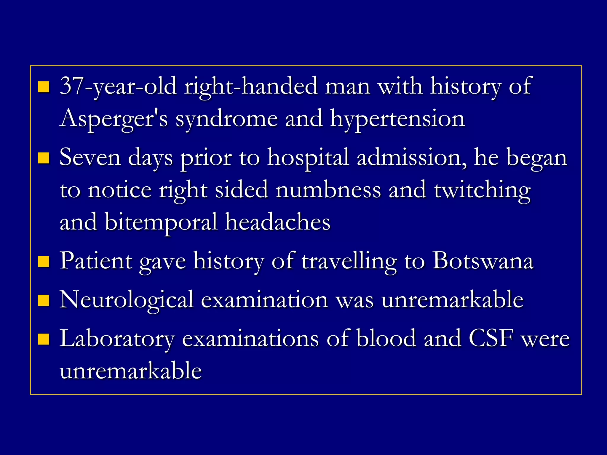
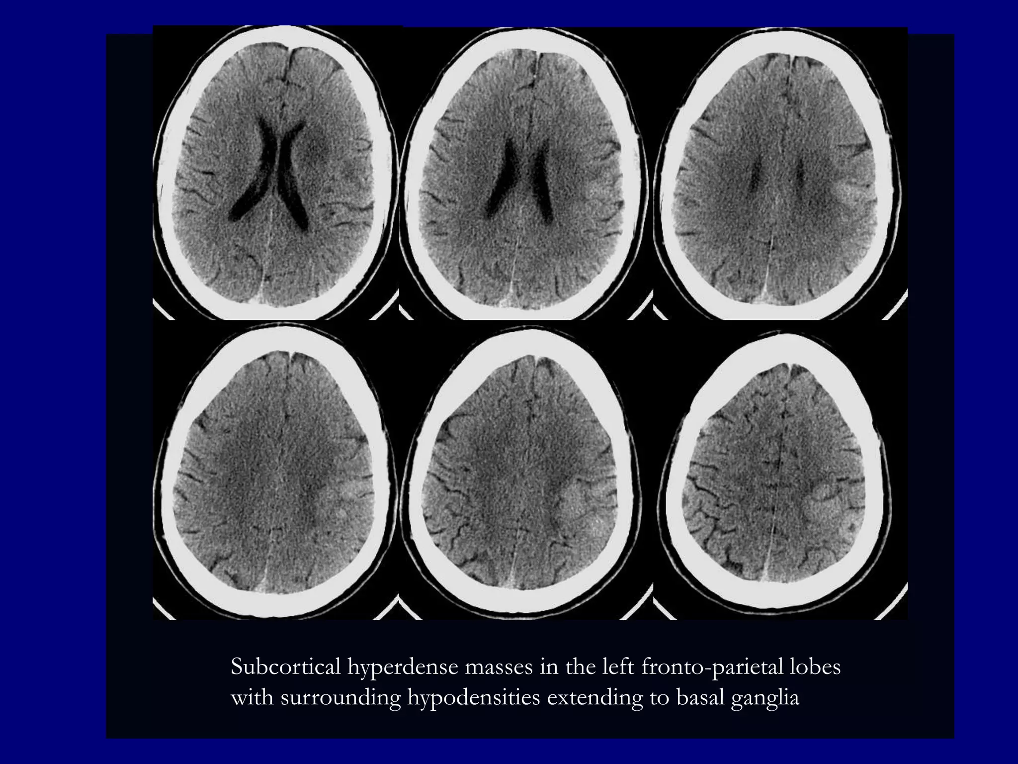
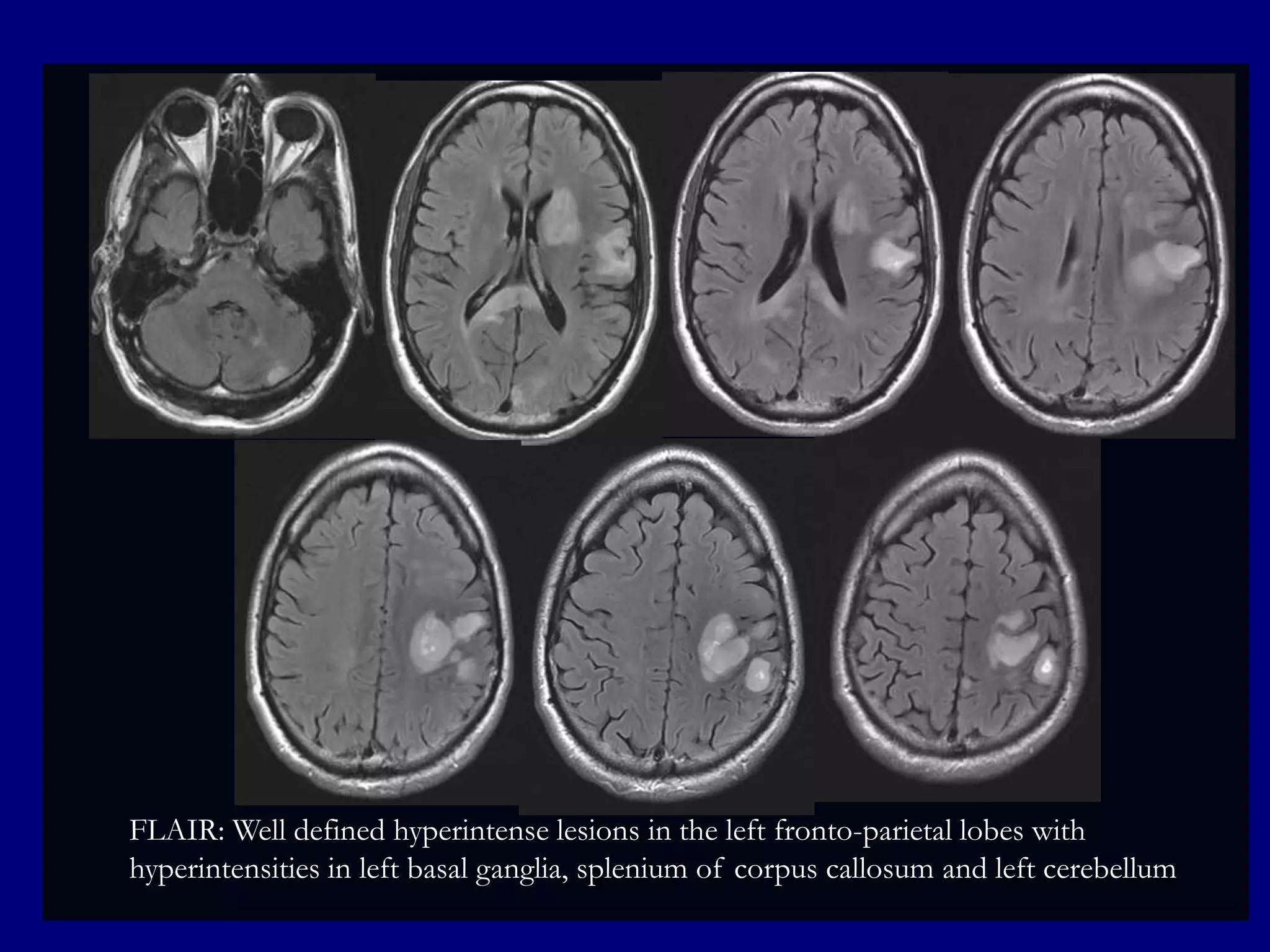
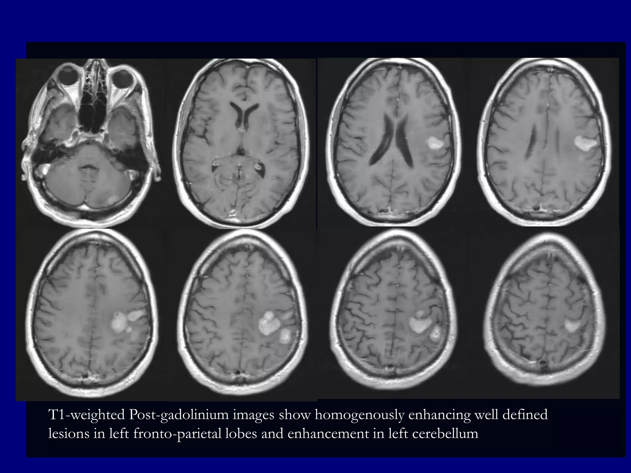
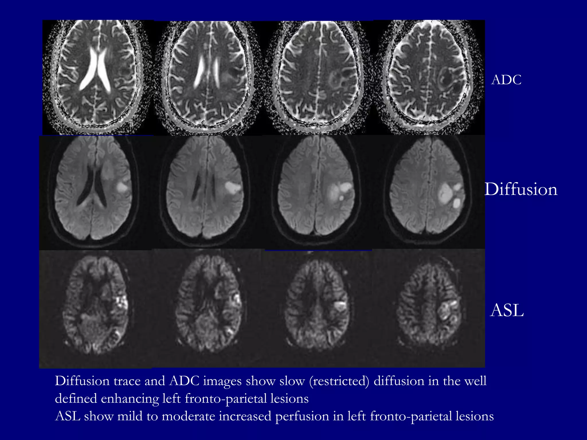
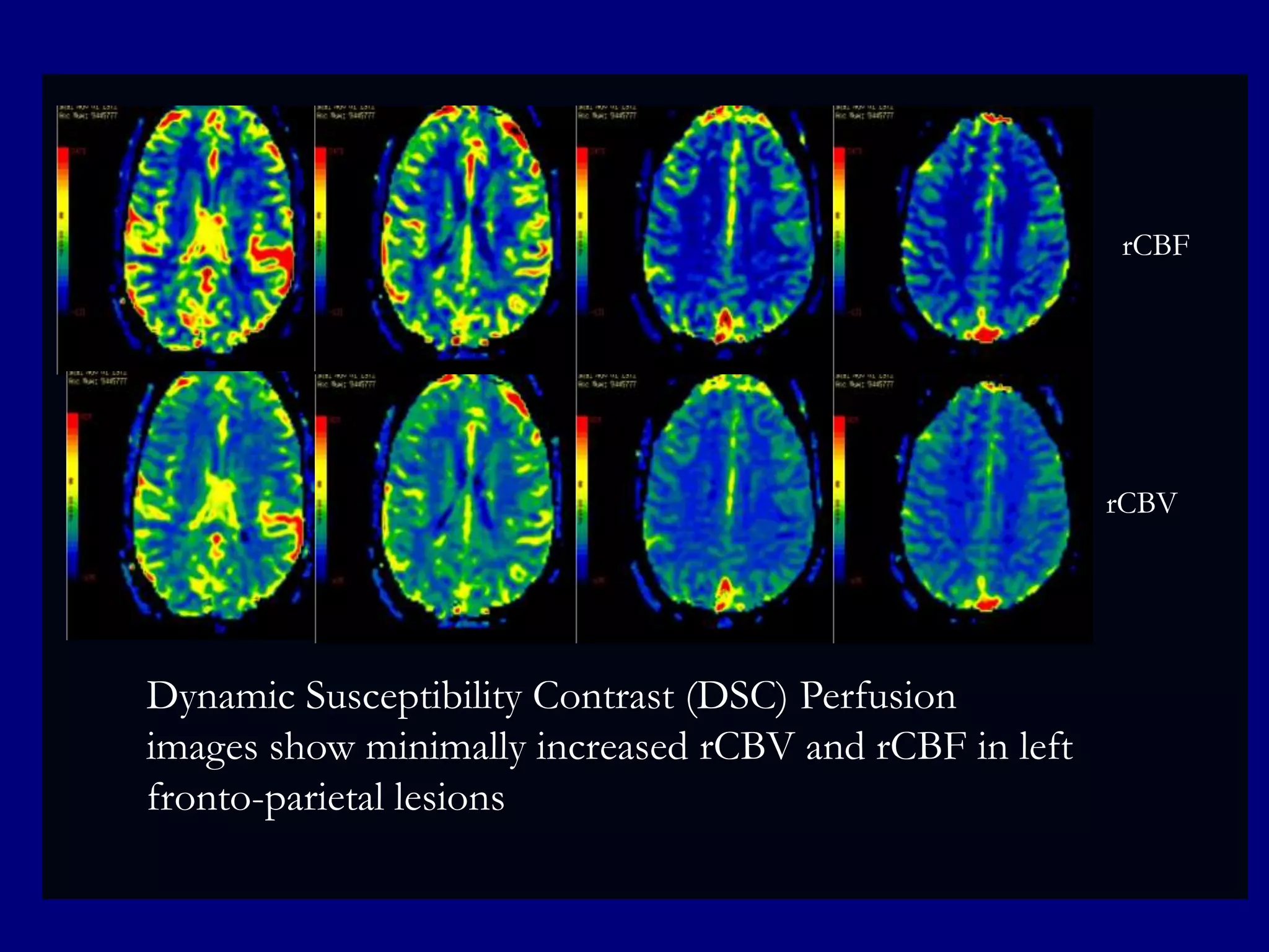
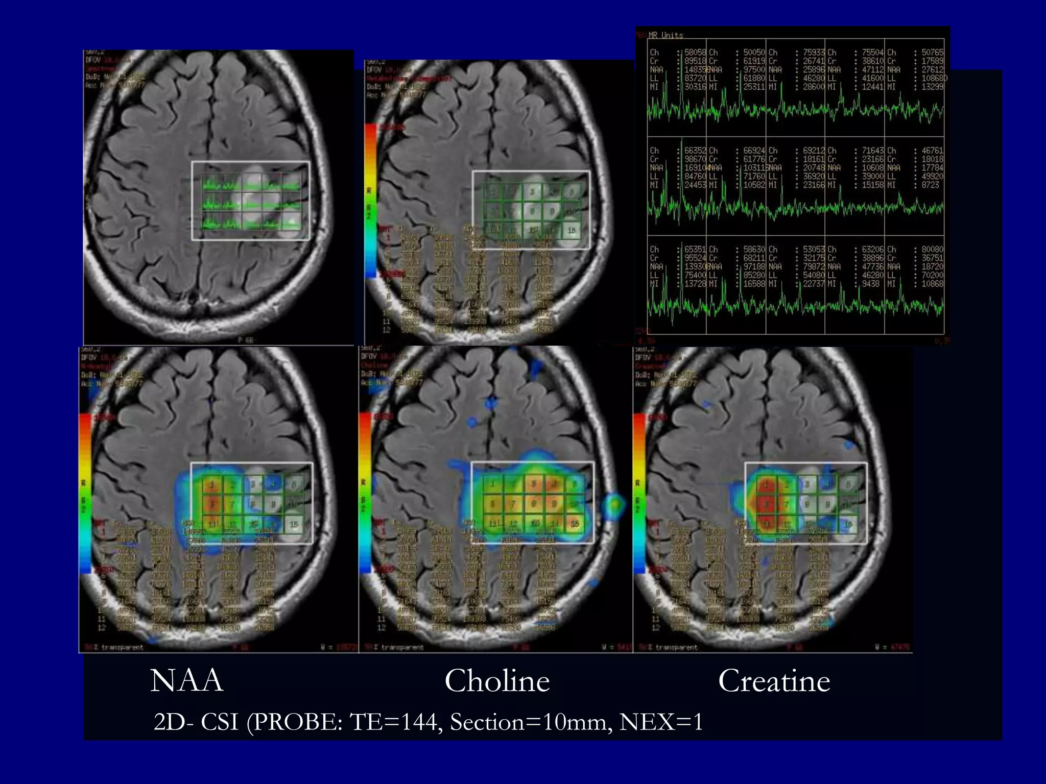
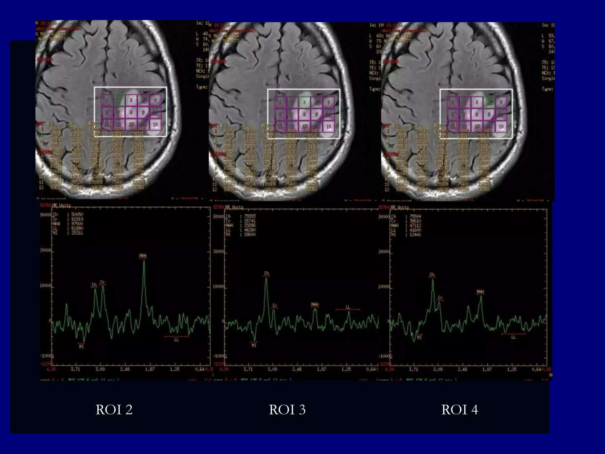
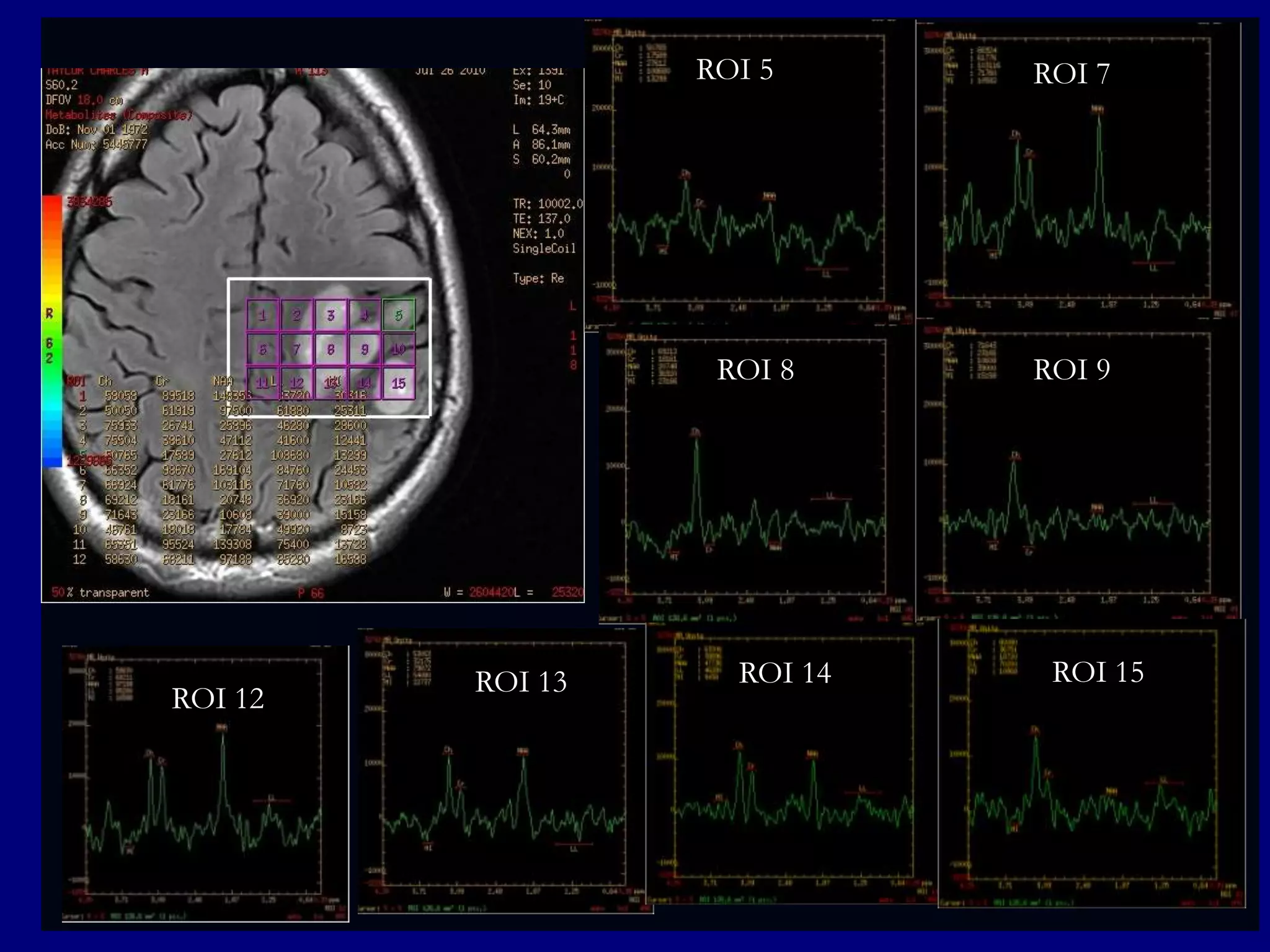
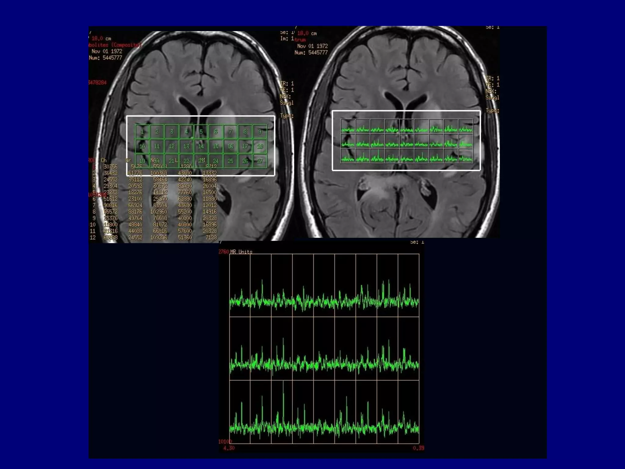
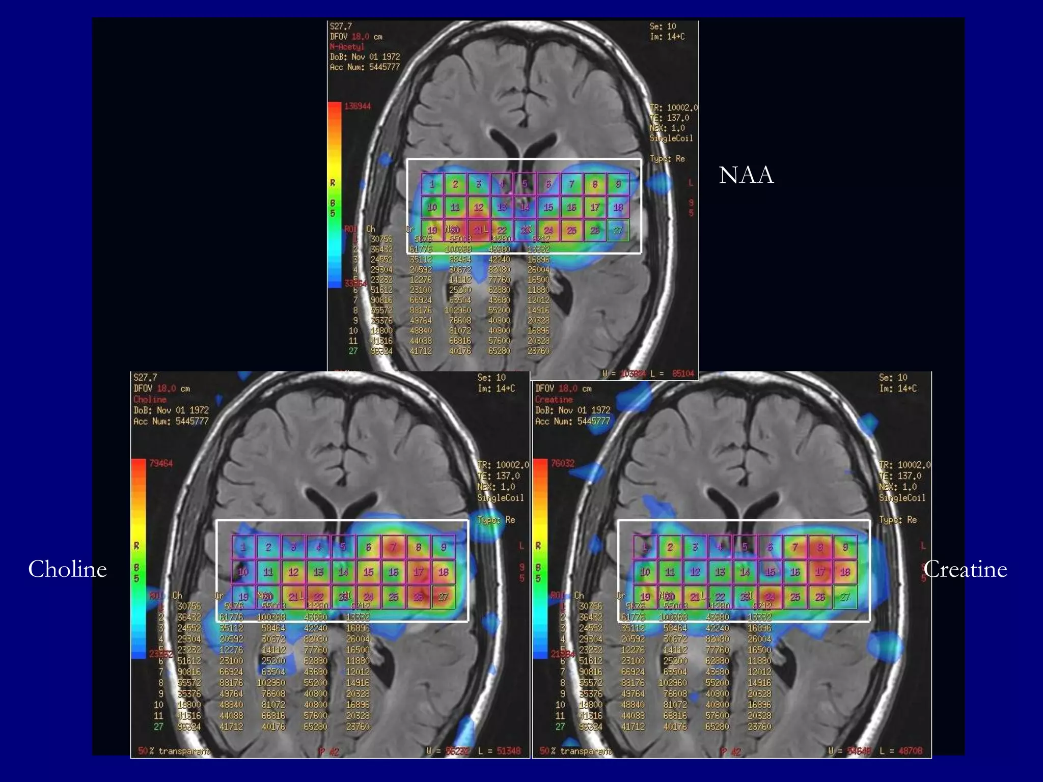
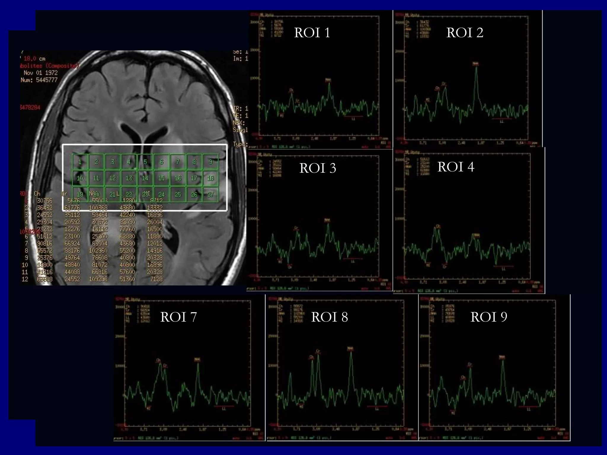
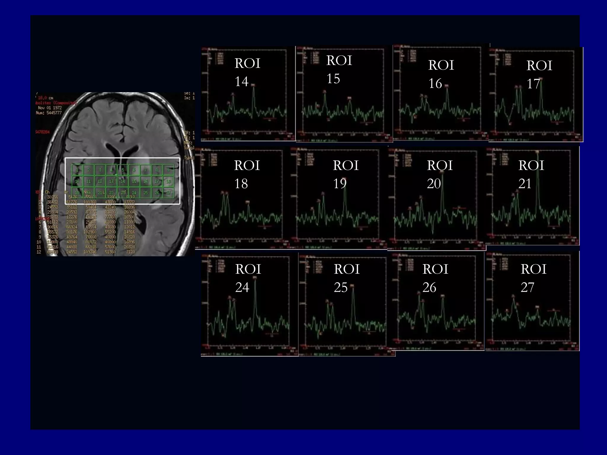
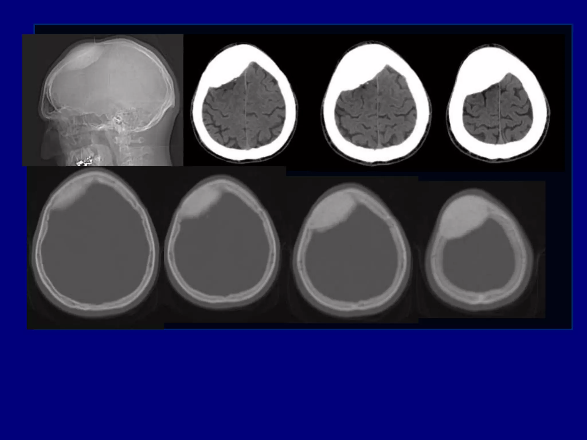
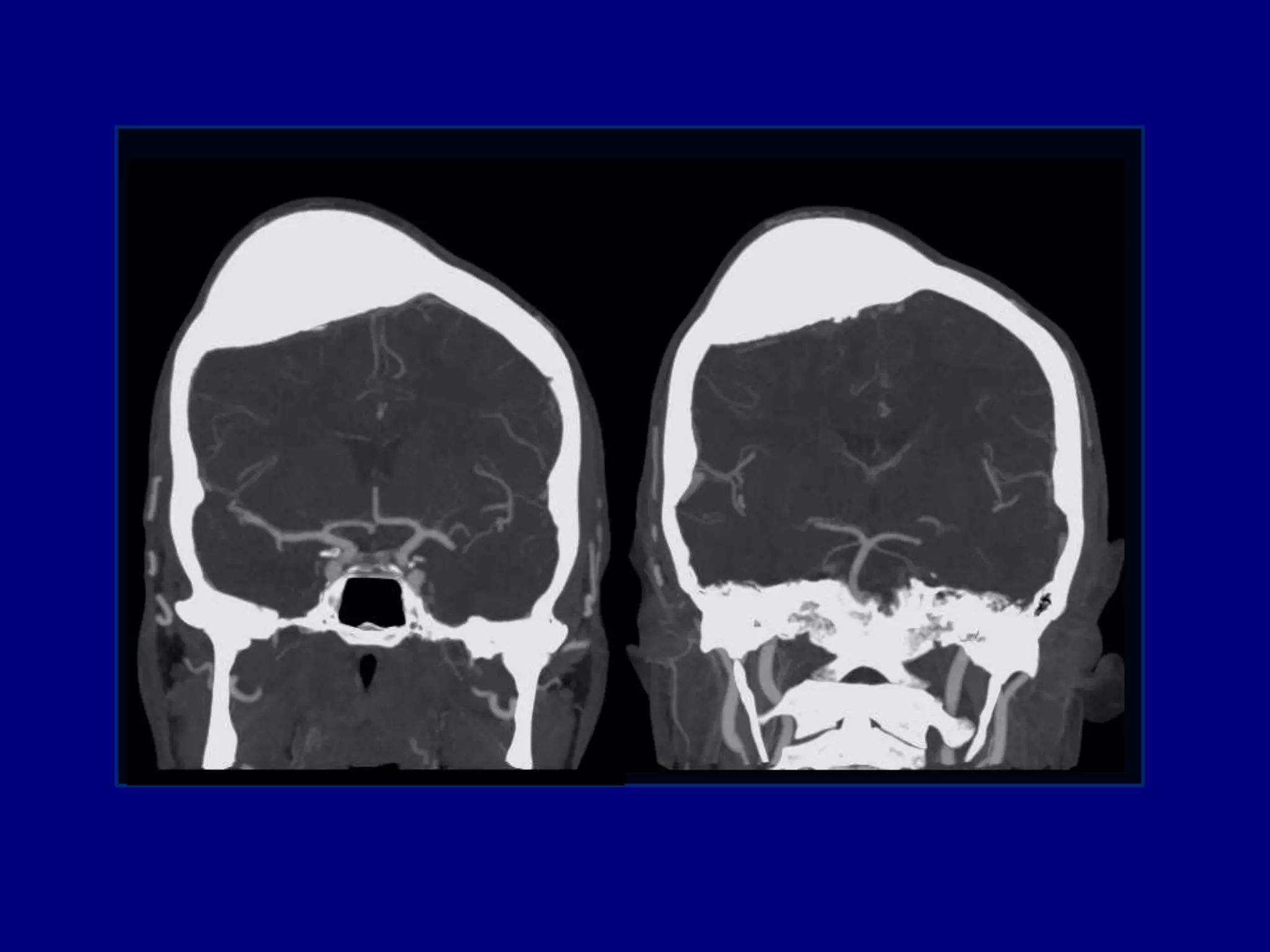
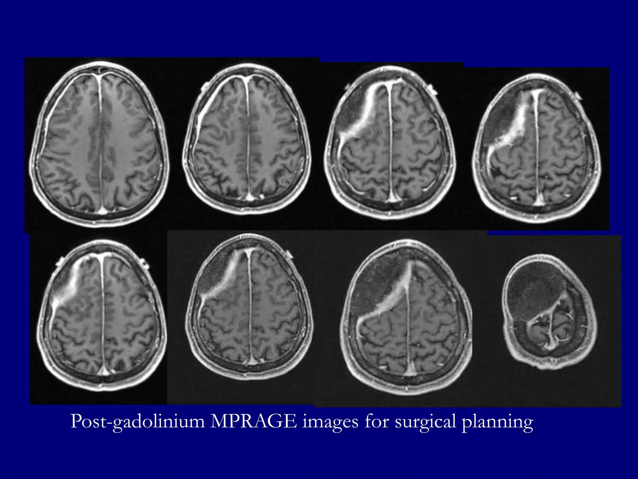
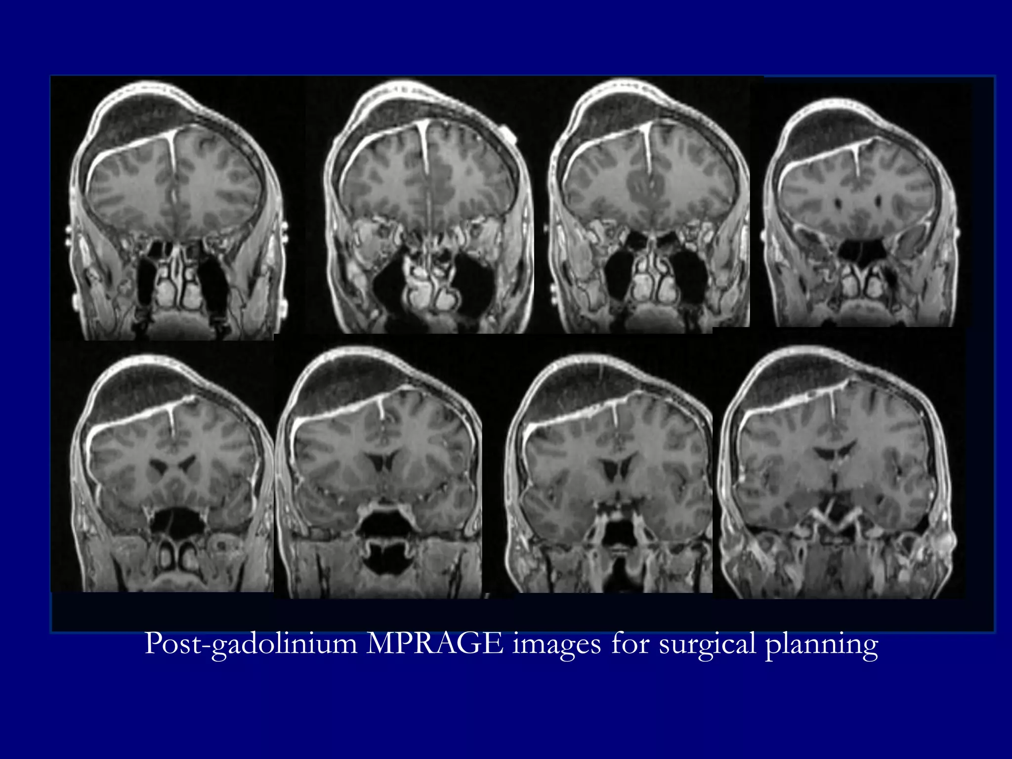
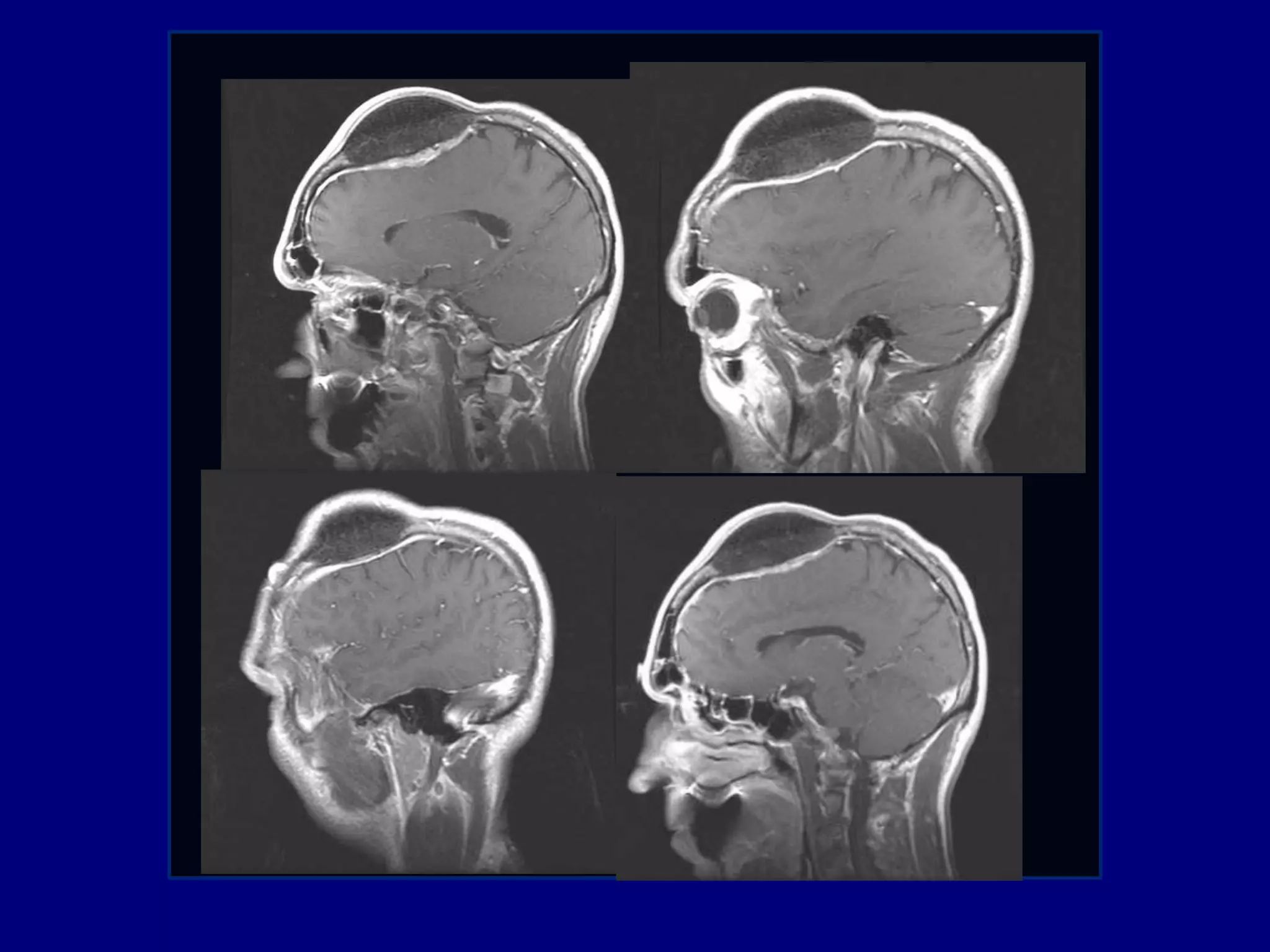
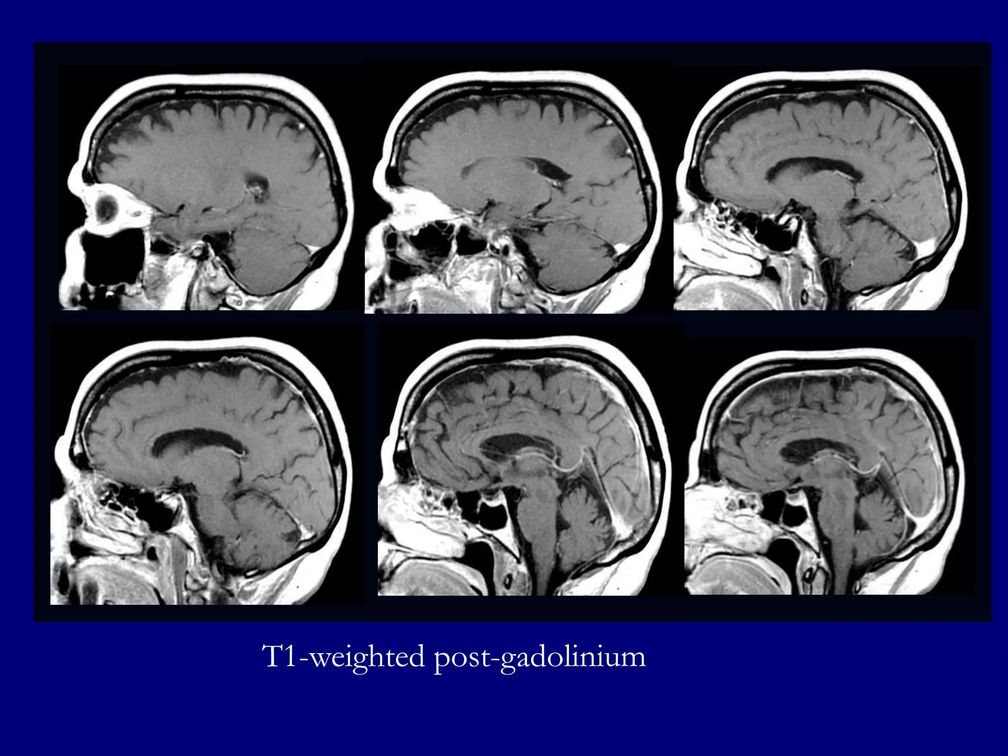
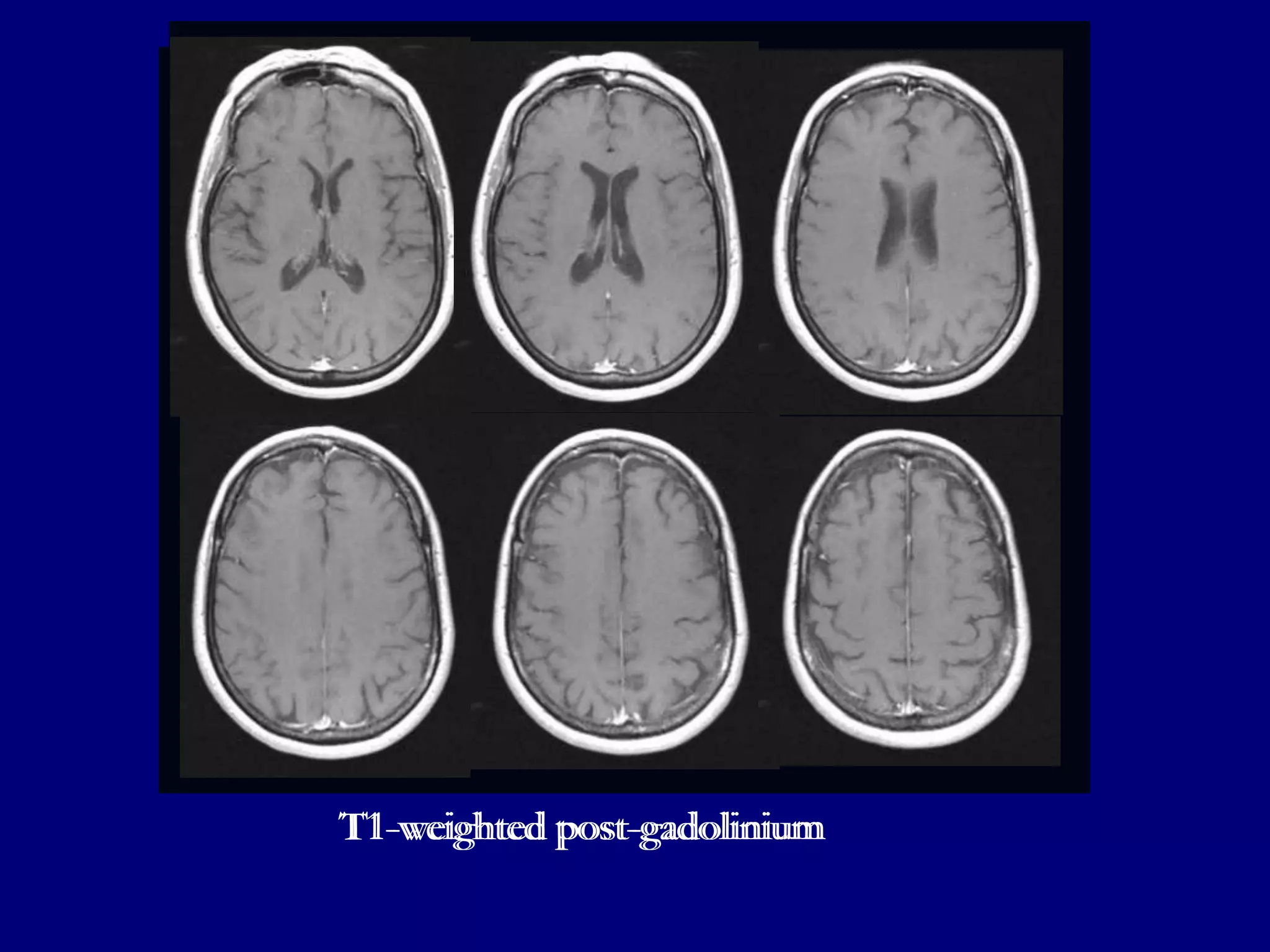
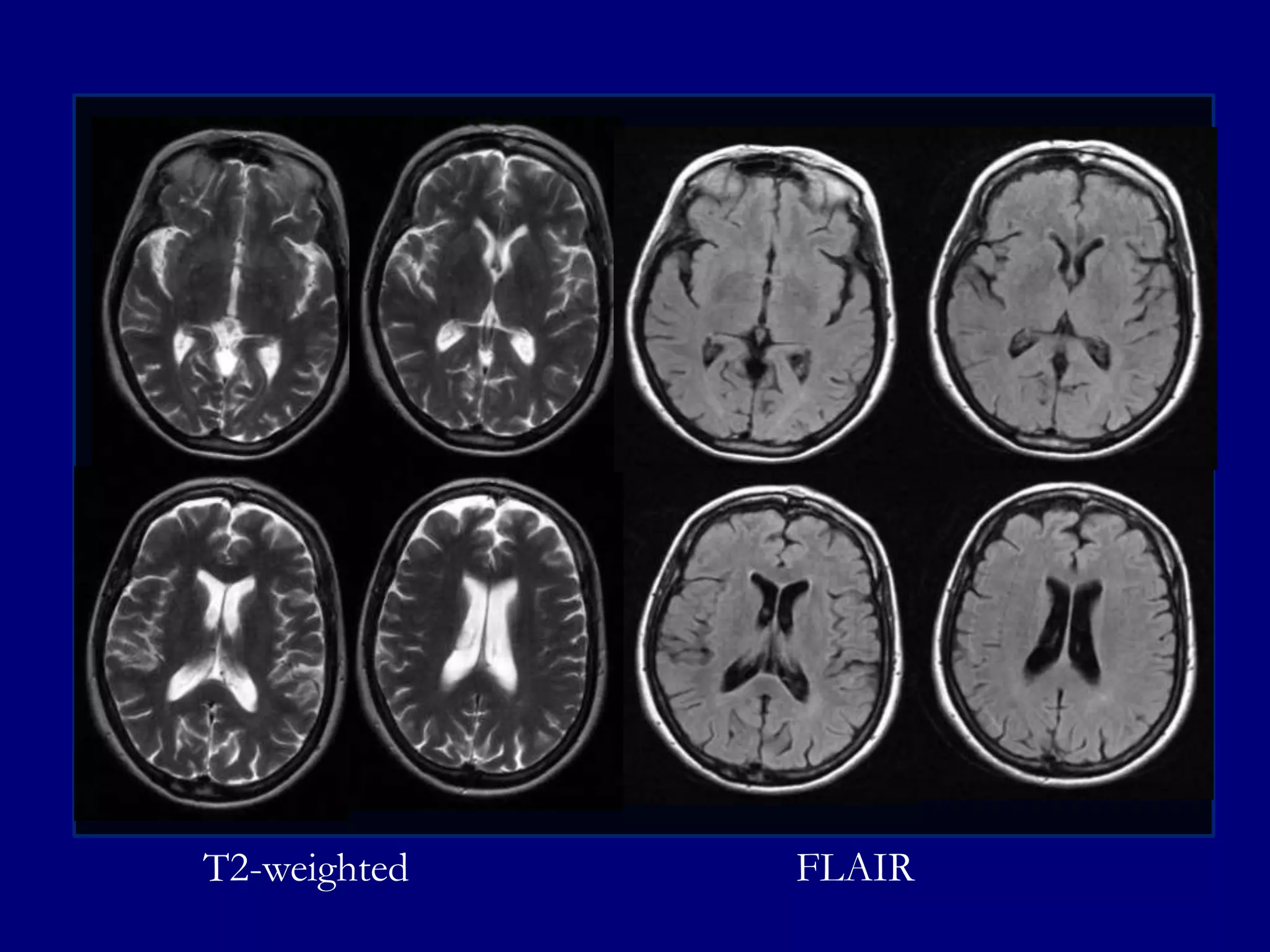
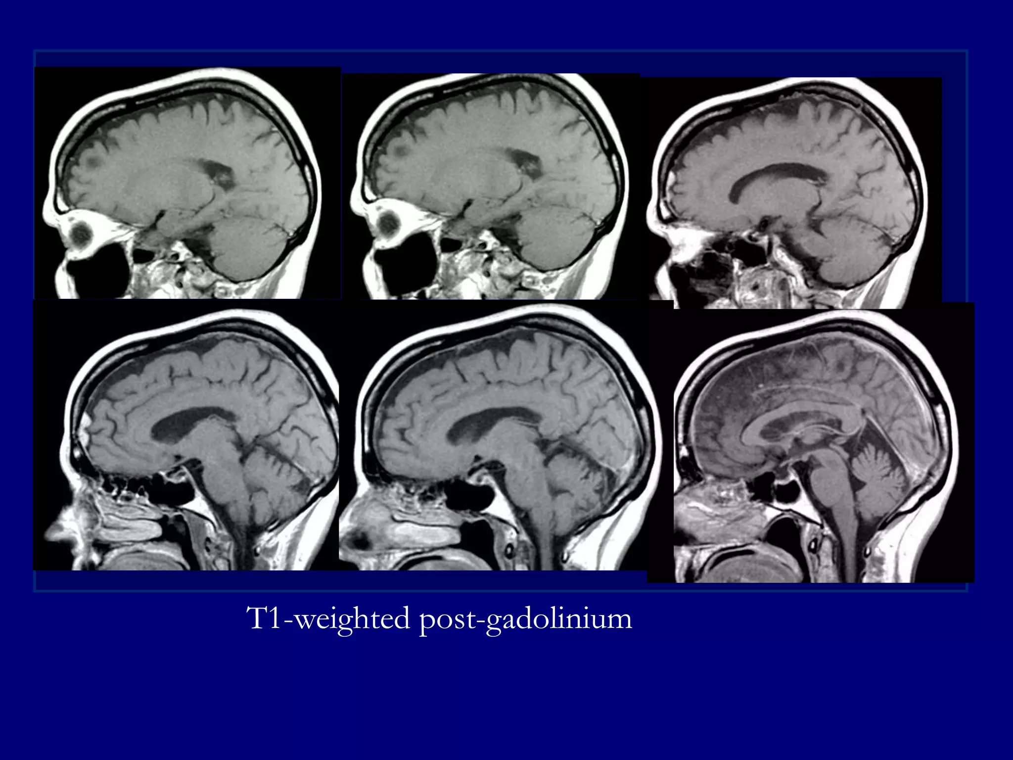
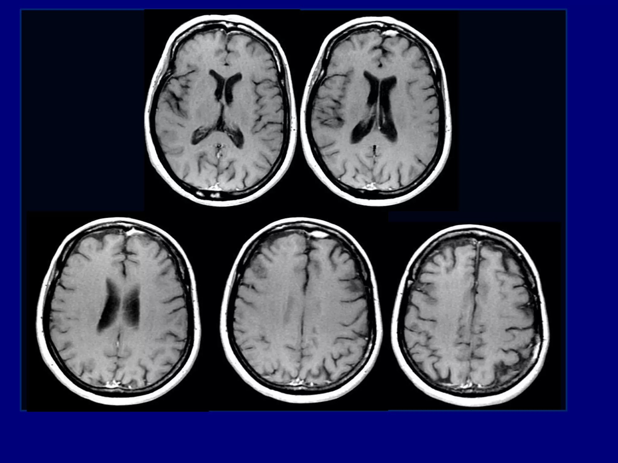
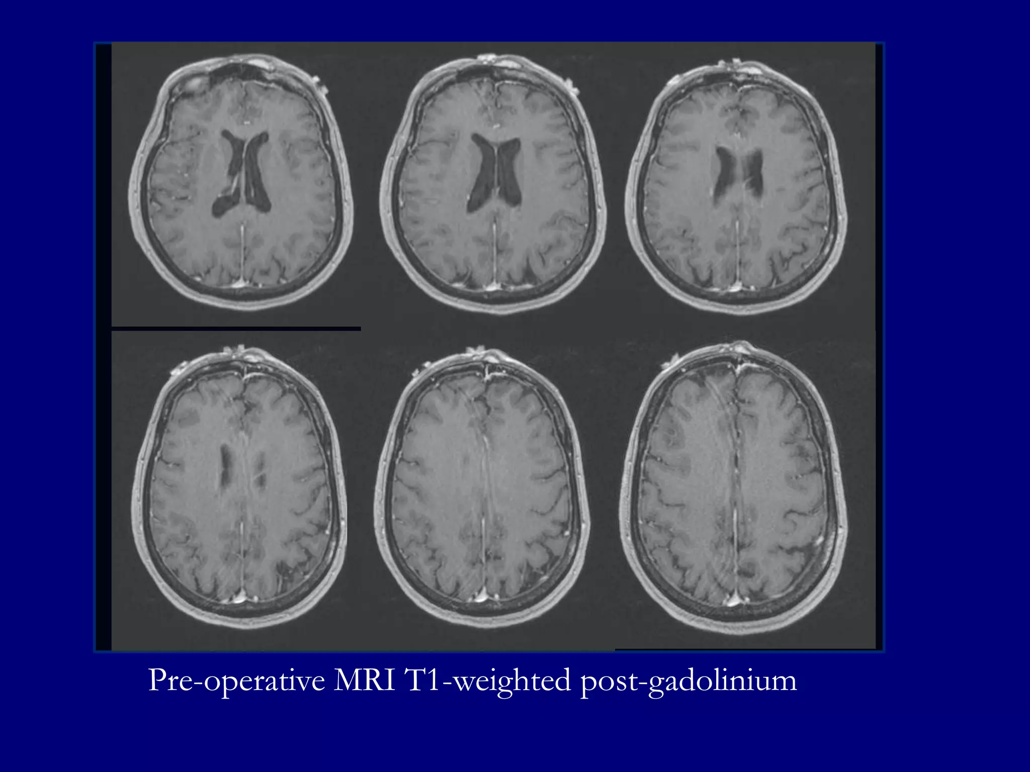
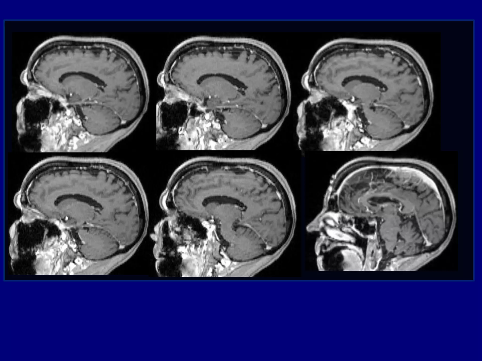
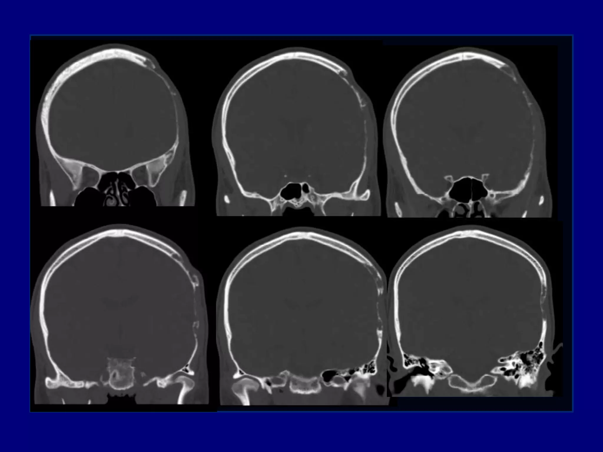
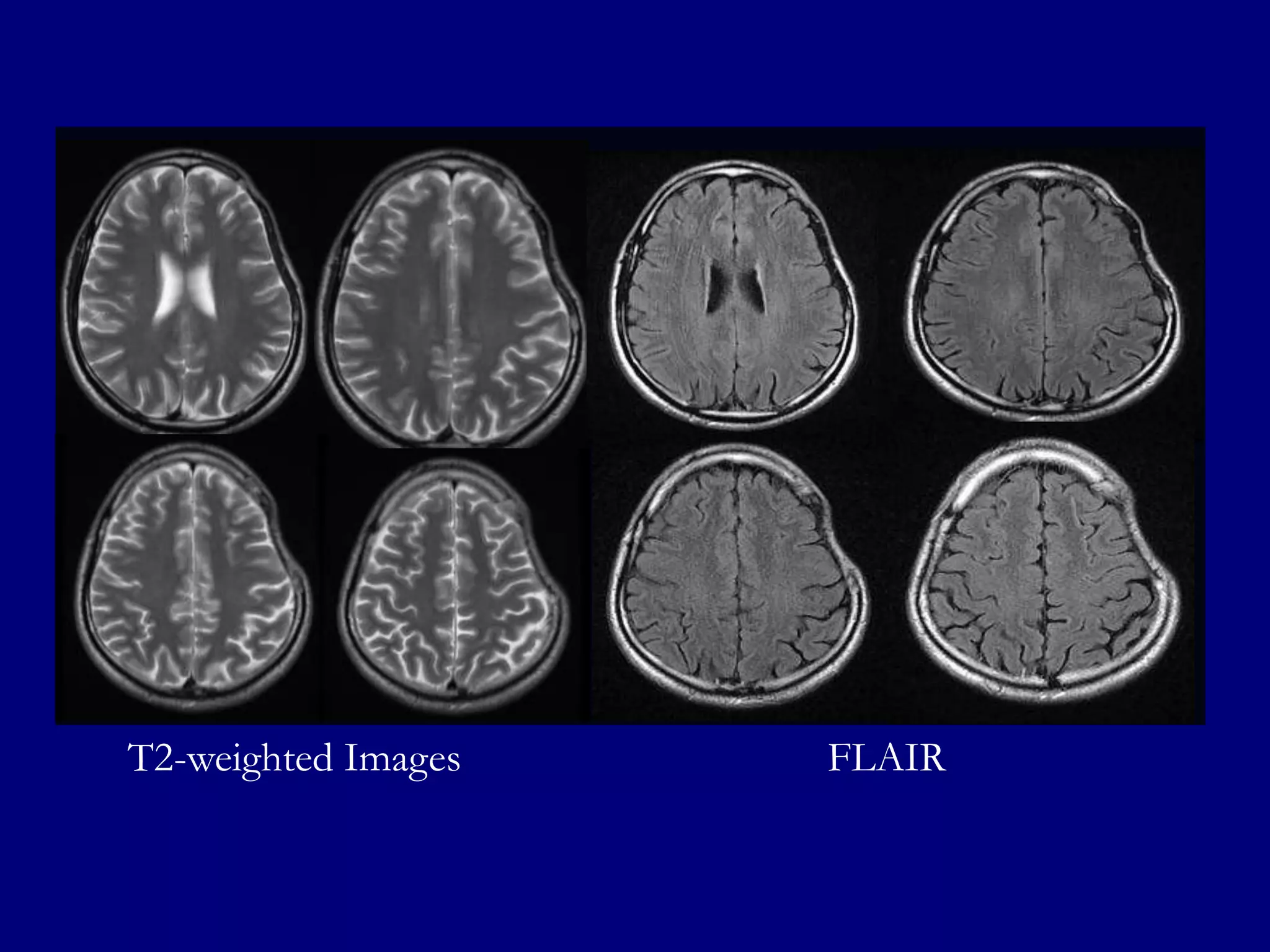
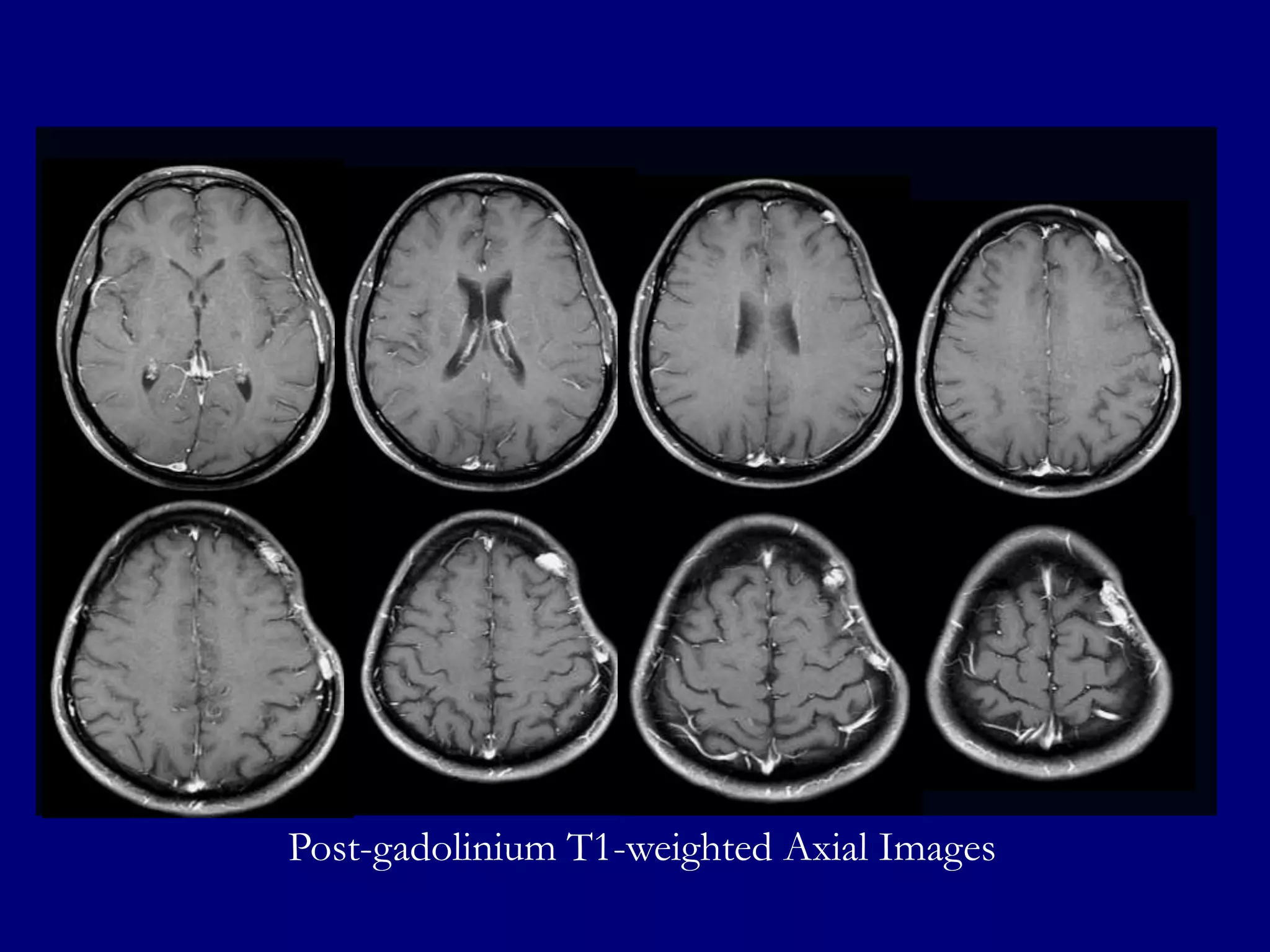
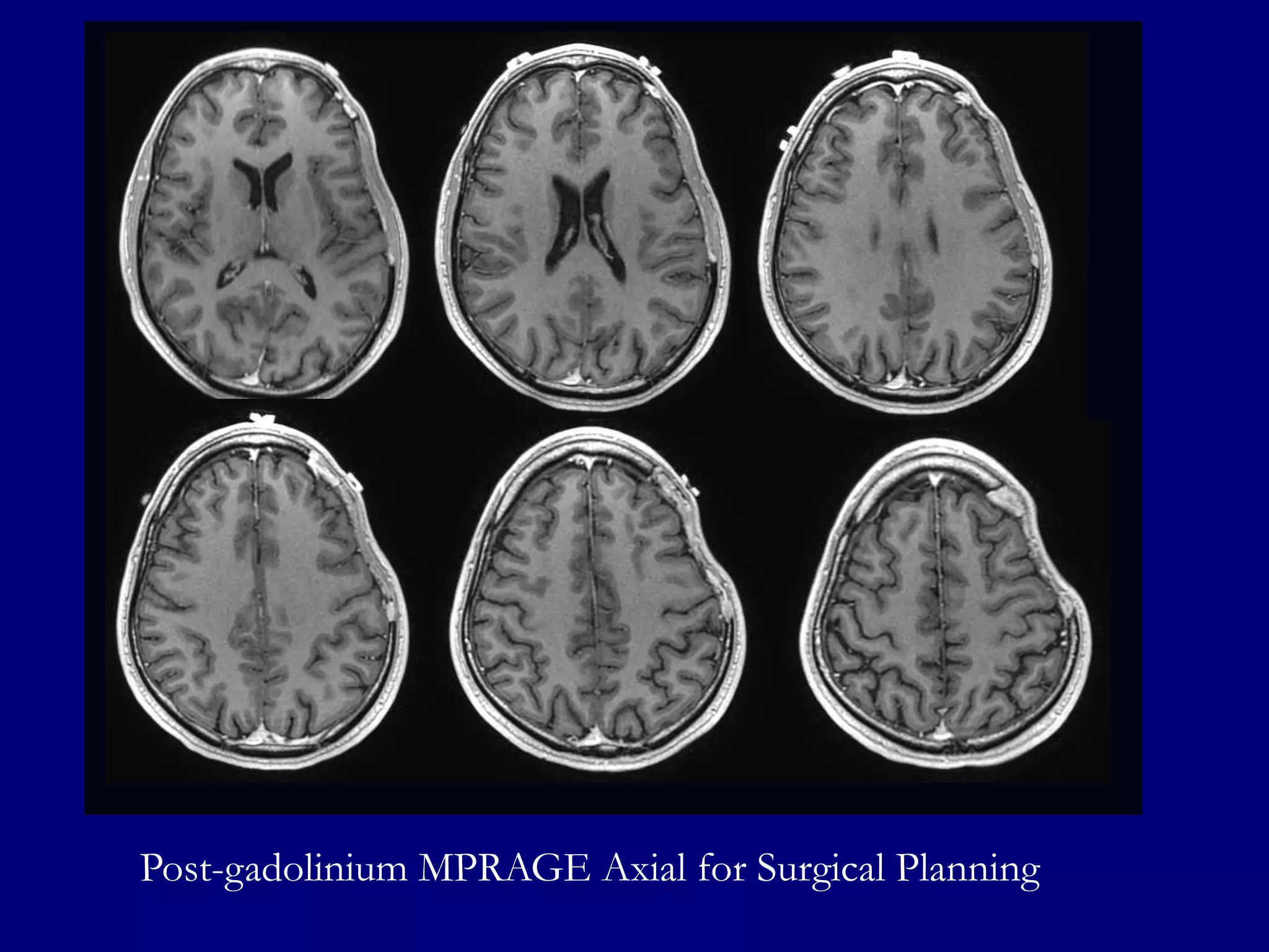
Unknown Cases
Rafeeque A. Bhadelia, MD
Associate Professor of Radiology
Harvard Medical School
Clinical Director, Neuroradiology
Beth Israel Deaconess Medical Center
HARVARD
MEDICAL
SCHOOL
Alexander Lin, PhD
Instructor in Radiology
Harvard Medical School
Clinical Spectroscopist
Center for Clinical Spectroscopy
Brigham and Women’s Hospital
Clinical Information








37-year-old right-handed man with history of
Asperger's syndrome and hypertension
Seven days prior to hospital admission, he began
to notice right sided numbness and twitching
and bitemporal headaches
Patient gave history of travelling to Botswana
Neurological examination was unremarkable
Laboratory examinations of blood and CSF were
unremarkable
Subcortical hyperdense masses in the left fronto-parietal lobes
with surrounding hypodensities extending to basal ganglia
FLAIR: Well defined hyperintense lesions in the left fronto-parietal lobes with
hyperintensities in left basal ganglia, splenium of corpus callosum and left cerebellum
T1-weighted Post-gadolinium images show homogenously enhancing well defined
lesions in left fronto-parietal lobes and enhancement in left cerebellum
ADC

Diffusion

ASL

Diffusion trace and ADC images show slow (restricted) diffusion in the well
defined enhancing left fronto-parietal lesions
ASL show mild to moderate increased perfusion in left fronto-parietal lesions
rCBF

rCBV

Dynamic Susceptibility Contrast (DSC) Perfusion
images show minimally increased rCBV and rCBF in left
fronto-parietal lesions
Differential Diagnosis







Glioblastoma Multiforme
Lymphoma
Metastasis
Tumefactive Multiple Sclerosis
Subacute Infarcts
NAA

Choline

2D- CSI (PROBE: TE=144, Section=10mm, NEX=1

Creatine
ROI 2

ROI 3

ROI 4
ROI 5

ROI 8

ROI 12

ROI 13

ROI 14

ROI 7

ROI 9

ROI 15
NAA

Choline

Creatine
ROI 1

ROI 3

ROI 7

ROI 8

ROI 2

ROI 4

ROI 9
ROI
14

ROI
15

ROI
16

ROI
17

ROI
18

ROI
19

ROI
20

ROI
21

ROI
24

ROI
25

ROI
26

ROI
27
Diagnosis ?
Diagnosis:
Primary CNS Lymphoma
Primary CNS Lymphoma (PCNLS)





PCNLS accounts for 1-5 % of brain tumors
PCNSL may have characteristic imaging
appearance due to hypercellularity and high
nuclear/cytoplasmic ratio
PCNLNS has predilection for the periventricular
and superficial regions and presents as multiple
masses predominantly in supra-tentorial regions




CT
 Hyperdense lesions ( 70%). Hemorrhage is uncommon
MRI
 FLAIR/T2-hyperintense homogenously enhancing
masses in high grade lesions. Enhancement may be
minimal or absent in low grade lesions
 Lesions may show slow(restricted diffusion) in high
grade lesions (more than in glioma or metastasis) due to
hypercellularity
 MR perfusion shows minimally increased (much less
than glioma), normal or decreased rCBV


Scintigraphy
Thallium201 scan can show increased uptake
 PET with FDG show very high uptake which may
help differentiation from glioma and metastasis




MR Spectroscopy
Large Choline peak
 High choline/creatine ratio
 Lactate peak may be seen



MRS Pitfalls
GBM: 2HG would have been definitive
 Metastasis: Lack of high Cho at the lesion
periphery
 Lymphoma: Short echo would show higher lipid
and lactate
 Tumefactive Multiple Sclerosis: Difficult to
differentiate with MRS
 Subacute Infarcts: Much higher lactate would have
been present

Asim Mian, MD
Assistant Professor of Radiology
Boston University School of Medicine
Neuroradiologist
Boston Medical Center
Clinical Information







59-year-old male with the history of a slow
growing bony tumor of his right calvarium
The mass has grown over several years
No similar masses elsewhere
No associated neurological symptoms or pain
Medical consultation for cosmetic reasons
Computed Tomography
Magnetic Resonance Imaging
Post-gadolinium MPRAGE images for surgical planning
Cerebral Angiography
Right Internal Carotid Artery Injection
Left External Carotid Artery Injection
Right External Carotid Artery Injection
Diagnosis ?
Diagnosis:
Primary Intra-osseous
Meningioma
Primary Intraosseous
Meningioma








Rare subtype of extra-dural meningioma arising
from the skull (convexity of skull base)
Most patients are >50-years with no sex
predilection
Presents as firm painless scalp mass with intact
skin and may be detected incidentally
<1% of all meningiomas
Osteoblastic 80%
 Osteolytic 20%



Plain Radiographs and CT




Focal area of hyperostosis in osteoblastic variety
with expansion of calvarium

MRI
Hypointense lesion on T1 and T2-weighted images
(or hyperintense on T2WI)
 May show intrinsic enhancement
 Enhancement of the underlying dura due to invasion
or dural irritation
 No “dural tail”



Angiography


Enlarged tortuous external carotid artery branch
supplying the lesion and dense tumor blush are
typical
Differential Diagnosis


Fibrous Dysplasia




Osteoma




No intrinsic enhancement or dural enhancement or tumor
blush

Osteosarcoma




Younger patient (stops growing after puberty). Ground glass
appearance. No dural enhancement or tumor blush

Irregular contour and soft-tissue mass. Heterogeneous signal
and enhancement

Paget’s Disease


Heterogeneous signal, no intrinsic or dural enhancement
Deepak Takhtani, MD
Associate Professor of Radiology
University of Massachusetts Medical School
Director
Neuroradiology Section
University of Massachusetts Medical Center
Clinical Information






65-year-old female, with a past medical history
significant for obesity, hypertension, and multiple
sclerosis
A routine follow-up MRI for multiple sclerosis at
an outside facility revealed a new dumbbellshaped mass in the frontal area on the left
Patient referred for biopsy with differential
diagnosis of plasmacytoma, lymphoma, or dural
based metastasis
2012

2013
MRI 2012
T1-weighted post-gadolinium
MRI 2013
T2-weighted

FLAIR
T1-weighted post-gadolinium
MRI 2013
Pre-operative Study
Pre-operative MRI T1-weighted post-gadolinium
Based on MR features,
surgery was cancelled and a
diagnostic Ultrasound was
performed
Transverse forehead
Resting

Transverse Forehead Valsalva
Color Doppler in resting

Color Doppler during Valsalva maneuver
Follow up MRI 2013
T2-weighted
Pre and post-gadolinium T1-weighted
Post-gadolinium MPRAGE
Diagnosis ?
Diagnosis:
Sinus Pericranii
Sinus Pericranii











A sub-periosteal blood-filled sac that communicates
with the intracranial veins through an emissary vein
Associated with Developmental Venous Anomaly
(DVA)
Mostly congenital but can be acquired
Usually located in frontal region
Represents 4% of palpable scalp masses
Can be seen at any age but mostly young adults
Increases in size in supine position or with Valsalva
maneuver. May completely disappear in supine position


CT




Slightly hyperdense sub-periosteal lesion which
enhances similar to other intra-cranial veins with
associated bony defect of emissary vein

MRI


Isointense on T1 and hyperintense on T2weighted images with intense enhancement
(puddling of contrast) in scalp lesion as well as of
the extra-axial venous component


Angiography




Arterial and capillary phases are normal with
accumulation of contrast adjacent to the
transcalvarial defect

Ultrasound
The scalp lesion changes size with upright position
and Valsalva maneuver
 Color Doppler shows change in flow direction with
Valsalva

Differential Diagnosis


Neonates and children
Cephalocele, meningocele
 Dermoid cyst
 Hemangioma
 Metastasis (neuroblastoma)




Adults
Sebaceous cyst
 Intracranial extra-axial mass (meningioma,
lymphoma)

Pamela W. Schaffer, MD
Associate Professor of Radiology
Harvard Medical School
Associate Director of Neuroradiology Clinical
Director MRI
Massachusetts General Hospital
Clinical Information


25-year old right handed man with complaints of
cranial defect left side of his head for three years



For the past 6 months he has had headaches in
the area of indentation, decreased hearing and
occasional visual problems



No history of fever, weight loss, systemic illness
or trauma
Computed Tomography
Magnetic Resonance Imaging
T2-weighted Images

FLAIR
Post-gadolinium T1-weighted Axial Images
Post-gadolinium MPRAGE Sagittal and Coronal
Post-gadolinium MPRAGE Axial for Surgical Planning
Diagnosis ?
Diagnosis:
Gorham Disease
(Vanishing Bone Disease)
Gorham Disease


Patients younger than 40-years in age



No sexual or familial predilection



Patient presents with progressive defect (depression) of
skull



Although medical and radiation therapy is attempted in
the past, surgical removal followed by artificial bone
replacement remains the treatment of choice
Pathology




Intra-osseous proliferation of hemangiomatous
or lymphangiomatous tissue resulting in
progressive osteolysis of the affected bone
Two stages of are described:
First stage: Vascular proliferation in connective
tissue consistent with hemangiomatosis
 Second stage: Fibrous tissue replaces the absorbed
bone without regeneration of bone matrix

Imaging


CT
Well defined skull defect with benign characteristics
 Thinning and disappearance of central portion and
absence of new bone formation




MRI
Low signal central portion on T1 and T2-weighted
images indicating fibrosis
 Enhancement at the margins suggestive of
hemangiomatous active component of the disease



Tc-99 bone imaging (not performed in this
case) shows absence of uptake in the central
portion with increased uptake at the margins

Lo et al AJNR 2004 25: 415-418
Thank You

HARVARD
MEDICAL
SCHOOL

NERRS Sep 2013 Neuroradiology Case Answers