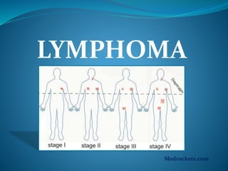
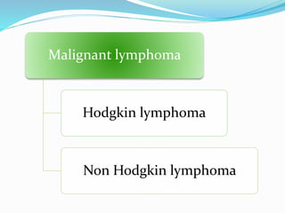
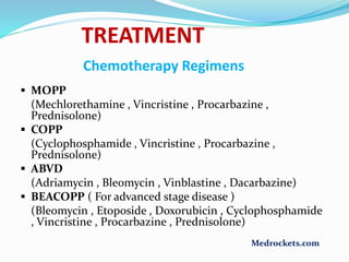
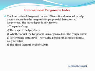
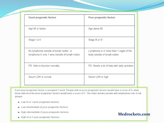
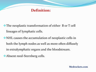
LYMPHOMA
Medrockets.com
• Malignant lymphoma is a term given to tumors of the lymphoid
system and specifically of lymphocytes and their precursor cells
• The name often refers to just the cancerous ones rather than all
such tumors.
• Many lymphomas are known to be due to specific genetic
mutations.
• 3rd most common cancer in children with incidence of 15 per
million children
LYMPHOMA
Medrockets.com
Malignant lymphoma
Hodgkin lymphoma
Non Hodgkin lymphoma
HODGKIN’S
LYMPHOMA
Medrockets.com
Hodgkin disease
 Definition:
A neoplastic transformation of lymphocytes particularly in lymph nodes.
Characterized by:
1) the presence of Reed-Sternberg cells on histology
2) spreading in an orderly fashion to contagious lymph nodes
( For example, Hodgkin lymphoma that starts in the cervical lymph nodes
may spread first to the supraclavicular nodes then to the axillary nodes )
 6% of childhood cancer
 5% of cancer in < 14 yr
 15% in person 15-19 yr
 Rare < 10 yr
Large cells ( >45um in diameter) with classically binucleate or
bilobed central nucleus each with a large acidophilic central
nucleoli surrounded by a clear halo. “owl’s eye appearance”
Red-Sternberg cells
Medrockets.com
Epidemiology of Hodgkin’s lymphoma
 Hodgkin disease has bimodal age distribution--one peak in the 20s
and 60s.
 Early peak middle to late 20s
 Second peak after 50 yr
 Sex Male : Female
4: 1 for 3-7 yr
3: 1 for 7-9 yr
1-3: 1 for > 10 yr
 100 folds risk for unaffected monozygotic twin of affected twin
 Associated with specific HLA antigen
 Infectious agents
Human herpes virus 6
CMV
Epstein – Barr virus
 Immunodeficiency
Medrockets.com
Etiology/Risk Factors
Doctors seldom know why one person develops
Hodgkin lymphoma and another does not. But
research shows that certain risk factors increase the
chance that a person will develop this disease.
Having one or more risk factors does not mean that a
person will develop Hodgkin lymphoma. Most people
who have risk factors never develop cancer.
Medrockets.com
Risk Factors
1) Certain viruses:
 Epstein-Barr virus (EBV)
 Human immunodeficiency virus (HIV)
2) Weakened immune system:
 inherited condition
 certain drugs used after an organ transplant
3) Age:
 Hodgkin lymphoma is most common among teens and adults aged 15 to
35 years and adults aged 55 years and older.
4) Family history:
 Family members, especially brothers and sisters, of a person with
Hodgkin lymphoma or other lymphomas may have an increased chance
of developing this disease.
Medrockets.com
Lymphocyte Predominant
10-15% of patients
More common in male
Younger patients
Localized disease
Has best prognosis
Mixed cellularity
30% of patients
< 10 yr of age
Advanced disease
Extranodal extension
Lymphocyte depletion
Rare in children
Common with HIV
Has worst prognosis
Nodular sclerosis
Most common
40% of younger patients
70% of adolescents
Classification
Rye Classification System
Medrockets.com
REAL Classification
( Revised European – American Classification of Lymphoid Neoplasms )
 Nodular lymphocyte predominance
 Classical Hodgkin lymphoma
 Lymphocyte rich
 Mix cellularity
 Nodular sclerosis
 Lymphocyte depletion
 Anaplastic large cell lymphoma Hodgkin like
Medrockets.com
 Enlarged, painless, rubbery, non- erythematous, nontender lymph nodes are
the hallmark of the disease.
 May become painful after drinking alcohol
 Hepatosplenomegaly
 Cough, dyspnea, hypoxia
 Pleural or pericardial effusion
 Heptocellular dysfunction
 B.M infiltration(Anemia, neutropenia, thrombocytopenia)
 25% have ''B'' symptoms
 Although pruritus is common in the disease it is not one of the ‘’B’’ symptoms.
 Cervical, supraclavicular and axillary lymphadenopathy are the most common
initial signs of the disease.
 Disease below diaphragm is rare (only3%)
Clinical presentation
Medrockets.com
Systemic Symptoms (B symptoms)
 Important in staging
 Unexplained fever > 390C
 Weight loss > 10% in 3m
 Drenching night sweats
Immune System abnormalities
 Anergy to delayed-hypersensitivity skin test
 Abnormal cellular immune response
 Decreased CD4:CD8 ratio
 Reduce natural killer cell cytotoxicity
Medrockets.com
Extralymphatic sites may be involved such as:
# Spleen
# Liver
# Bone marrow
# Lung
# CNS
Extralymphatic involvement is more common with
non-hodgkin lymphoma.
Emergency presentation:
Infections
SVC obstruction ( facial edema, increased JVP
and Dyspnea)
Medrockets.com
The doctor considers the following to determine the
stage of Hodgkin lymphoma:
The number of lymph nodes affected.
Whether these lymph nodes are on one or both sides of
the diaphragm.
Whether the disease has spread to the bone marrow,
spleen, liver, or lung.
Each stage is divided into A or B symptoms according to
the presence of systemic symptoms.
Staging of Hodgkin’s Lymphoma
Medrockets.com
Ann Arbor Staging Classification for Hodgkin
Disease
 Stage I
Involvement of a single lymph node (1)
or of a single extra lymphatic site or organ(1f)
 Stage II
Involvement of two or more lymph node regions on the same side of
the diaphragm(II)
or localised involvement of an extra lymphatic site
or organ and one or more lymph node regions on the same side of the
diaphragm (IIf)
Medrockets.com
Stage III
Involvement of lymph node regions on both sides of the
diaphragm (III) which may be accompanied by the involvement of
spleen (IIIS) or by localized involvement of an extra lymphatic site or
organ ( IIIf) or both ( IIIsf)
Stage IV
Diffuse or disseminated involvement of one or more
extra lymphatic organs or tissues with or without associated lymph node
involvement.
The absence or presence of fever > 38C for three consecutive days ,
drenching night sweats , or unexplained loss of > 10% body weight in
the 6 months preceding admission are to be denoted in all cases by
the suffice letters A & B respectively.
Medrockets.com
Medrockets.com
DIAGNOSIS
 An excisional lymph node biopsy is the essential first step in diagnosis.
 A biopsy is the only sure way to diagnose Hodgkin lymphoma.
 Excisional Biopsy
 Light Microscopy
 Immunocytochemistry
 Molecular Studies
 Chest X – Ray
 Mediastinal Mass
 CT Scan
 Chest
 Abdomen
 Pelvis
 Blood CP & ESR
 LFT’s
 Bone Marrow Aspiration
 Serum Copper & Ferritin
 Bone Scan
 Gallium 67 Scan / FDG/PET
Medrockets.com
TREATMENT
 Treatment depends on :
 Stage of the disease
 Age at diagnosis
 Presence / absence of B symptoms
 Presence of hilar lymphadenopathy
 Presence of bulky nodal disease
 Current Treatment Regimen
 Combined chemotherapy with or without low dose involved
field radiation therapy
Medrockets.com
Chemotherapy Regimens
 MOPP
(Mechlorethamine , Vincristine , Procarbazine ,
Prednisolone)
 COPP
(Cyclophosphamide , Vincristine , Procarbazine ,
Prednisolone)
 ABVD
(Adriamycin , Bleomycin , Vinblastine , Dacarbazine)
 BEACOPP ( For advanced stage disease )
(Bleomycin , Etoposide , Doxorubicin , Cyclophosphamide
, Vincristine , Procarbazine , Prednisolone)
TREATMENT
Medrockets.com
 Therapy is entirely based on the stage.
 Localized disease ( stage IA and IIA ) is managed predominantly with
radiation.
 All patients with evidence of ‘’B’’ symptoms as well as stage III and IV
are managed with chemotherapy.
 The most effective combination chemotherapeutic regimen for
Hodgkin lymphoma is ABVD ( adriamycin, bleomycin, vinblastin and
dacarbazine).
 ABVD is superior to MOP (meclorethamine, vincristin(oncovin) ,
prednisolone and procarbazine) because ABVD has fewer side effects
such as:
1) Permanent sterility
2) Secondary cancer formation
3) Aplastic anemia
4) Peripheral neuropathy
TREATMENT
Medrockets.com
International Prognostic Index
 The International Prognostic Index (IPI) was first developed to help
doctors determine the prognosis for people with fast-growing
lymphomas. The index depends on 5 factors:
1) The patient’s age
2) The stage of the lymphoma
3) Whether or not the lymphoma is in organs outside the lymph system
4) Performance status (PS) – how well a person can complete normal
daily activities
5) The blood (serum) level of (LDH)
Medrockets.com
Medrockets.com
LONG TERM COMPLICATIONS
 Secondary malignancy
 Acute Myelogenous Leukemia
 Non Hodgkin lymphoma
 Carcinomas of breast , lungs & thyroid
 Short stature
 Hypothyroidism
 Sterility
 Dental caries
 Sub clinical pulmonary dysfunction
 Ischemic heart disease
Medrockets.com
NON-HODGKIN’S
LYMPHOMA
Medrockets.com
The neoplastic transformation of either B or T cell
lineages of lymphatic cells.
NHL causes the accumulation of neoplastic cells in
both the lymph nodes as well as more often diffusely
in extralymphatic organs and the bloodstream.
Absent reed-Sternberg cells.
Definition:
Medrockets.com
 60% of all lymphomas in children
 8-10% of all malignancies in children between 5-19
yrs of age
 Secondary causes of NHL include;
 Inherited / acquired immune deficiencies
 Viruses
 HIV
 EBV
 Genetic Syndromes
 Ataxia Telangiectasia
 Bloom syndrome
EPIDEMIOLOGY
Medrockets.com
Risk factors
INFECTIONS:
Human immunodeficiency virus (HIV)
 Epstein-Barr virus (EBV): linked to Burkitt lymphoma.
Helicobacter pylori: Extranodal tissues generating
lymphoma include MALT ( Mucosa associated lymphoid
tissue)
Human T-cell leukemia/lymphoma virus( HTLV-1)
Hepatitis C virus
Age:
Most people with non-Hodgkin lymphoma are older than
60.
Medrockets.com
 Burkitt Lymphoma
40% of NHL
B Cell Origin
 Lymphoblastic Lymphoma
30% of NHL
80% T Cell Origin & 20% B Cell Origin
 Diffuse Large B Cell Lymphoma
20% of NHL
B Cell Origin
 Anaplastic Large Cell Lymphoma
10% of NHL
70% T Cell Origin
PATHOLOGICAL SUB TYPES OF NHL
Medrockets.com
Burkitt Lymphoma
Clinical Presentation
Clinical presentation is the same as for Hodgkin
lymphoma.
The difference is that Hodgkin is localized to
cervical and supraclavicular nodes 80%-90% of
the time. Whereas NHL is localized 10-20% of the
time.
CNS involvement is more common with NHL.
HIV positive patients often have CNS involvement.
Medrockets.com
 Burkitt Lymphoma
 Abdominal Tumor
 Head & Neck Disease
 Involvement of bone marrow & CNS
 Lymphoblastic Lymphoma
 Intrathoracic / mediastinal supradiaphragmatic mass
 Involvement of bone marrow & CNS
 Diffuse Large B Cell Lymphoma
 Abdominal Mass
 Mediastinal Mass
 Anaplastic Large Cell Lymphoma
 Primary cutaneous manifestation
 Systemic disease ( fever , weight loss)
 Dissemination to liver , spleen , lung , mediastinum & skin
Clinical Presentation
Medrockets.com
 Other clinical features include;
 Lymphadenopathy
 Superior vena cava syndrome
 Dyspnea
 Abdominal Mass
 Intestinal obstruction / intussusception
 Ascites
 Nasal Stuffiness
 Earache
 Tonsil enlargement
 Localised bone involvement
 Acute paraplegia secondary to CNS / spinal cord compression
 Tumor Lysis Syndrome
Clinical Presentation
Medrockets.com
Staging system for Non-Hodgkin
lymphoma
Stage Description
I A single tumor (extranodal) or single anatomic
area (nodal) with the exclusion of mediastinum or
abdomen
II A single tumor (extranodal) with regional node
involvement
two or more nodes areas on the same side of
diaphragm
Two single (extranodal) tumors with or without the
regional node involvement on same side of diaphragm
A primary gastrointestinal tract tumor usually in the
ileocecal area, with or without involvement of associated mesenteric nodes,
which may must bemgrossly ( > 90%) resected
Medrockets.com
Stage III Two single tumors (extranodal) on opposite
side of the diaphragm
Two more nodal areas above and below the
diaphragm
Any primary intarthoracic tumor (mediastinal,
pleural, or thymic)
Any extensive primary intra – abdominal
disease
IV Any of the above, with initial involvement of
central nervous system or bone marrow t time
of diagnosis
Medrockets.com
DIFFERENTIAL DIAGNOSIS
 Hodgkin Disease
 Leukemia
 Germ Cell Tumor
 Wilms Tumor
 Neuroblastoma
 Rhabdomyosarcoma
 Reactive lymphadenitis
Medrockets.com
 Tissue biopsy for;
 Flow cytometry
 Karyotyping
 Bone marrow biopsy is more central in the initial staging of NHL
 Complete Blood Count
 Serum Electrolytes, Calcium , Phosphorus , Uric acid
 LFT’s & RFT’s
 Bone Marrow Aspiration & Biopsy
 CSF Examination
 Chest X Ray
 CT Scan
 Head & Neck
 Chest
 Abdomen & Pelvis
 PET Scan & Bone Scan
Diagnosis
Medrockets.com
Grades
NHL divided into Low and high grade
A high grade lymphoma has cells which look quite
different from normal cells.
They tend to grow fast (aggressive).usually look
follicular. Incurable. Wider dissemination at
presentation.
Low grade lymphomas have cells which look much like
normal cells and multiply slowly(indolent).usually
look diffuse. Long term treatment maybe achievable.
Medrockets.com
Many low-grade lymphomas remain indolent for many
years.
Treatment of the non-symptomatic patient is often
Avoided.
 In this case watchful waiting is often the initial course of
action. This is carried out because the harms and risks of
treatment outweigh the benefits.
If a low-grade lymphoma is becoming symptomatic,
radiotherapy or chemotherapy are the treatments of
choice.
They don’t cure the lymphoma, they can alleviate the
symptoms.
Patients with these types of lymphoma can live near-
normal lifespans, but the disease is Incurable.
Low-grade lymphomas
Medrockets.com
High-grade lymphomas
Treatment of the aggressive, forms of lymphoma can
result in a cure in the majority of cases.
However, the prognosis for patients with a poor
response to therapy is worse.
Treatment for these types of lymphoma typically
consists of aggressive chemotherapy, including
the CHOP or R-CHOP regimen.
Medrockets.com
Treatment
The initial chemotherapeutic regimen is CHOP
 ( cyclophosmamide, hydroxy-adriamycine, oncovin
and prednisolone).
CNS lymphoma is often treated with radiation in
addition to CHOP.
Relapses can be controlled with BM transplantation.
Some pts express CD-20 antigen in greater amount. In
this case, monoclonal antibody Rituximab should be
used.
Rituximab is an anti-CD20 antibody that has limited
toxicity and add survival benefit to the use of CHOP.
Medrockets.com
Duration of Treatment
 Burkitt Lymphoma & Diffuse Large B Cell Lymphoma
………. 6 weeks to 6 months
 Lymphoblastic Lymphoma …..24 months
Medrockets.com
COMPLICATIONS
 Infections
 Mucositis
 Pancytopenia
 Electrolyte imbalance
 Poor nutrition
 Growth retardation
 Cardiac Toxicity
 Gonadal Toxicity with Infertility
 Secondary malignancies
Medrockets.com
Thank you
Medrockets.com

Lymphoma

  • 1.
  • 2.
    • Malignant lymphomais a term given to tumors of the lymphoid system and specifically of lymphocytes and their precursor cells • The name often refers to just the cancerous ones rather than all such tumors. • Many lymphomas are known to be due to specific genetic mutations. • 3rd most common cancer in children with incidence of 15 per million children LYMPHOMA Medrockets.com
  • 3.
  • 4.
  • 5.
    Hodgkin disease  Definition: Aneoplastic transformation of lymphocytes particularly in lymph nodes. Characterized by: 1) the presence of Reed-Sternberg cells on histology 2) spreading in an orderly fashion to contagious lymph nodes ( For example, Hodgkin lymphoma that starts in the cervical lymph nodes may spread first to the supraclavicular nodes then to the axillary nodes )  6% of childhood cancer  5% of cancer in < 14 yr  15% in person 15-19 yr  Rare < 10 yr
  • 6.
    Large cells (>45um in diameter) with classically binucleate or bilobed central nucleus each with a large acidophilic central nucleoli surrounded by a clear halo. “owl’s eye appearance” Red-Sternberg cells Medrockets.com
  • 7.
    Epidemiology of Hodgkin’slymphoma  Hodgkin disease has bimodal age distribution--one peak in the 20s and 60s.  Early peak middle to late 20s  Second peak after 50 yr  Sex Male : Female 4: 1 for 3-7 yr 3: 1 for 7-9 yr 1-3: 1 for > 10 yr  100 folds risk for unaffected monozygotic twin of affected twin  Associated with specific HLA antigen  Infectious agents Human herpes virus 6 CMV Epstein – Barr virus  Immunodeficiency Medrockets.com
  • 8.
    Etiology/Risk Factors Doctors seldomknow why one person develops Hodgkin lymphoma and another does not. But research shows that certain risk factors increase the chance that a person will develop this disease. Having one or more risk factors does not mean that a person will develop Hodgkin lymphoma. Most people who have risk factors never develop cancer. Medrockets.com
  • 9.
    Risk Factors 1) Certainviruses:  Epstein-Barr virus (EBV)  Human immunodeficiency virus (HIV) 2) Weakened immune system:  inherited condition  certain drugs used after an organ transplant 3) Age:  Hodgkin lymphoma is most common among teens and adults aged 15 to 35 years and adults aged 55 years and older. 4) Family history:  Family members, especially brothers and sisters, of a person with Hodgkin lymphoma or other lymphomas may have an increased chance of developing this disease. Medrockets.com
  • 10.
    Lymphocyte Predominant 10-15% ofpatients More common in male Younger patients Localized disease Has best prognosis Mixed cellularity 30% of patients < 10 yr of age Advanced disease Extranodal extension Lymphocyte depletion Rare in children Common with HIV Has worst prognosis Nodular sclerosis Most common 40% of younger patients 70% of adolescents Classification Rye Classification System Medrockets.com
  • 11.
    REAL Classification ( RevisedEuropean – American Classification of Lymphoid Neoplasms )  Nodular lymphocyte predominance  Classical Hodgkin lymphoma  Lymphocyte rich  Mix cellularity  Nodular sclerosis  Lymphocyte depletion  Anaplastic large cell lymphoma Hodgkin like Medrockets.com
  • 12.
     Enlarged, painless,rubbery, non- erythematous, nontender lymph nodes are the hallmark of the disease.  May become painful after drinking alcohol  Hepatosplenomegaly  Cough, dyspnea, hypoxia  Pleural or pericardial effusion  Heptocellular dysfunction  B.M infiltration(Anemia, neutropenia, thrombocytopenia)  25% have ''B'' symptoms  Although pruritus is common in the disease it is not one of the ‘’B’’ symptoms.  Cervical, supraclavicular and axillary lymphadenopathy are the most common initial signs of the disease.  Disease below diaphragm is rare (only3%) Clinical presentation Medrockets.com
  • 13.
    Systemic Symptoms (Bsymptoms)  Important in staging  Unexplained fever > 390C  Weight loss > 10% in 3m  Drenching night sweats Immune System abnormalities  Anergy to delayed-hypersensitivity skin test  Abnormal cellular immune response  Decreased CD4:CD8 ratio  Reduce natural killer cell cytotoxicity Medrockets.com
  • 14.
    Extralymphatic sites maybe involved such as: # Spleen # Liver # Bone marrow # Lung # CNS Extralymphatic involvement is more common with non-hodgkin lymphoma. Emergency presentation: Infections SVC obstruction ( facial edema, increased JVP and Dyspnea) Medrockets.com
  • 15.
    The doctor considersthe following to determine the stage of Hodgkin lymphoma: The number of lymph nodes affected. Whether these lymph nodes are on one or both sides of the diaphragm. Whether the disease has spread to the bone marrow, spleen, liver, or lung. Each stage is divided into A or B symptoms according to the presence of systemic symptoms. Staging of Hodgkin’s Lymphoma Medrockets.com
  • 16.
    Ann Arbor StagingClassification for Hodgkin Disease  Stage I Involvement of a single lymph node (1) or of a single extra lymphatic site or organ(1f)  Stage II Involvement of two or more lymph node regions on the same side of the diaphragm(II) or localised involvement of an extra lymphatic site or organ and one or more lymph node regions on the same side of the diaphragm (IIf) Medrockets.com
  • 17.
    Stage III Involvement oflymph node regions on both sides of the diaphragm (III) which may be accompanied by the involvement of spleen (IIIS) or by localized involvement of an extra lymphatic site or organ ( IIIf) or both ( IIIsf) Stage IV Diffuse or disseminated involvement of one or more extra lymphatic organs or tissues with or without associated lymph node involvement. The absence or presence of fever > 38C for three consecutive days , drenching night sweats , or unexplained loss of > 10% body weight in the 6 months preceding admission are to be denoted in all cases by the suffice letters A & B respectively. Medrockets.com
  • 18.
  • 19.
    DIAGNOSIS  An excisionallymph node biopsy is the essential first step in diagnosis.  A biopsy is the only sure way to diagnose Hodgkin lymphoma.  Excisional Biopsy  Light Microscopy  Immunocytochemistry  Molecular Studies  Chest X – Ray  Mediastinal Mass  CT Scan  Chest  Abdomen  Pelvis  Blood CP & ESR  LFT’s  Bone Marrow Aspiration  Serum Copper & Ferritin  Bone Scan  Gallium 67 Scan / FDG/PET Medrockets.com
  • 20.
    TREATMENT  Treatment dependson :  Stage of the disease  Age at diagnosis  Presence / absence of B symptoms  Presence of hilar lymphadenopathy  Presence of bulky nodal disease  Current Treatment Regimen  Combined chemotherapy with or without low dose involved field radiation therapy Medrockets.com
  • 21.
    Chemotherapy Regimens  MOPP (Mechlorethamine, Vincristine , Procarbazine , Prednisolone)  COPP (Cyclophosphamide , Vincristine , Procarbazine , Prednisolone)  ABVD (Adriamycin , Bleomycin , Vinblastine , Dacarbazine)  BEACOPP ( For advanced stage disease ) (Bleomycin , Etoposide , Doxorubicin , Cyclophosphamide , Vincristine , Procarbazine , Prednisolone) TREATMENT Medrockets.com
  • 22.
     Therapy isentirely based on the stage.  Localized disease ( stage IA and IIA ) is managed predominantly with radiation.  All patients with evidence of ‘’B’’ symptoms as well as stage III and IV are managed with chemotherapy.  The most effective combination chemotherapeutic regimen for Hodgkin lymphoma is ABVD ( adriamycin, bleomycin, vinblastin and dacarbazine).  ABVD is superior to MOP (meclorethamine, vincristin(oncovin) , prednisolone and procarbazine) because ABVD has fewer side effects such as: 1) Permanent sterility 2) Secondary cancer formation 3) Aplastic anemia 4) Peripheral neuropathy TREATMENT Medrockets.com
  • 23.
    International Prognostic Index The International Prognostic Index (IPI) was first developed to help doctors determine the prognosis for people with fast-growing lymphomas. The index depends on 5 factors: 1) The patient’s age 2) The stage of the lymphoma 3) Whether or not the lymphoma is in organs outside the lymph system 4) Performance status (PS) – how well a person can complete normal daily activities 5) The blood (serum) level of (LDH) Medrockets.com
  • 24.
  • 25.
    LONG TERM COMPLICATIONS Secondary malignancy  Acute Myelogenous Leukemia  Non Hodgkin lymphoma  Carcinomas of breast , lungs & thyroid  Short stature  Hypothyroidism  Sterility  Dental caries  Sub clinical pulmonary dysfunction  Ischemic heart disease Medrockets.com
  • 26.
  • 27.
    The neoplastic transformationof either B or T cell lineages of lymphatic cells. NHL causes the accumulation of neoplastic cells in both the lymph nodes as well as more often diffusely in extralymphatic organs and the bloodstream. Absent reed-Sternberg cells. Definition: Medrockets.com
  • 28.
     60% ofall lymphomas in children  8-10% of all malignancies in children between 5-19 yrs of age  Secondary causes of NHL include;  Inherited / acquired immune deficiencies  Viruses  HIV  EBV  Genetic Syndromes  Ataxia Telangiectasia  Bloom syndrome EPIDEMIOLOGY Medrockets.com
  • 29.
    Risk factors INFECTIONS: Human immunodeficiencyvirus (HIV)  Epstein-Barr virus (EBV): linked to Burkitt lymphoma. Helicobacter pylori: Extranodal tissues generating lymphoma include MALT ( Mucosa associated lymphoid tissue) Human T-cell leukemia/lymphoma virus( HTLV-1) Hepatitis C virus Age: Most people with non-Hodgkin lymphoma are older than 60. Medrockets.com
  • 30.
     Burkitt Lymphoma 40%of NHL B Cell Origin  Lymphoblastic Lymphoma 30% of NHL 80% T Cell Origin & 20% B Cell Origin  Diffuse Large B Cell Lymphoma 20% of NHL B Cell Origin  Anaplastic Large Cell Lymphoma 10% of NHL 70% T Cell Origin PATHOLOGICAL SUB TYPES OF NHL Medrockets.com
  • 31.
  • 32.
    Clinical Presentation Clinical presentationis the same as for Hodgkin lymphoma. The difference is that Hodgkin is localized to cervical and supraclavicular nodes 80%-90% of the time. Whereas NHL is localized 10-20% of the time. CNS involvement is more common with NHL. HIV positive patients often have CNS involvement. Medrockets.com
  • 33.
     Burkitt Lymphoma Abdominal Tumor  Head & Neck Disease  Involvement of bone marrow & CNS  Lymphoblastic Lymphoma  Intrathoracic / mediastinal supradiaphragmatic mass  Involvement of bone marrow & CNS  Diffuse Large B Cell Lymphoma  Abdominal Mass  Mediastinal Mass  Anaplastic Large Cell Lymphoma  Primary cutaneous manifestation  Systemic disease ( fever , weight loss)  Dissemination to liver , spleen , lung , mediastinum & skin Clinical Presentation Medrockets.com
  • 34.
     Other clinicalfeatures include;  Lymphadenopathy  Superior vena cava syndrome  Dyspnea  Abdominal Mass  Intestinal obstruction / intussusception  Ascites  Nasal Stuffiness  Earache  Tonsil enlargement  Localised bone involvement  Acute paraplegia secondary to CNS / spinal cord compression  Tumor Lysis Syndrome Clinical Presentation Medrockets.com
  • 35.
    Staging system forNon-Hodgkin lymphoma Stage Description I A single tumor (extranodal) or single anatomic area (nodal) with the exclusion of mediastinum or abdomen II A single tumor (extranodal) with regional node involvement two or more nodes areas on the same side of diaphragm Two single (extranodal) tumors with or without the regional node involvement on same side of diaphragm A primary gastrointestinal tract tumor usually in the ileocecal area, with or without involvement of associated mesenteric nodes, which may must bemgrossly ( > 90%) resected Medrockets.com
  • 36.
    Stage III Twosingle tumors (extranodal) on opposite side of the diaphragm Two more nodal areas above and below the diaphragm Any primary intarthoracic tumor (mediastinal, pleural, or thymic) Any extensive primary intra – abdominal disease IV Any of the above, with initial involvement of central nervous system or bone marrow t time of diagnosis Medrockets.com
  • 37.
    DIFFERENTIAL DIAGNOSIS  HodgkinDisease  Leukemia  Germ Cell Tumor  Wilms Tumor  Neuroblastoma  Rhabdomyosarcoma  Reactive lymphadenitis Medrockets.com
  • 38.
     Tissue biopsyfor;  Flow cytometry  Karyotyping  Bone marrow biopsy is more central in the initial staging of NHL  Complete Blood Count  Serum Electrolytes, Calcium , Phosphorus , Uric acid  LFT’s & RFT’s  Bone Marrow Aspiration & Biopsy  CSF Examination  Chest X Ray  CT Scan  Head & Neck  Chest  Abdomen & Pelvis  PET Scan & Bone Scan Diagnosis Medrockets.com
  • 39.
    Grades NHL divided intoLow and high grade A high grade lymphoma has cells which look quite different from normal cells. They tend to grow fast (aggressive).usually look follicular. Incurable. Wider dissemination at presentation. Low grade lymphomas have cells which look much like normal cells and multiply slowly(indolent).usually look diffuse. Long term treatment maybe achievable. Medrockets.com
  • 40.
    Many low-grade lymphomasremain indolent for many years. Treatment of the non-symptomatic patient is often Avoided.  In this case watchful waiting is often the initial course of action. This is carried out because the harms and risks of treatment outweigh the benefits. If a low-grade lymphoma is becoming symptomatic, radiotherapy or chemotherapy are the treatments of choice. They don’t cure the lymphoma, they can alleviate the symptoms. Patients with these types of lymphoma can live near- normal lifespans, but the disease is Incurable. Low-grade lymphomas Medrockets.com
  • 41.
    High-grade lymphomas Treatment ofthe aggressive, forms of lymphoma can result in a cure in the majority of cases. However, the prognosis for patients with a poor response to therapy is worse. Treatment for these types of lymphoma typically consists of aggressive chemotherapy, including the CHOP or R-CHOP regimen. Medrockets.com
  • 42.
    Treatment The initial chemotherapeuticregimen is CHOP  ( cyclophosmamide, hydroxy-adriamycine, oncovin and prednisolone). CNS lymphoma is often treated with radiation in addition to CHOP. Relapses can be controlled with BM transplantation. Some pts express CD-20 antigen in greater amount. In this case, monoclonal antibody Rituximab should be used. Rituximab is an anti-CD20 antibody that has limited toxicity and add survival benefit to the use of CHOP. Medrockets.com
  • 43.
    Duration of Treatment Burkitt Lymphoma & Diffuse Large B Cell Lymphoma ………. 6 weeks to 6 months  Lymphoblastic Lymphoma …..24 months Medrockets.com
  • 44.
    COMPLICATIONS  Infections  Mucositis Pancytopenia  Electrolyte imbalance  Poor nutrition  Growth retardation  Cardiac Toxicity  Gonadal Toxicity with Infertility  Secondary malignancies Medrockets.com
  • 45.