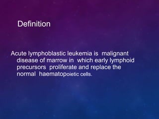
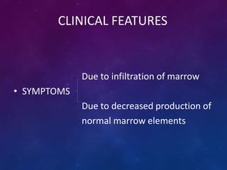
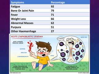
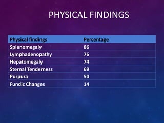
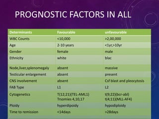
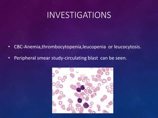
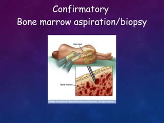
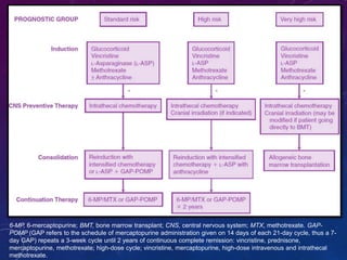
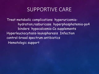
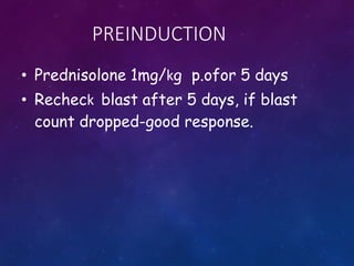
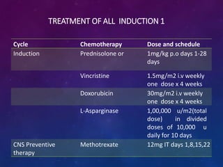
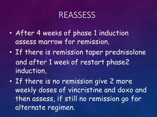
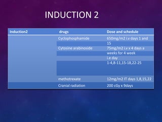
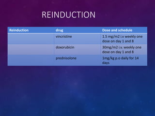
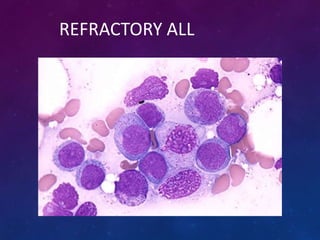
ACUTE
LYMPHOBLASTIC
LEUKEMIA
By
Dr. Ayush Garg
Acute lymphoblastic leukemia is malignant
disease of marrow in which early lymphoid
precursors proliferate and replace the
normal haematopoietic cells.
Definition
• Commonest form of malignancy in childhood.
• Majority are children of 2-10 yrs
• Peak incidence at 4 – 5 yrs of age.
• Acute onset with short history of duration.
• 85% are B cell
15% are T cell
• 5 times more frequent in childhood than AML
EPIDEMIOLOGY
CHROMOSOMAL ABNORMALITIES IN ALL
ABNORMALITIES ADULTS(%) CHILDREN(%)
Normal karyotype 16-34 9
Hypodiploidy 4-9 1
Hyperdiploidy 2-9 25
t (9;22) 11-30 4
t (4;11) 3-7 6
t (10;14) 4-6 4
t (8;14) 4 2
t ( 1;19) 3 5
9p abnormality 5-16 7-13
6q abnormality 2-6 4-6
12p abnormality 4-5 22
FACTORS PREDISPOSING ALL
GENETIC ENVRONMENTAL
Downs,turner, klinefelter Ionising Radiation
Fanconi,diamond blackfan Drugs
NF Type1 Alkylating Agents
Ataxia telengiectasia Nitrosourea
SCID Epipodophyllotoxin
PNH Benzene Exposure
Li-fraumeni syndrome Advanced Maternal Age
Blooms syndrome Paternal Smoking
BLAST CELLS
• ALL is characterized by presence of immature cells – blast cells
of >20% in the -blood picture
-bone marrow examination
• Features of blast cells
- very high N:C ratio
- large cells with large nuclei
- absence of cytoplasmic granules
- presence of round or convoluted nuclei
NORMAL MARROW
ENTIRE MARROW
REPLACED BY BLAST
MARROW SHOWING
BLASTS
FAB CLASSIFICATION OF ALL
Cytologic
Features
L1 L2 L3
Cell Size Small Cells
Predominate,
Homo Genous
Large,
heterogenous In
Size
Large Homogenous
Cytoplasm Scanty Variable,often
Moderately
Abundant
Moderately
Abundant
Nucleoli Small One Or More, often
Large
One Or More,
prominent
Nuclear Shape Homogenous Variable,
Heterogenous
Stippled,
Homogenous
Nuclear Shape Regular Irregular Clefts Regular
Cyt.Basophilia Variable Variable Intensely Basophilic
Cyt.Vacuolation Variable Variable Prominent
Immunologic
Subtype
% Of Cases FAB Subtype Cytogenetic
Abnormalites
Pre B ALL 75 L1,L2 t(9;22),t(4;11)
t(1;19)
T cell ALL 20 L1,L2 14q11 Or
7q34
Mature Bcell
All(burkitt
Leukemia)
5 L3 t(8;14)
CLASSIFICATION OF ALL(WHO)
PRE B ALL(L1)
SMALL BLASTS WTH THIN RIM OF
CYTOPLASM
T CELL ALL(L1)
T Cell ALL (L2)
Irregular clefts of nucleus
B CELL ALL (85%)
Type Tdt Calla Surface Ig
Early Pro B
ALL
Positive Negative Negative
Pre Bcell
ALL
Positive Positive Negative
Mature B
ALL
Negative Positive Positive
T CELL ALL(15%)
• Early subtype
•
•
CD3 -, CD4-,CD8- or
CD3-,CD4+,CD8+.
• Later subtype
• CD3+ with CD4+ or CD8+
T CELL ALL(CD3 POSITIVE)
CLINICAL FEATURES
Due to infiltration of marrow
• SYMPTOMS
Due to decreased production of
normal marrow elements
Symptoms Percentage
Fatigue 92
Bone Or Joint Pain 79
Fever 71
Weight Loss 66
Abnormal Masses 62
Purpura 51
Other Haemorrhage 27
Infection 17
PHYSICAL FINDINGS
Physical findings Percentage
Splenomegaly 86
Lymphadenopathy 76
Hepatomegaly 74
Sternal Tenderness 69
Purpura 50
Fundic Changes 14
PROGNOSTIC FACTORS IN ALL
Determinants Favourable unfavourable
WBC Counts <10,000 >2,00,000
Age 2-10 years <1yr,>10yr
Gender female male
Ethnicity white blac
Node,liver,splenomegaly absent massive
Testicular enlargement absent present
CNS involvement absent Csf blast and pleocytosis
FAB Type L1 L2
Cytogenetics T(12;21)(TEL-AML1)
Trsomies 4,10,17
t(9;22)(bcr-abl)
t(4;11)(MLL-AF4)
Ploidy hyperdipoidy hypodiploidy
Time to remission <14days >28days
INVESTIGATIONS
• CBC-Anemia,thrombocytopenia,leucopenia or leucocytosis.
• Peripheral smear study-circulating blast can be seen.
Confirmatory
Bone marrow aspiration/biopsy
CRITERIA FOR DIAGNOSIS
• Bone marrow or peripheral smear showing
Aleast 30% blast(FAB) Atleast
20%blast (WHO)
• MPO Negative, TDT positive is hallmark of Lymphoblast,
however in L3 TDT is negative
INVESTIGATIONS
• Cytogenetics.
• Flow cytometry.
• LDH,Serum uric acid
• Coagulation profile
• LFT,RFT
• Chest xray,CT chest
• Blood culture
• Baseline Echo,ECG
TREATMENT
Pre Chemotherapy supportive care
Chemotherapy
Preinduction
Remission induction-phase 1 & 2
Reinduction
CNS preventive therapy
consolidation
Maintenance therapy
Allogenic stem cell transplantation
Newer drugs
Supportive care
Treatment of relapse
Effects of treatment
6-MP, 6-mercaptopurine; BMT, bone marrow transplant; CNS, central nervous system; MTX, methotrexate. GAP-
POMP (GAP refers to the schedule of mercaptopurine administration given on 14 days of each 21-day cycle, thus a 7-
day GAP) repeats a 3-week cycle until 2 years of continuous complete remission: vincristine, prednisone,
mercaptopurine, methotrexate; high-dose cycle; vincristine, mercaptopurine, high-dose intravenous and intrathecal
methotrexate.
SUPPORTIVE CARE
Treat metabolic complications hyperuricemia-
hydration,rasburicase hyperphosphatemia-po4
binders hypocalcemia-Ca supplements
Hyperleuckocytosis-leukopharesis Infection
control-broad spectrum antibiotics
Hematologic support
PREINDUCTION
• Prednisolone 1mg/kg p.ofor 5 days
• Recheck blast after 5 days, if blast
count dropped-good response.
TREATMENT OF ALL INDUCTION 1
Cycle Chemotherapy Dose and schedule
Induction Prednisolone or 1mg/kg p.o days 1-28
days
Vincristine 1.5mg/m2 i.v weekly
one dose x 4 weeks
Doxorubicin 30mg/m2 i.v weekly
one dose x 4 weeks
L-Asparginase 1,00,000 u/m2(total
dose) in divided
doses of 10,000 u
daily for 10 days
CNS Preventive
therapy
Methotrexate 12mg IT days 1,8,15,22
REASSESS
• After 4 weeks of phase 1 induction
assess marrow for remission.
• If there is remission taper prednisolone
and after 1 week of restart phase2
induction,
• If there is no remission give 2 more
weekly doses of vincristine and doxo and
then assess, if still no remission go for
alternate regimen.
INDUCTION 2
Induction2 drugs Dose and schedule
Cyclophosphamide 650mg/m2 i.v days 1 and
15
Cytosine arabinoside 75mg/m2 i.v x 4 days a
weeks for 4 week
i.e day
1-4,8-11,15-18,22-25
methotrexate 12mg/m2 IT days 1,8,15,22
Cranial radiation 200 cGy x 9days
REINDUCTION
Reinduction drug Dose and schedule
vincristine 1.5 mg/m2 i.v weekly one
dose on day 1 and 8
doxorubicin 30mg/m2 i.v. weekly one
dose on day 1 and 8
prednisolone 1mg/kg p.o daily for 14
days
CONSOLIDATION
Consoldation Drugs Dose and schedule
cyclophosphamide 750/m2 .i.v days 1 and 15
Cytosine arabinoside 75mg/m2 doses days 1-4
and 15-18
MAINTENANCE PHASE
DURATION- UPTO 2 YEARS
Maintenance Drug Dose and schedule
1st month methotrexate 12.5mg i.t on day 1
vincristine 1.4mg/m2 .v day 1
prednisolone 1mg/kg p.o daily day 1-7
6 mercaptopurine 60mg/m2 p.o. daily for
next 3 weeks
methotrexate 15mg/m2 p.o. once a
week for 3 weeks.
2nd month 6 MCP and T.Methotxerate
for 4 weeks.
CNS PROPHYLAXIS
• In most regimens, CNS prophylaxis for patients at lower risk is
achieved with systemic and intrathecal chemotherapy without cranial
irradiation.
• Children with high-risk features are at an increased risk of CNS relapse
and, historically, have received prophylactic cranial irradiation.
• These features include a presenting WBC count of 50,000/μL or
greater; those with WBC counts over 100,000/μL are at particularly
high risk of CNS relapse.
• Additional high-risk features that are indications on some treatment
protocols for cranial irradiation are T-cell phenotype, Ph
chromosome–positive ALL, and the presence of t(4;11).
• Infants younger than age 12 months with 11q23 abnormalities are at
high risk of CNS relapse but because of their young age are usually
treated without cranial irradiation, using intensified systemic and
intrathecal chemotherapy to treat the CNS.
• Historically, the standard dose for high-risk ALL cranial
prophylaxis had been 1800 cGy; however, published trials
from the Berlin-Frankfurt-Munster group used 1200 cGy in
patients with CNS-1 disease with good results. The
standard dose for prophylactic cranial irradiation in those
patients with high-risk disease still treated with irradiation
is now 1200 cGy.
• Patients with an isolated CNS relapse occurring 18 months
or more after initial diagnosis have an event-free survival
of approximately 80% when treated with intensive
systemic chemotherapy and cranial or craniospinal
irradiation. A dose of 1800 cGy to the cranial field has been
shown to be effective in such patients.
TECHNIQUE OF BRAIN RT
The technique for cranial
irradiation must ensure
coverage of the cranial
meninges as well as other
areas of potential access to
the CNS such as
- the cribriform plate,
- the posterior retina and
posterior globe of the eye,
- the exit regions of cranial
nerves III, IV, V, and VI, and
- the inferior extent of the
temporal meninges.
The field extends to the C1-C2 interspace unless a clinical field arrangement is used,
in which case the field would be brought to the C2-C3 interspace.
• To ensure adequate dose to the meninges, the entire
calvaria is covered and the energy should be 4 MV or 6 MV
photons.
• Custom blocks are used to shield normal tissues.
• Parallel opposed lateral fields are used.
• Immobilization is critical to ensure precise treatment and
reproducibility and use of immobilization devices is
recommended when possible.
• Sedation may be required for younger patients who are
unable to fully cooperate.
CRANIAL IRRADIATION TECHNIQUE
 Every field should be treated in every sessions
 Daily Single Dose Is 1.5 Gy. This is administered in 5 sessions per
week until the total dose has been applied
 Angulation of the beam (3-5 deg posterior),half beam- to avoid
opthalmological complications
 Fields-lateral parallel opposed cranial fields,postero-anterior spinal
fields
CRANIAL IRRADIATION DOSAGE
 CHILDREN LESS THAN ONE YEAR- NO IRRADIATION
 ONE TO TWO YEARS- 12 GY
 MORE THAN 2 YEARS- 18 GY
 ADULTS 24-30Gy
ALL IC BFM 2002
CRANIAL PROPHYLAXIS - ALL
 ALL-BFM 83- 12 Gy of preventive CRT was as effective as
18 Gy OF HIGH- SRG
 ALL-BFM 90- Reducton Of Long Term Morbidity In PRED-GR
patients by limiting radiation dose -12 Gy to MR-ALL AND
HR
 ALL -86 TO 90-In critical groups incidence of CNS relapse
was less than 5%. Especially With HD-MTX and MTX -IT
incidence was less than 3 %.
 ALL-BFM-90-12 Gy instead of 18 Gy provided equally
efficient CNS prophylaxis in high risk groups had PGR
ALL-BFM 95
CRANIAL PROPHYLAXIS-ADULTS
Central Nervous
System
Recurrence Rate in
Adult Acute
Lymphoblastic
Leukemia- Cancer
May 15, 2010
PROTOCOL CNS
RELAPSE
PROPHYLAXIS
HYPERCVAD
4%
NO RT
BFM
1% 18GY
AUG BFM
1% 18GY
CALGB
11% 24GY
NEURAXIS IRRADIATION
 INDICATIONS- OVERT CNS LEUKEMIA IN ADULTS:
Unsuitable for chemotherapy, isolated CNS relapse.
 DOSAGE- TO THE CRANIUM -24 TO 30 Gy.
TO THE SPINE-15 to 18Gy.
1.5 TO 1.8 Gy /#
The Role of Craniospinal Irradiation in Adults with a Central Nervous System Recurrence of
Leukemia- Cancer 2004;100:2176–80.
FOLLOW UP
• If the patient completes chemotherapy for 2 years without
relapse-stop chemo and follow up.
• No relapse within 5 years-can be declared as cured.
REFRACTORY ALL
ALLOGENIC STEM CELL TRANSPANTATION
• Usually done in second remission.
• Can be done in first remission in high
risk patients
WBC>25000
philadelphia chromosome positive
poor initial response to remission
induction
NEWER DRUGS
Monoclonal antibodies Rituximab(cd20),
Epratuzumab(CD22), Alemtuzumab(CD52),
Gemtuzumab(CD33)
Antimetabolites
Clofarabine, Nelarabine
Tyrosine kinase inhibitor
Imatinib, Nilotinib, Dasatinib
Vornistat, Sirolimus, Everolimus,
Oblimersen,
LATE EFFECTS OF TREATMENT
Cranial irradiation-cognitive and
intellectual impairment,cns neoplaysms
Chemotherapeutic drugs-secondary AML
Endorine dysfunctions-short
stature,obesity,growth retardation
Anthracycline-cardiotoxicity
Steroid-avascular necrosis of bone.
RELAPSE
Reappearance of blast at any site in the body after initial remisson
during chemotherapy or after comleting chemo.
Marrow relapse-poor outcome
Hyper CVAD regimen
allogenic BM transplant
CNS relapse -Triple IT –alternate days till csf clears,then twice
weekly 6 doses.then one dose every week 6 doses.
- cranial irradiation
Testicular relapse
-chemotherapy plus b/l testicular
radiation
THANK YOU

Acute Lymphoblastic Leukemia

  • 1.
  • 2.
    Acute lymphoblastic leukemiais malignant disease of marrow in which early lymphoid precursors proliferate and replace the normal haematopoietic cells. Definition
  • 3.
    • Commonest formof malignancy in childhood. • Majority are children of 2-10 yrs • Peak incidence at 4 – 5 yrs of age. • Acute onset with short history of duration. • 85% are B cell 15% are T cell • 5 times more frequent in childhood than AML EPIDEMIOLOGY
  • 4.
    CHROMOSOMAL ABNORMALITIES INALL ABNORMALITIES ADULTS(%) CHILDREN(%) Normal karyotype 16-34 9 Hypodiploidy 4-9 1 Hyperdiploidy 2-9 25 t (9;22) 11-30 4 t (4;11) 3-7 6 t (10;14) 4-6 4 t (8;14) 4 2 t ( 1;19) 3 5 9p abnormality 5-16 7-13 6q abnormality 2-6 4-6 12p abnormality 4-5 22
  • 5.
    FACTORS PREDISPOSING ALL GENETICENVRONMENTAL Downs,turner, klinefelter Ionising Radiation Fanconi,diamond blackfan Drugs NF Type1 Alkylating Agents Ataxia telengiectasia Nitrosourea SCID Epipodophyllotoxin PNH Benzene Exposure Li-fraumeni syndrome Advanced Maternal Age Blooms syndrome Paternal Smoking
  • 6.
    BLAST CELLS • ALLis characterized by presence of immature cells – blast cells of >20% in the -blood picture -bone marrow examination • Features of blast cells - very high N:C ratio - large cells with large nuclei - absence of cytoplasmic granules - presence of round or convoluted nuclei
  • 7.
    NORMAL MARROW ENTIRE MARROW REPLACEDBY BLAST MARROW SHOWING BLASTS
  • 8.
    FAB CLASSIFICATION OFALL Cytologic Features L1 L2 L3 Cell Size Small Cells Predominate, Homo Genous Large, heterogenous In Size Large Homogenous Cytoplasm Scanty Variable,often Moderately Abundant Moderately Abundant Nucleoli Small One Or More, often Large One Or More, prominent Nuclear Shape Homogenous Variable, Heterogenous Stippled, Homogenous Nuclear Shape Regular Irregular Clefts Regular Cyt.Basophilia Variable Variable Intensely Basophilic Cyt.Vacuolation Variable Variable Prominent
  • 9.
    Immunologic Subtype % Of CasesFAB Subtype Cytogenetic Abnormalites Pre B ALL 75 L1,L2 t(9;22),t(4;11) t(1;19) T cell ALL 20 L1,L2 14q11 Or 7q34 Mature Bcell All(burkitt Leukemia) 5 L3 t(8;14) CLASSIFICATION OF ALL(WHO)
  • 10.
    PRE B ALL(L1) SMALLBLASTS WTH THIN RIM OF CYTOPLASM
  • 11.
  • 12.
    T Cell ALL(L2) Irregular clefts of nucleus
  • 13.
    B CELL ALL(85%) Type Tdt Calla Surface Ig Early Pro B ALL Positive Negative Negative Pre Bcell ALL Positive Positive Negative Mature B ALL Negative Positive Positive
  • 14.
    T CELL ALL(15%) •Early subtype • • CD3 -, CD4-,CD8- or CD3-,CD4+,CD8+. • Later subtype • CD3+ with CD4+ or CD8+
  • 15.
    T CELL ALL(CD3POSITIVE)
  • 16.
    CLINICAL FEATURES Due toinfiltration of marrow • SYMPTOMS Due to decreased production of normal marrow elements
  • 17.
    Symptoms Percentage Fatigue 92 BoneOr Joint Pain 79 Fever 71 Weight Loss 66 Abnormal Masses 62 Purpura 51 Other Haemorrhage 27 Infection 17
  • 18.
    PHYSICAL FINDINGS Physical findingsPercentage Splenomegaly 86 Lymphadenopathy 76 Hepatomegaly 74 Sternal Tenderness 69 Purpura 50 Fundic Changes 14
  • 19.
    PROGNOSTIC FACTORS INALL Determinants Favourable unfavourable WBC Counts <10,000 >2,00,000 Age 2-10 years <1yr,>10yr Gender female male Ethnicity white blac Node,liver,splenomegaly absent massive Testicular enlargement absent present CNS involvement absent Csf blast and pleocytosis FAB Type L1 L2 Cytogenetics T(12;21)(TEL-AML1) Trsomies 4,10,17 t(9;22)(bcr-abl) t(4;11)(MLL-AF4) Ploidy hyperdipoidy hypodiploidy Time to remission <14days >28days
  • 20.
    INVESTIGATIONS • CBC-Anemia,thrombocytopenia,leucopenia orleucocytosis. • Peripheral smear study-circulating blast can be seen.
  • 21.
  • 22.
    CRITERIA FOR DIAGNOSIS •Bone marrow or peripheral smear showing Aleast 30% blast(FAB) Atleast 20%blast (WHO) • MPO Negative, TDT positive is hallmark of Lymphoblast, however in L3 TDT is negative
  • 23.
    INVESTIGATIONS • Cytogenetics. • Flowcytometry. • LDH,Serum uric acid • Coagulation profile • LFT,RFT • Chest xray,CT chest • Blood culture • Baseline Echo,ECG
  • 24.
    TREATMENT Pre Chemotherapy supportivecare Chemotherapy Preinduction Remission induction-phase 1 & 2 Reinduction CNS preventive therapy consolidation Maintenance therapy Allogenic stem cell transplantation Newer drugs Supportive care Treatment of relapse Effects of treatment
  • 25.
    6-MP, 6-mercaptopurine; BMT,bone marrow transplant; CNS, central nervous system; MTX, methotrexate. GAP- POMP (GAP refers to the schedule of mercaptopurine administration given on 14 days of each 21-day cycle, thus a 7- day GAP) repeats a 3-week cycle until 2 years of continuous complete remission: vincristine, prednisone, mercaptopurine, methotrexate; high-dose cycle; vincristine, mercaptopurine, high-dose intravenous and intrathecal methotrexate.
  • 26.
    SUPPORTIVE CARE Treat metaboliccomplications hyperuricemia- hydration,rasburicase hyperphosphatemia-po4 binders hypocalcemia-Ca supplements Hyperleuckocytosis-leukopharesis Infection control-broad spectrum antibiotics Hematologic support
  • 27.
    PREINDUCTION • Prednisolone 1mg/kgp.ofor 5 days • Recheck blast after 5 days, if blast count dropped-good response.
  • 28.
    TREATMENT OF ALLINDUCTION 1 Cycle Chemotherapy Dose and schedule Induction Prednisolone or 1mg/kg p.o days 1-28 days Vincristine 1.5mg/m2 i.v weekly one dose x 4 weeks Doxorubicin 30mg/m2 i.v weekly one dose x 4 weeks L-Asparginase 1,00,000 u/m2(total dose) in divided doses of 10,000 u daily for 10 days CNS Preventive therapy Methotrexate 12mg IT days 1,8,15,22
  • 29.
    REASSESS • After 4weeks of phase 1 induction assess marrow for remission. • If there is remission taper prednisolone and after 1 week of restart phase2 induction, • If there is no remission give 2 more weekly doses of vincristine and doxo and then assess, if still no remission go for alternate regimen.
  • 30.
    INDUCTION 2 Induction2 drugsDose and schedule Cyclophosphamide 650mg/m2 i.v days 1 and 15 Cytosine arabinoside 75mg/m2 i.v x 4 days a weeks for 4 week i.e day 1-4,8-11,15-18,22-25 methotrexate 12mg/m2 IT days 1,8,15,22 Cranial radiation 200 cGy x 9days
  • 31.
    REINDUCTION Reinduction drug Doseand schedule vincristine 1.5 mg/m2 i.v weekly one dose on day 1 and 8 doxorubicin 30mg/m2 i.v. weekly one dose on day 1 and 8 prednisolone 1mg/kg p.o daily for 14 days
  • 32.
    CONSOLIDATION Consoldation Drugs Doseand schedule cyclophosphamide 750/m2 .i.v days 1 and 15 Cytosine arabinoside 75mg/m2 doses days 1-4 and 15-18
  • 33.
    MAINTENANCE PHASE DURATION- UPTO2 YEARS Maintenance Drug Dose and schedule 1st month methotrexate 12.5mg i.t on day 1 vincristine 1.4mg/m2 .v day 1 prednisolone 1mg/kg p.o daily day 1-7 6 mercaptopurine 60mg/m2 p.o. daily for next 3 weeks methotrexate 15mg/m2 p.o. once a week for 3 weeks. 2nd month 6 MCP and T.Methotxerate for 4 weeks.
  • 34.
    CNS PROPHYLAXIS • Inmost regimens, CNS prophylaxis for patients at lower risk is achieved with systemic and intrathecal chemotherapy without cranial irradiation. • Children with high-risk features are at an increased risk of CNS relapse and, historically, have received prophylactic cranial irradiation. • These features include a presenting WBC count of 50,000/μL or greater; those with WBC counts over 100,000/μL are at particularly high risk of CNS relapse. • Additional high-risk features that are indications on some treatment protocols for cranial irradiation are T-cell phenotype, Ph chromosome–positive ALL, and the presence of t(4;11). • Infants younger than age 12 months with 11q23 abnormalities are at high risk of CNS relapse but because of their young age are usually treated without cranial irradiation, using intensified systemic and intrathecal chemotherapy to treat the CNS.
  • 35.
    • Historically, thestandard dose for high-risk ALL cranial prophylaxis had been 1800 cGy; however, published trials from the Berlin-Frankfurt-Munster group used 1200 cGy in patients with CNS-1 disease with good results. The standard dose for prophylactic cranial irradiation in those patients with high-risk disease still treated with irradiation is now 1200 cGy. • Patients with an isolated CNS relapse occurring 18 months or more after initial diagnosis have an event-free survival of approximately 80% when treated with intensive systemic chemotherapy and cranial or craniospinal irradiation. A dose of 1800 cGy to the cranial field has been shown to be effective in such patients.
  • 36.
    TECHNIQUE OF BRAINRT The technique for cranial irradiation must ensure coverage of the cranial meninges as well as other areas of potential access to the CNS such as - the cribriform plate, - the posterior retina and posterior globe of the eye, - the exit regions of cranial nerves III, IV, V, and VI, and - the inferior extent of the temporal meninges. The field extends to the C1-C2 interspace unless a clinical field arrangement is used, in which case the field would be brought to the C2-C3 interspace.
  • 37.
    • To ensureadequate dose to the meninges, the entire calvaria is covered and the energy should be 4 MV or 6 MV photons. • Custom blocks are used to shield normal tissues. • Parallel opposed lateral fields are used. • Immobilization is critical to ensure precise treatment and reproducibility and use of immobilization devices is recommended when possible. • Sedation may be required for younger patients who are unable to fully cooperate.
  • 38.
    CRANIAL IRRADIATION TECHNIQUE Every field should be treated in every sessions  Daily Single Dose Is 1.5 Gy. This is administered in 5 sessions per week until the total dose has been applied  Angulation of the beam (3-5 deg posterior),half beam- to avoid opthalmological complications  Fields-lateral parallel opposed cranial fields,postero-anterior spinal fields
  • 39.
    CRANIAL IRRADIATION DOSAGE CHILDREN LESS THAN ONE YEAR- NO IRRADIATION  ONE TO TWO YEARS- 12 GY  MORE THAN 2 YEARS- 18 GY  ADULTS 24-30Gy ALL IC BFM 2002
  • 40.
    CRANIAL PROPHYLAXIS -ALL  ALL-BFM 83- 12 Gy of preventive CRT was as effective as 18 Gy OF HIGH- SRG  ALL-BFM 90- Reducton Of Long Term Morbidity In PRED-GR patients by limiting radiation dose -12 Gy to MR-ALL AND HR  ALL -86 TO 90-In critical groups incidence of CNS relapse was less than 5%. Especially With HD-MTX and MTX -IT incidence was less than 3 %.  ALL-BFM-90-12 Gy instead of 18 Gy provided equally efficient CNS prophylaxis in high risk groups had PGR ALL-BFM 95
  • 41.
    CRANIAL PROPHYLAXIS-ADULTS Central Nervous System RecurrenceRate in Adult Acute Lymphoblastic Leukemia- Cancer May 15, 2010 PROTOCOL CNS RELAPSE PROPHYLAXIS HYPERCVAD 4% NO RT BFM 1% 18GY AUG BFM 1% 18GY CALGB 11% 24GY
  • 42.
    NEURAXIS IRRADIATION  INDICATIONS-OVERT CNS LEUKEMIA IN ADULTS: Unsuitable for chemotherapy, isolated CNS relapse.  DOSAGE- TO THE CRANIUM -24 TO 30 Gy. TO THE SPINE-15 to 18Gy. 1.5 TO 1.8 Gy /# The Role of Craniospinal Irradiation in Adults with a Central Nervous System Recurrence of Leukemia- Cancer 2004;100:2176–80.
  • 43.
    FOLLOW UP • Ifthe patient completes chemotherapy for 2 years without relapse-stop chemo and follow up. • No relapse within 5 years-can be declared as cured.
  • 44.
  • 45.
    ALLOGENIC STEM CELLTRANSPANTATION • Usually done in second remission. • Can be done in first remission in high risk patients WBC>25000 philadelphia chromosome positive poor initial response to remission induction
  • 46.
    NEWER DRUGS Monoclonal antibodiesRituximab(cd20), Epratuzumab(CD22), Alemtuzumab(CD52), Gemtuzumab(CD33) Antimetabolites Clofarabine, Nelarabine Tyrosine kinase inhibitor Imatinib, Nilotinib, Dasatinib Vornistat, Sirolimus, Everolimus, Oblimersen,
  • 47.
    LATE EFFECTS OFTREATMENT Cranial irradiation-cognitive and intellectual impairment,cns neoplaysms Chemotherapeutic drugs-secondary AML Endorine dysfunctions-short stature,obesity,growth retardation Anthracycline-cardiotoxicity Steroid-avascular necrosis of bone.
  • 48.
    RELAPSE Reappearance of blastat any site in the body after initial remisson during chemotherapy or after comleting chemo. Marrow relapse-poor outcome Hyper CVAD regimen allogenic BM transplant CNS relapse -Triple IT –alternate days till csf clears,then twice weekly 6 doses.then one dose every week 6 doses. - cranial irradiation Testicular relapse -chemotherapy plus b/l testicular radiation
  • 49.

Editor's Notes

  • #41 SRG- Standard risk group, PGR- prednisolone good responders