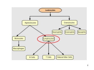
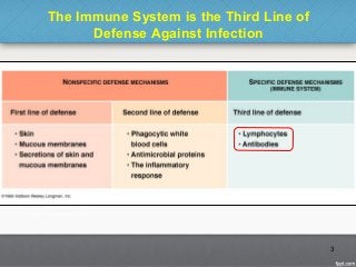
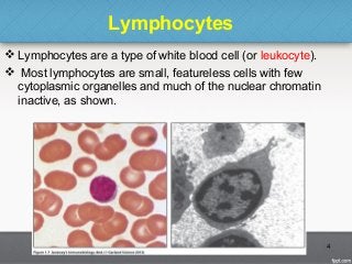
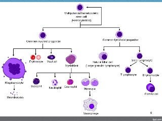
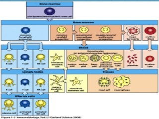
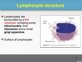
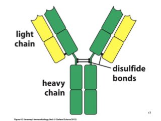
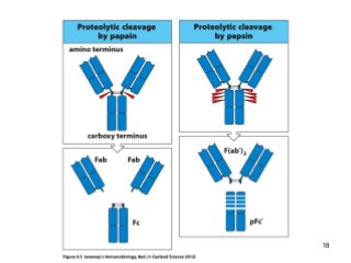
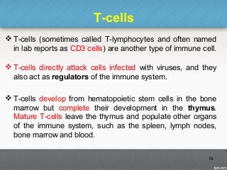
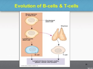
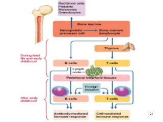
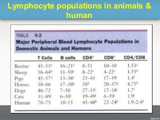
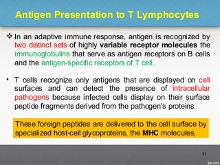
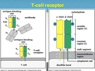
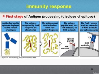
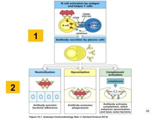
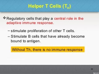
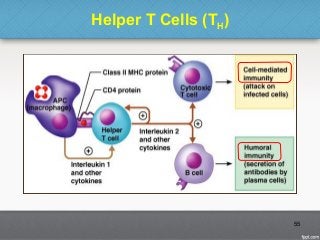
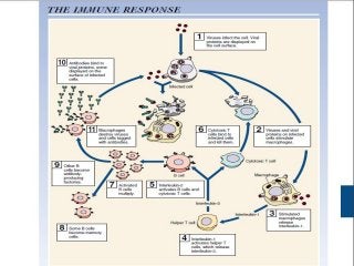
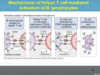
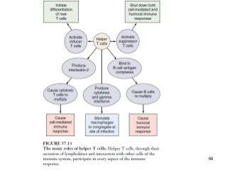
 Lymphocytes
T-cells & B-cells
Urmia University
Supervisor: Dr. Najafi
Presenter: M. Behroozlak
May 2014
In the name of God
2
The Immune System is the Third Line of
Defense Against Infection
3
Lymphocytes
 Lymphocytes are a type of white blood cell (or leukocyte).
 Most lymphocytes are small, featureless cells with few
cytoplasmic organelles and much of the nuclear chromatin
inactive, as shown.
4
Lymphocytes
Lymphocytes can be divided into three main
types:
• Large granular lymphocytes: these are part of the innate
immune system, and are natural killer cells (NKCs).
• Small lymphocytes: these lymphocytes are the main
agents of the aquired immune system.
The two main types are:
• T cells and
• B cells.
5
6
7
Lymphocyte structure
 Lymphocytes are
surrounded by a thin
cytoplasm containig some
mitochondria, free
ribosomes and a small
golgi apparatus.
 Surface of lymphocytes
8
Distribution of lymphocytes in the body
Lymphocytes are
mainly found in
lymphoid organs, in
blood and scattered
under mocusal
surfaces.
9
Distribution of lymphoid tissues in the
body
 Central lymphoid
organs (yellow)
 Secondary
lymphoid organs
(blue)
10
Types of lymphocytes
 There are two major types of lymphocyte:
 B-cells & T-cells
 B lymphocytes or B cells (sometimes called B-lymphocytes
and often named on lab reports as CD19 or CD20 cells)
which when activated differentiate into plasma cells that
secrete antibodies.
 B-cells develop in the bone marrow from hematopoietic
stem cells. When mature, B-cells can be found in the bone
marrow, lymph nodes, spleen, some areas of the intestine,
and the bloodstream.
11
B-Cells
 When B-cells encounter foreign material (antigens), they
respond by maturing into another cell type called plasma
cells.
 B-cells can also mature into memory cells, which allows a
rapid response if the same infection is encountered again.
Plasma cells are the mature cells that actually produce the
antibodies.
 Antibodies, the major product of plasma cells, find their way
into the bloodstream, tissues, respiratory secretions,
intestinal secretions, and even tears.
12
B-Cells
 For every foreign antigen, there are antibody molecules
specifically designed to fit that antigen, like a lock and key.
 When antibody molecules recognize a microorganism as
foreign, they physically attach to it and set off a complex
chain of events involving other components of the immune
system that work to eventually destroy the germ.
 Antibodies vary with respect to their specialized functions in
the body. These variations are determined by the
antibody’s chemical structure, which in turn determines the
class of the antibody (or immunoglobulin).
13
14
B-Cells
There are five major classes of antibodies:
(IgG, IgA, IgM, IgD and IgE).
• IgG has four different subclasses (IgG1, IgG2, IgG3,
IgG4).
• IgA has two subclasses (IgA1 and IgA2).
• Each immunoglobulin class has distinct chemical
characteristics that provide it with specific functions
15
16
17
18
T-cells
 T-cells (sometimes called T-lymphocytes and often named
in lab reports as CD3 cells) are another type of immune cell.
 T-cells directly attack cells infected with viruses, and they
also act as regulators of the immune system.
 T-cells develop from hematopoietic stem cells in the bone
marrow but complete their development in the thymus.
Mature T-cells leave the thymus and populate other organs
of the immune system, such as the spleen, lymph nodes,
bone marrow and blood.
19
Evolution of B-cells & T-cells
20
21
T-cells
Each T-cell reacts with a specific antigen, just as each
antibody molecule reacts with a specific antigen.
 In fact, T-cells have molecules on their surfaces that
are similar to antibodies are called TCR & BCR.
The variety of different T-cells is so extensive that the
body has T-cells that can react against virtually any
antigen.
22
Types of T Cells
Helper T cells (Th) often denoted in lab reports as
CD4 T-cells)
“Killer” or cytotoxic T-cells (Tc)
(often denoted in lab reports as CD8 T-cells),
Suppressor T-cells or Regulatory T-cells (Treg).
23
Each has a different role to play in the immune system.
Functions of T-cells
 Helper T-cells assist B-cells to produce antibodies and assist
killer T-cells in their attack on foreign substances.
 Killer, or cytotoxic, T-cells perform the actual destruction of
infected cells. Killer T-cells protect the body from certain
bacteria and viruses that have the ability to survive and even
reproduce within the body’s own cells.
 Regulatory T-cells suppress or turn off other T-lymphocytes.
Without regulatory cells, the immune system would keep
working even after an infection has been cured.
24
Lymphocyte populations in animals &
human
25
Natural killer cells (NK Cells)
 Natural killer (NK) cells are so named because they easily
kill cells infected with viruses.
 They are said to be “natural killer” cells as they do not
require the same thymic education that T-cells require.
 NK cells are derived from the bone marrow and are present
in relatively low numbers in the bloodstream and in tissues.
 They are important in defending against viruses and
possibly preventing cancer as well and are thought to be
important in the innate immune defense against
intracellular pathogens..
26
27
Lymphocyte Mitogens
A mitogen is a chemical substance that encourages
a cell to commence cell division, triggering mitosis. A
mitogen is usually some form of a protein.
B cells can enter mitosis when they encounter an
antigen matching their immunoglobulin.
Mitogens are often used to stimulate lymphocytes and
therefore assess immune function.
28
Lymphocyte Mitogens
 Lectines are proteins that bind to cell surface glycoproteins
and so trigger cell division.
 Not all lymphocytes respond equally well to all lectines.
29
Species of lectin Acts upon T-cells Acts upon B-cells
phytohaemagglutini
n (PHA) yes no
concanavalin A
(conA) yes no
pokeweed mitogen
(PWM) yes yes
lipopolysaccharide
(LPS) no yes
Types of adaptive imunity
30
Antigen Presentation to T Lymphocytes
 In an adaptive immune response, antigen is recognized by
two distinct sets of highly variable receptor molecules the
immunoglobulins that serve as antigen receptors on B cells
and the antigen-specific receptors of T cell.
• T cells recognize only antigens that are displayed on cell
surfaces and can detect the presence of intracellular
pathogens because infected cells display on their surface
peptide fragments derived from the pathogen's proteins.
31
These foreign peptides are delivered to the cell surface by
specialized host-cell glycoproteins, the MHC molecules,
T-cell receptor
32
Antigen presenting cells (APC)
 Major APCs are dendritic cells (DCs), macrophages, and
activated B cells.
 The major initiators of adaptive immunity are DCs, which
actively migrate to the lymph nodes and secondary
lymphoid organs and present antigens to T and B cells.
 Macrophages are specialized to internalize extracellular
pathogens, especially after they have been coated with
antibody, and to present their antigens.
 B cells have antigen-specific receptors that enable them to
internalize large amounts of specific antigen, process it, and
present it.
33
34
immunity response
 First stage of Antigen processing (disclose of epitope)
35
Epitope (index of antigen)
36
immunity response
 Second stage of antigen processing:
presenting of antigen by MHC to T-cells.
 T-cell receptor differs from the B-cell receptor in an
important way:
 it does not recognize and bind antigen directly, but
instead recognizes short peptide fragments of pathogen
protein antigens, which are bound to MHC molecules on
the surfaces of other cells.
37
Types of MHC
Other names of MHC in:
Human (HLA)
Cow (BoLA)
Horse (ELA)
Sheep (OLA)
Mice (H-2 Complex)
38
Both types of MHC proteins are
important to T cell activation
MHC Proteins
Class I MHC proteins
– Always recognized by CD8 T cells
– Display peptides from endogenous antigens are egraded
by proteases and enter the endoplasmic reticulum
– Transported via TAP (transporter associated with antigen
processing)
– Loaded onto class I MHC molecules
– Displayed on the cell surface in association with a class I
MHC molecule
39
Class I MHC Proteins
40
MHC Proteins
Class II MHC proteins
– Class II MHC proteins are found only on mature B cells,
some T cells, and antigen-presenting cells
– Always recognized by CD4 T cells
– A phagosome containing pathogens (with exogenous
antigens) merges with a lysosome
– Invariant protein prevents class II MHC proteins from
binding to peptides in the endoplasmic reticulum
– CLIP removed by HLA-DM
41
Class II MHC Proteins
42
Pathways of antigen processing
43
Co-receptors of MHC class I & II
• Th (T-Helper) contains
specific molecule
called CD4.
• Tc (T-Cytotoxic) & Treg
contains specific
molecule called CD8.
44
T Cell Activation
T Cell Activation reguired two independent
signals:
 Binding of the peptide:MHC complex by the T-cell receptor
and, the CD4 co-receptor, transmits a signal to the T cell
that antigen has been encountered (major signal).
 Activation of naive T cells requires a second signal, the co-
stimulatory signal, to be delivered by the same antigen-
presenting cell.
 Signals that plays in role in differentiation of T-cells.
45
T Cell Activation
Binding of the T-cell receptor
(TCR) and its co-receptor CD4
to the peptide:MHC class II
complex on the antigen-
presenting cell (APC) delivers
a signal that can induce the
clonal expansion of T cells
only when the co-stimulatory
signal is given by binding of
CD28 to B7 molecules.
46
47
48
B- Cell Activation
 The humoral immune response is mediated by antibody
molecules that are secreted by plasma cells.
 First stage:
• Antigen that binds to the BCR signals B cells and is, at the
same time, internalized and processed into peptides that
activate armed helper T cells.
• Signals from the bound antigen and from the helper T cell
induce the B cell to proliferate and differentiate into a plasma
cell secreting specific antibody.
49
50
Armed helper T cells stimulate the proliferation and
then the differentiation of antigen-binding B-cells
 The specific interaction of an antigen-binding B cell with an armed
helper T cell leads to the expression of the B-cell stimulatory molecule
CD40 ligand (CD40L/CD154) on the helper T-cell surface and to the
secretion of the B-cell stimulatory cytokines IL-4, IL-5, and IL-6, which
drive the proliferation and differentiation of the B cell into antibody-
secreting plasma cells.
51
B- Cell Activation
 Second stage:
 These antibodies protect the host from infection in three
main ways:
 They can inhibit the toxic effects or infectivity of pathogens
by binding to them: this is termed neutralization
 By coating the pathogens, they can enable accessory cells
that recognize the Fc portions of arrays of antibodies to
ingest and kill the pathogen, a process called opsonization
 Antibodies can also trigger activation of the complement
system. Complement proteins can strongly enhance
opsonization, and can directly kill some bacterial cells
52
53
1
2
Helper T Cells (TH)
Regulatory cells that play a central role in the
adaptive immune response.
– stimulate proliferation of other T cells.
– Stimulate B cells that have already become
bound to antigen.
54
Without Th, there is no immune response
Helper T Cells (TH)
55
56
57
Mechanisms of helper T cell-mediated
activation of B lymphocytes
58
59

Lymphocytes: T-cells & B-cells

  • 1.
     Lymphocytes T-cells &B-cells Urmia University Supervisor: Dr. Najafi Presenter: M. Behroozlak May 2014 In the name of God
  • 2.
  • 3.
    The Immune Systemis the Third Line of Defense Against Infection 3
  • 4.
    Lymphocytes  Lymphocytes area type of white blood cell (or leukocyte).  Most lymphocytes are small, featureless cells with few cytoplasmic organelles and much of the nuclear chromatin inactive, as shown. 4
  • 5.
    Lymphocytes Lymphocytes can bedivided into three main types: • Large granular lymphocytes: these are part of the innate immune system, and are natural killer cells (NKCs). • Small lymphocytes: these lymphocytes are the main agents of the aquired immune system. The two main types are: • T cells and • B cells. 5
  • 6.
  • 7.
  • 8.
    Lymphocyte structure  Lymphocytesare surrounded by a thin cytoplasm containig some mitochondria, free ribosomes and a small golgi apparatus.  Surface of lymphocytes 8
  • 9.
    Distribution of lymphocytesin the body Lymphocytes are mainly found in lymphoid organs, in blood and scattered under mocusal surfaces. 9
  • 10.
    Distribution of lymphoidtissues in the body  Central lymphoid organs (yellow)  Secondary lymphoid organs (blue) 10
  • 11.
    Types of lymphocytes There are two major types of lymphocyte:  B-cells & T-cells  B lymphocytes or B cells (sometimes called B-lymphocytes and often named on lab reports as CD19 or CD20 cells) which when activated differentiate into plasma cells that secrete antibodies.  B-cells develop in the bone marrow from hematopoietic stem cells. When mature, B-cells can be found in the bone marrow, lymph nodes, spleen, some areas of the intestine, and the bloodstream. 11
  • 12.
    B-Cells  When B-cellsencounter foreign material (antigens), they respond by maturing into another cell type called plasma cells.  B-cells can also mature into memory cells, which allows a rapid response if the same infection is encountered again. Plasma cells are the mature cells that actually produce the antibodies.  Antibodies, the major product of plasma cells, find their way into the bloodstream, tissues, respiratory secretions, intestinal secretions, and even tears. 12
  • 13.
    B-Cells  For everyforeign antigen, there are antibody molecules specifically designed to fit that antigen, like a lock and key.  When antibody molecules recognize a microorganism as foreign, they physically attach to it and set off a complex chain of events involving other components of the immune system that work to eventually destroy the germ.  Antibodies vary with respect to their specialized functions in the body. These variations are determined by the antibody’s chemical structure, which in turn determines the class of the antibody (or immunoglobulin). 13
  • 14.
  • 15.
    B-Cells There are fivemajor classes of antibodies: (IgG, IgA, IgM, IgD and IgE). • IgG has four different subclasses (IgG1, IgG2, IgG3, IgG4). • IgA has two subclasses (IgA1 and IgA2). • Each immunoglobulin class has distinct chemical characteristics that provide it with specific functions 15
  • 16.
  • 17.
  • 18.
  • 19.
    T-cells  T-cells (sometimescalled T-lymphocytes and often named in lab reports as CD3 cells) are another type of immune cell.  T-cells directly attack cells infected with viruses, and they also act as regulators of the immune system.  T-cells develop from hematopoietic stem cells in the bone marrow but complete their development in the thymus. Mature T-cells leave the thymus and populate other organs of the immune system, such as the spleen, lymph nodes, bone marrow and blood. 19
  • 20.
  • 21.
  • 22.
    T-cells Each T-cell reactswith a specific antigen, just as each antibody molecule reacts with a specific antigen.  In fact, T-cells have molecules on their surfaces that are similar to antibodies are called TCR & BCR. The variety of different T-cells is so extensive that the body has T-cells that can react against virtually any antigen. 22
  • 23.
    Types of TCells Helper T cells (Th) often denoted in lab reports as CD4 T-cells) “Killer” or cytotoxic T-cells (Tc) (often denoted in lab reports as CD8 T-cells), Suppressor T-cells or Regulatory T-cells (Treg). 23 Each has a different role to play in the immune system.
  • 24.
    Functions of T-cells Helper T-cells assist B-cells to produce antibodies and assist killer T-cells in their attack on foreign substances.  Killer, or cytotoxic, T-cells perform the actual destruction of infected cells. Killer T-cells protect the body from certain bacteria and viruses that have the ability to survive and even reproduce within the body’s own cells.  Regulatory T-cells suppress or turn off other T-lymphocytes. Without regulatory cells, the immune system would keep working even after an infection has been cured. 24
  • 25.
    Lymphocyte populations inanimals & human 25
  • 26.
    Natural killer cells(NK Cells)  Natural killer (NK) cells are so named because they easily kill cells infected with viruses.  They are said to be “natural killer” cells as they do not require the same thymic education that T-cells require.  NK cells are derived from the bone marrow and are present in relatively low numbers in the bloodstream and in tissues.  They are important in defending against viruses and possibly preventing cancer as well and are thought to be important in the innate immune defense against intracellular pathogens.. 26
  • 27.
  • 28.
    Lymphocyte Mitogens A mitogenis a chemical substance that encourages a cell to commence cell division, triggering mitosis. A mitogen is usually some form of a protein. B cells can enter mitosis when they encounter an antigen matching their immunoglobulin. Mitogens are often used to stimulate lymphocytes and therefore assess immune function. 28
  • 29.
    Lymphocyte Mitogens  Lectinesare proteins that bind to cell surface glycoproteins and so trigger cell division.  Not all lymphocytes respond equally well to all lectines. 29 Species of lectin Acts upon T-cells Acts upon B-cells phytohaemagglutini n (PHA) yes no concanavalin A (conA) yes no pokeweed mitogen (PWM) yes yes lipopolysaccharide (LPS) no yes
  • 30.
  • 31.
    Antigen Presentation toT Lymphocytes  In an adaptive immune response, antigen is recognized by two distinct sets of highly variable receptor molecules the immunoglobulins that serve as antigen receptors on B cells and the antigen-specific receptors of T cell. • T cells recognize only antigens that are displayed on cell surfaces and can detect the presence of intracellular pathogens because infected cells display on their surface peptide fragments derived from the pathogen's proteins. 31 These foreign peptides are delivered to the cell surface by specialized host-cell glycoproteins, the MHC molecules,
  • 32.
  • 33.
    Antigen presenting cells(APC)  Major APCs are dendritic cells (DCs), macrophages, and activated B cells.  The major initiators of adaptive immunity are DCs, which actively migrate to the lymph nodes and secondary lymphoid organs and present antigens to T and B cells.  Macrophages are specialized to internalize extracellular pathogens, especially after they have been coated with antibody, and to present their antigens.  B cells have antigen-specific receptors that enable them to internalize large amounts of specific antigen, process it, and present it. 33
  • 34.
  • 35.
    immunity response  Firststage of Antigen processing (disclose of epitope) 35
  • 36.
    Epitope (index ofantigen) 36
  • 37.
    immunity response  Secondstage of antigen processing: presenting of antigen by MHC to T-cells.  T-cell receptor differs from the B-cell receptor in an important way:  it does not recognize and bind antigen directly, but instead recognizes short peptide fragments of pathogen protein antigens, which are bound to MHC molecules on the surfaces of other cells. 37
  • 38.
    Types of MHC Othernames of MHC in: Human (HLA) Cow (BoLA) Horse (ELA) Sheep (OLA) Mice (H-2 Complex) 38 Both types of MHC proteins are important to T cell activation
  • 39.
    MHC Proteins Class IMHC proteins – Always recognized by CD8 T cells – Display peptides from endogenous antigens are egraded by proteases and enter the endoplasmic reticulum – Transported via TAP (transporter associated with antigen processing) – Loaded onto class I MHC molecules – Displayed on the cell surface in association with a class I MHC molecule 39
  • 40.
    Class I MHCProteins 40
  • 41.
    MHC Proteins Class IIMHC proteins – Class II MHC proteins are found only on mature B cells, some T cells, and antigen-presenting cells – Always recognized by CD4 T cells – A phagosome containing pathogens (with exogenous antigens) merges with a lysosome – Invariant protein prevents class II MHC proteins from binding to peptides in the endoplasmic reticulum – CLIP removed by HLA-DM 41
  • 42.
    Class II MHCProteins 42
  • 43.
    Pathways of antigenprocessing 43
  • 44.
    Co-receptors of MHCclass I & II • Th (T-Helper) contains specific molecule called CD4. • Tc (T-Cytotoxic) & Treg contains specific molecule called CD8. 44
  • 45.
    T Cell Activation TCell Activation reguired two independent signals:  Binding of the peptide:MHC complex by the T-cell receptor and, the CD4 co-receptor, transmits a signal to the T cell that antigen has been encountered (major signal).  Activation of naive T cells requires a second signal, the co- stimulatory signal, to be delivered by the same antigen- presenting cell.  Signals that plays in role in differentiation of T-cells. 45
  • 46.
    T Cell Activation Bindingof the T-cell receptor (TCR) and its co-receptor CD4 to the peptide:MHC class II complex on the antigen- presenting cell (APC) delivers a signal that can induce the clonal expansion of T cells only when the co-stimulatory signal is given by binding of CD28 to B7 molecules. 46
  • 47.
  • 48.
  • 49.
    B- Cell Activation The humoral immune response is mediated by antibody molecules that are secreted by plasma cells.  First stage: • Antigen that binds to the BCR signals B cells and is, at the same time, internalized and processed into peptides that activate armed helper T cells. • Signals from the bound antigen and from the helper T cell induce the B cell to proliferate and differentiate into a plasma cell secreting specific antibody. 49
  • 50.
  • 51.
    Armed helper Tcells stimulate the proliferation and then the differentiation of antigen-binding B-cells  The specific interaction of an antigen-binding B cell with an armed helper T cell leads to the expression of the B-cell stimulatory molecule CD40 ligand (CD40L/CD154) on the helper T-cell surface and to the secretion of the B-cell stimulatory cytokines IL-4, IL-5, and IL-6, which drive the proliferation and differentiation of the B cell into antibody- secreting plasma cells. 51
  • 52.
    B- Cell Activation Second stage:  These antibodies protect the host from infection in three main ways:  They can inhibit the toxic effects or infectivity of pathogens by binding to them: this is termed neutralization  By coating the pathogens, they can enable accessory cells that recognize the Fc portions of arrays of antibodies to ingest and kill the pathogen, a process called opsonization  Antibodies can also trigger activation of the complement system. Complement proteins can strongly enhance opsonization, and can directly kill some bacterial cells 52
  • 53.
  • 54.
    Helper T Cells(TH) Regulatory cells that play a central role in the adaptive immune response. – stimulate proliferation of other T cells. – Stimulate B cells that have already become bound to antigen. 54 Without Th, there is no immune response
  • 55.
  • 56.
  • 57.
  • 58.
    Mechanisms of helperT cell-mediated activation of B lymphocytes 58
  • 59.