Immunity
 Immunity is body's ability to resist or eliminate potentially harmful foreign materials
or abnormal cells.
 Immunology is the study of the bodies defense mechanism against foreign disease
causing agents.
 Consists of following activities:
– Defence against invading pathogens or microbes
– Removal of 'worn-out' cells & tissue debris
– Identification & destruction of abnormal or mutant cells
– Rejection of 'foreign' cells (e.g., organ transplant)
 Inappropriate responses:
Allergies - response to normally harmless substances
Autoimmune diseases
Differences b/n Innate & Acquired immunity
A. Innate immunity : is the inborn
capacity of body to resist
pathogens.
 By chance, if the organisms enter the
body, innate immunity eliminates
them before development of a
disease.
 It is otherwise called the natural or
non-specific immunity.
 This type of immunity represents the
first line of defense against any type
of pathogens..
B. Acquired immunity :is the resistance
developed in the body against any
specific foreign body like bacteria,
viruses, toxins, vaccines or transplanted
tissues.
So, this type of immunity is also
known as specific immunity.

It is the most powerful immune
mechanism that protects the body from
the invading organisms or toxic
substances.

Lymphocytes are responsible for
acquired immunity
Innate Immune System…
Innate Immunity…
ANTIGENS
 „ Antigens are the substances (chemicals) which induce specific immune reactions
in the body.
 Antigens are of two types:
1. Autoantigens (self antigens) : present on the body’s own cells such as ‘A’ antigen
and ‘B’ antigen in RBCs.
2. Foreign antigen s (non-self antigens) : that enter the body from outside.
 Following are non-self antigens:
 Chemicals (proteins, lipoproteins, and polysaccharides ) located on the surfaces of
foreign bodies.
 Toxins from microbial organisms.
 Materials from transplanted organs or incompatible blood cells.
 Allergens or allergic substances like pollen grains called haptens
ANTIGENS…
Types of Non-self Antigens
• Non-self antigens are classified into two types,
depending upon the response developed against
them in the body:
1. Antigens, which induce the development of
immunity or production of antibodies
(immunogenicity).
2. Antigens, which react with specific antibodies and
produce allergic reactions (allergic reactivity).
Antigen-Presenting Cells
 Antigen-presenting cells: are the special type of cells in the
body, which induce the release of antigenic materials from
invading organisms and later present these materials to the
helper T cells.
 Types of Antigen-Presenting Cells
 Antigen-presenting cells are of three types:
1. Macrophages: major antigen-presenting cells.
2. Dendritic cells: from spleen, skin & lymph nodes.
3. B lymphocytes: antigen presenting cells & antigen receiving cells
Antigen-Presenting Cells…
 Major histocompatiblility complex: are large molecule situated on the
surface of the antigen presenting cells. MHC are two types.
1. Class I MHC molecule: It is found on every cell in human body.
 It is specifically responsible for presentation of endogenous antigens
(antigens produced intracellularly such as viral proteins and tumor
antigens) to cytotoxic T cells.
2. Class II MHC molecule: It is found on B cells, macrophages and other
antigen-presenting cells.
 It is responsible for presenting the exogenous antigens (antigens of
bacteria or viruses which are engulfed by antigen-presenting cells) to
helper T cell.
Antigen-Presenting Cells…
 White blood cells and all other nucleated cells in the
body have proteins, called major histocompatibility
(MHC) antigens, protruding from their plasma
membrane into the extracellular fluid.
 These “cell identity markers” are unique for each
person (except identical twins).
 Although RBCs possess blood group antigens, they
lack the MHC antigens.
Figure : Antigen Processing and Presentation
Antigen-Presenting Cells…
Types of Acquired immunity & their Development
1. Cellular immunity
 Cellular immunity develops from T-lymphocytic
cells.
 “T” stands for Thymus gland located at the
lower portion of the trachea.
 T-cells normally form in the stem cells of the
bone marrow and migrate to the thymus gland
b/s the thymus is a pre-processing or maturation
area of T-cells.
• Thymosin and cytokines help in the maturation
of T-cells
• Cell-mediated immunity does not involve
antibodies.
2. Humoral immunity
 Humoral immunity develops from B-
lymphocytic cells.
 B stands for “ Bursa of Fabricius”
from Birds.
 B-cells emerge from the stem cells of
the bone marrow and are pre-
processed in the liver (early fetal life)
and as well as in the bone marrow
after birth.
 Then B-Lymphocytes develop to
plasma cells that in turn secrete
different types of circulating
Origin & Maturation of Lymphocytes
Development of Cellular immunity
 During fetal development, lymphocytes are produce from committed stem
cells in the bone marrow. Stem cells can not however activate T-cells
 Therefore, T-cells migrate to thymus to be pre-processed. Here, T-cells
proliferate and diversify and learn to recognize their self antigens, so that
they do not destroy their own tissue
 In the thymus, dangerous T-cells that attack self tissues are destroyed and
phagocytised (thymic selection).
 Then, T-cells move to different lymphoid tissues through the blood and
stay in these lymphoid tissues for long until they meet or contact specific
antigen.
Types of T-lymphocytes (Cells)
 During the processing, T lymphocytes are transformed into four types:
1. Helper T cells or inducer T cells: These cells are also called CD4 cells
b/se of the presence of molecules called CD4 on their surface.
2. Cytotoxic T cells or killer T cells: These cells are also called CD8 cells
b/se of presence of molecules called CD8 on their surface.
3. Suppressor T cells or regulatory T cells: suppress the activities of the
killer T cells destroying the body’s own tissues along with invaded
organisms.
4. Memory T cells: When the body is exposed to the same organism for
the second time, the memory cells identify the organism and
immediately activate the other T cells.
Figure: Differentiation of T Cells within the Thymus : Thymocytes enter the thymus and go through a series of
developmental stages that ensures both function and tolerance before they leave and become
functional components of the adaptive immune response.
Helper T-lymphocytes and their functions
 Helper T-cells : important to know their functions b/s AIDS
(Acquired Immunodeficiency Syndrome) destroys mainly the
T-helper cells.
 T-helper cells are the most numerous and produce
lymphokines
that produce different chemicals called interleukins (IL-2 to 6).
 General Functions of Helper T-cells include:
 Stimulation of T-cytotoxic and T-suppressor cells.
 Stimulation of B-cells to form plasma cells.
 Activation of the macrophage system etc.
Release
Antigen-presenting cells
present the antigenic products
bound with human leukocyte
anti gen (HLA) (which is
present in class II MHC
molecule) to helper T cells.
This activates helper T cells
through series of events.
Fig: Clonal Selection and Expansion of T Lymphocytes :Stem cells differentiate into T cells with specific
receptors, called clones. The clones with receptors specific for antigens on the pathogen are selected for and
expanded.
Cytotoxic T-cell and their functions
 Cytotoxic -T-cells kill invaders by
direct attack (apoptosis) through
the following methods:
 Bore a hole through the membrane,
so that electrolytes and fluid enters
& burst the microbes.
2. Release toxic substances and kill
invaders
3. Destroy all types of foreign cells
(e.g., cancer cells, transplant cells)
Figure: Pathogen Presentation (a) CD4 is associated with helper and regulatory T cells. An extracellular
pathogen is processed and presented in the binding cleft of a class II MHC molecule, and this interaction is
strengthened by the CD4 molecule. (b) CD8 is associated with cytotoxic T cells. An intracellular pathogen
is presented by a class I MHC molecule, and CD8 interacts with it.
Development of Humoral immunity
 B-lymphocytes also originate from stem cells in bone marrow.
 They migrate to lymphoid tissues and become sensitized by specific
antigens. After sensitization, they multiply forming clones of identical
cells.
 The blood and lymph are the body fluids (humours or humors in
Latin). Since the B lymphocytes provide immunity through humors,
this type of immunity is called humoral immunity.
 B lymphocytes are transformed into two types:
1. Plasma cells: produce four types of antibodies
2. Memory cells: fight back when the same type of antigen comes into
contact in a 2nd
exposure.
Cooperation between T and B- cells
 Helper T-cells support or help the B-cells by
producing chemicals known as Lymphokines or
(cytokines)
 These cytokines can stimulate B- lymphocytes
and other immune cells (e.g, macrophages) to
perform their specific functions.
 Without the helper T-cells, the amount of
antibodies produced by B-cells is not adequate
to combat infections.
 HIV, the virus that causes AIDS normally infects
helper T-cells & inactivates immune response
Figure: Clonal Selection of B Cells: During a primary B cell immune response, both antibody-secreting
plasma cells and memory B cells are produced. These memory cells lead to the differentiation of more
plasma cells and memory B cells during secondary responses.
Structure of Antibodies
 There are 2-heavy and 2-light
chains, which have constant
and variable portions.
A. Constant portion = provides
an attachment surface on
tissues
B. Variable = is a place where
antigens attach to antibodies
specifically
Figure: Antibody and IgG2 Structures :The typical four chain structure of a generic antibody
(a) and the corresponding three-dimensional structure of the antibody IgG2 (b). (credit b:
modification of work by Tim Vickers
Functions of Different Antibodies
 An antibody is defined as a protein that is produced by B
lymphocytes in response to the presence of an antigen.
 Antibody is gamma globulin in nature and it is also called
immunoglobulin (Ig).
1. IgA : plays a role in localized defense mechanism found in
saliva , tears, colostrum, GIT.
2. IgD: : found mainly on the surface B-cells and involved in
recognition of the antigen by B cells.
3. IgE: involved in allergic reactions (bind to mast & basophils)
4. IgG: most abundant type & is responsible for passive
immunity to the fetus and complement fixation
Fig: Types of
antibodies
Mechanism of action of antibodies
A. Direct attack
B. Through complement System
A. By direct attack: inactivate the invaders by the following methods
1. Agglutination: involves the clumping or binding of bacterial
antigens to each other, so that they become dysfunctional.
2. Precipitation : The complex antigen-antibody reaction is made
insoluble and precipitates down.
3. Neutralization: The antibodies cover the active sites of the
invader and inactivate the toxic sites of the antigens.
4. Lyses: antibodies rupture the cell membrane of the organisms and
then destroy them.
Mechanism of action of Immunoglobulins
The Complement System
 Actions of Antibodies through Complement System
 The indirect actions of antibodies are stronger than the
direct actions and play more important role in defense
mechanism of the body than the direct actions.
 Complement system is the one that enhances or accelerates
various activities during fight against invading organisms.
 It is a system of plasma enzymes, which are identified by
numbers from C1 to C9.
The Complement System…
Mechanism of action
Phagocytosis
Lysis (rupture)
Agglutination and
Neutralization
Chemotaxis
 Activation of mast cells
and basophiles
These are inactive enzymes (11 proteins) in plasma. Activated by antigen-antibody
reactions by the so called “classic pathway” and amplify the previous actions.
Fig: Complement cascade
(IgG, IgM)
C42b
C4 has 2 components:
1. C4b-binding
2. C4a-away (not bind)
C3-convertase
Membrane Attack Complex (M.A.C)
(Disturb physical integrity of bacterial
membrane)→lysis of cells
Early component of
complement system
Terminal
component of
complement system
(Phagocytosis) Amplification of phagocytosis process
Figure 21.13 Complement Cascade and Function The classical pathway, used during adaptive
immune responses, occurs when C1 reacts with antibodies that have bound an antigen.
Mechanism of Self Tolerance
 Self tolerance is a condition of self-recognition of the bodies antigens
that prevents self-destruction of the bodies own cells.
 The immunity system does not form antibodies or sensitized
lymphocytes against its own antigens.
 Normally, self cells have a specific antigen which stands for each
individual. This specific antigen is called human leukocyte antigen
(HLA). This HLA is expressed by a group of genes called major
histocompatability complex (MHC).
 The importance of MHC proteins is that they allow T-cells to distinguish
self from non-self.
 In other words, T-cell receptors recognize antigenic peptides in
association with MHC molecules.
Autoimmune Diseases
 Autoimmune disease: is defined as a condition in which the immune
system mistakenly attacks body’s own cells and tissues.
 Normally, an antigen induces the immune response in the body. The
condition in which the immune system fails to give response to an
antigen is called tolerance.
 This is true with respect to body’s own antigens that are called self
antigens or autoantigens.
 Normally, body has the tolerance against self antigen.
 However, in some occasions, the tolerance fails or becomes
incomplete against self antigen. This state is called autoimmunity
and it leads to the activation of T-lymphocytes.
Immunization
 Immunization :is defined as the procedure by
which the body is prepared to fight against a
specific disease.
 It is used to induce the immune resistance of the
body to a specific disease.
 Active immunity = production of antibodies as
a result of exposure to an antigen (vaccine). Can
be natural or aquired
 Passive immunity = direct transfer of
antibodies formed by another person (or
animal), e.g., transfer of IgG antibodies from
mother to fetus across placenta or in colostrum
IgA('first milk).

Immune System....................... ppt.pptx

  • 1.
    Immunity  Immunity isbody's ability to resist or eliminate potentially harmful foreign materials or abnormal cells.  Immunology is the study of the bodies defense mechanism against foreign disease causing agents.  Consists of following activities: – Defence against invading pathogens or microbes – Removal of 'worn-out' cells & tissue debris – Identification & destruction of abnormal or mutant cells – Rejection of 'foreign' cells (e.g., organ transplant)  Inappropriate responses: Allergies - response to normally harmless substances Autoimmune diseases
  • 5.
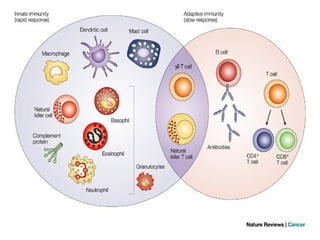
    Differences b/n Innate& Acquired immunity A. Innate immunity : is the inborn capacity of body to resist pathogens.  By chance, if the organisms enter the body, innate immunity eliminates them before development of a disease.  It is otherwise called the natural or non-specific immunity.  This type of immunity represents the first line of defense against any type of pathogens.. B. Acquired immunity :is the resistance developed in the body against any specific foreign body like bacteria, viruses, toxins, vaccines or transplanted tissues. So, this type of immunity is also known as specific immunity.  It is the most powerful immune mechanism that protects the body from the invading organisms or toxic substances.  Lymphocytes are responsible for acquired immunity
  • 6.
  • 7.
  • 8.
    ANTIGENS  „ Antigensare the substances (chemicals) which induce specific immune reactions in the body.  Antigens are of two types: 1. Autoantigens (self antigens) : present on the body’s own cells such as ‘A’ antigen and ‘B’ antigen in RBCs. 2. Foreign antigen s (non-self antigens) : that enter the body from outside.  Following are non-self antigens:  Chemicals (proteins, lipoproteins, and polysaccharides ) located on the surfaces of foreign bodies.  Toxins from microbial organisms.  Materials from transplanted organs or incompatible blood cells.  Allergens or allergic substances like pollen grains called haptens
  • 9.
    ANTIGENS… Types of Non-selfAntigens • Non-self antigens are classified into two types, depending upon the response developed against them in the body: 1. Antigens, which induce the development of immunity or production of antibodies (immunogenicity). 2. Antigens, which react with specific antibodies and produce allergic reactions (allergic reactivity).
  • 10.
    Antigen-Presenting Cells  Antigen-presentingcells: are the special type of cells in the body, which induce the release of antigenic materials from invading organisms and later present these materials to the helper T cells.  Types of Antigen-Presenting Cells  Antigen-presenting cells are of three types: 1. Macrophages: major antigen-presenting cells. 2. Dendritic cells: from spleen, skin & lymph nodes. 3. B lymphocytes: antigen presenting cells & antigen receiving cells
  • 11.
    Antigen-Presenting Cells…  Majorhistocompatiblility complex: are large molecule situated on the surface of the antigen presenting cells. MHC are two types. 1. Class I MHC molecule: It is found on every cell in human body.  It is specifically responsible for presentation of endogenous antigens (antigens produced intracellularly such as viral proteins and tumor antigens) to cytotoxic T cells. 2. Class II MHC molecule: It is found on B cells, macrophages and other antigen-presenting cells.  It is responsible for presenting the exogenous antigens (antigens of bacteria or viruses which are engulfed by antigen-presenting cells) to helper T cell.
  • 12.
    Antigen-Presenting Cells…  Whiteblood cells and all other nucleated cells in the body have proteins, called major histocompatibility (MHC) antigens, protruding from their plasma membrane into the extracellular fluid.  These “cell identity markers” are unique for each person (except identical twins).  Although RBCs possess blood group antigens, they lack the MHC antigens.
  • 13.
    Figure : AntigenProcessing and Presentation
  • 14.
  • 15.
    Types of Acquiredimmunity & their Development 1. Cellular immunity  Cellular immunity develops from T-lymphocytic cells.  “T” stands for Thymus gland located at the lower portion of the trachea.  T-cells normally form in the stem cells of the bone marrow and migrate to the thymus gland b/s the thymus is a pre-processing or maturation area of T-cells. • Thymosin and cytokines help in the maturation of T-cells • Cell-mediated immunity does not involve antibodies. 2. Humoral immunity  Humoral immunity develops from B- lymphocytic cells.  B stands for “ Bursa of Fabricius” from Birds.  B-cells emerge from the stem cells of the bone marrow and are pre- processed in the liver (early fetal life) and as well as in the bone marrow after birth.  Then B-Lymphocytes develop to plasma cells that in turn secrete different types of circulating
  • 16.
    Origin & Maturationof Lymphocytes
  • 17.
    Origin & Maturationof Lymphocytes
  • 18.
    Development of Cellularimmunity  During fetal development, lymphocytes are produce from committed stem cells in the bone marrow. Stem cells can not however activate T-cells  Therefore, T-cells migrate to thymus to be pre-processed. Here, T-cells proliferate and diversify and learn to recognize their self antigens, so that they do not destroy their own tissue  In the thymus, dangerous T-cells that attack self tissues are destroyed and phagocytised (thymic selection).  Then, T-cells move to different lymphoid tissues through the blood and stay in these lymphoid tissues for long until they meet or contact specific antigen.
  • 19.
    Types of T-lymphocytes(Cells)  During the processing, T lymphocytes are transformed into four types: 1. Helper T cells or inducer T cells: These cells are also called CD4 cells b/se of the presence of molecules called CD4 on their surface. 2. Cytotoxic T cells or killer T cells: These cells are also called CD8 cells b/se of presence of molecules called CD8 on their surface. 3. Suppressor T cells or regulatory T cells: suppress the activities of the killer T cells destroying the body’s own tissues along with invaded organisms. 4. Memory T cells: When the body is exposed to the same organism for the second time, the memory cells identify the organism and immediately activate the other T cells.
  • 20.
    Figure: Differentiation ofT Cells within the Thymus : Thymocytes enter the thymus and go through a series of developmental stages that ensures both function and tolerance before they leave and become functional components of the adaptive immune response.
  • 21.
    Helper T-lymphocytes andtheir functions  Helper T-cells : important to know their functions b/s AIDS (Acquired Immunodeficiency Syndrome) destroys mainly the T-helper cells.  T-helper cells are the most numerous and produce lymphokines that produce different chemicals called interleukins (IL-2 to 6).  General Functions of Helper T-cells include:  Stimulation of T-cytotoxic and T-suppressor cells.  Stimulation of B-cells to form plasma cells.  Activation of the macrophage system etc.
  • 22.
    Release Antigen-presenting cells present theantigenic products bound with human leukocyte anti gen (HLA) (which is present in class II MHC molecule) to helper T cells. This activates helper T cells through series of events.
  • 23.
    Fig: Clonal Selectionand Expansion of T Lymphocytes :Stem cells differentiate into T cells with specific receptors, called clones. The clones with receptors specific for antigens on the pathogen are selected for and expanded.
  • 24.
    Cytotoxic T-cell andtheir functions  Cytotoxic -T-cells kill invaders by direct attack (apoptosis) through the following methods:  Bore a hole through the membrane, so that electrolytes and fluid enters & burst the microbes. 2. Release toxic substances and kill invaders 3. Destroy all types of foreign cells (e.g., cancer cells, transplant cells)
  • 26.
    Figure: Pathogen Presentation(a) CD4 is associated with helper and regulatory T cells. An extracellular pathogen is processed and presented in the binding cleft of a class II MHC molecule, and this interaction is strengthened by the CD4 molecule. (b) CD8 is associated with cytotoxic T cells. An intracellular pathogen is presented by a class I MHC molecule, and CD8 interacts with it.
  • 27.
    Development of Humoralimmunity  B-lymphocytes also originate from stem cells in bone marrow.  They migrate to lymphoid tissues and become sensitized by specific antigens. After sensitization, they multiply forming clones of identical cells.  The blood and lymph are the body fluids (humours or humors in Latin). Since the B lymphocytes provide immunity through humors, this type of immunity is called humoral immunity.  B lymphocytes are transformed into two types: 1. Plasma cells: produce four types of antibodies 2. Memory cells: fight back when the same type of antigen comes into contact in a 2nd exposure.
  • 28.
    Cooperation between Tand B- cells  Helper T-cells support or help the B-cells by producing chemicals known as Lymphokines or (cytokines)  These cytokines can stimulate B- lymphocytes and other immune cells (e.g, macrophages) to perform their specific functions.  Without the helper T-cells, the amount of antibodies produced by B-cells is not adequate to combat infections.  HIV, the virus that causes AIDS normally infects helper T-cells & inactivates immune response
  • 30.
    Figure: Clonal Selectionof B Cells: During a primary B cell immune response, both antibody-secreting plasma cells and memory B cells are produced. These memory cells lead to the differentiation of more plasma cells and memory B cells during secondary responses.
  • 31.
    Structure of Antibodies There are 2-heavy and 2-light chains, which have constant and variable portions. A. Constant portion = provides an attachment surface on tissues B. Variable = is a place where antigens attach to antibodies specifically
  • 32.
    Figure: Antibody andIgG2 Structures :The typical four chain structure of a generic antibody (a) and the corresponding three-dimensional structure of the antibody IgG2 (b). (credit b: modification of work by Tim Vickers
  • 33.
    Functions of DifferentAntibodies  An antibody is defined as a protein that is produced by B lymphocytes in response to the presence of an antigen.  Antibody is gamma globulin in nature and it is also called immunoglobulin (Ig). 1. IgA : plays a role in localized defense mechanism found in saliva , tears, colostrum, GIT. 2. IgD: : found mainly on the surface B-cells and involved in recognition of the antigen by B cells. 3. IgE: involved in allergic reactions (bind to mast & basophils) 4. IgG: most abundant type & is responsible for passive immunity to the fetus and complement fixation Fig: Types of antibodies
  • 34.
    Mechanism of actionof antibodies A. Direct attack B. Through complement System A. By direct attack: inactivate the invaders by the following methods 1. Agglutination: involves the clumping or binding of bacterial antigens to each other, so that they become dysfunctional. 2. Precipitation : The complex antigen-antibody reaction is made insoluble and precipitates down. 3. Neutralization: The antibodies cover the active sites of the invader and inactivate the toxic sites of the antigens. 4. Lyses: antibodies rupture the cell membrane of the organisms and then destroy them.
  • 35.
    Mechanism of actionof Immunoglobulins
  • 36.
    The Complement System Actions of Antibodies through Complement System  The indirect actions of antibodies are stronger than the direct actions and play more important role in defense mechanism of the body than the direct actions.  Complement system is the one that enhances or accelerates various activities during fight against invading organisms.  It is a system of plasma enzymes, which are identified by numbers from C1 to C9.
  • 37.
    The Complement System… Mechanismof action Phagocytosis Lysis (rupture) Agglutination and Neutralization Chemotaxis  Activation of mast cells and basophiles These are inactive enzymes (11 proteins) in plasma. Activated by antigen-antibody reactions by the so called “classic pathway” and amplify the previous actions. Fig: Complement cascade (IgG, IgM) C42b C4 has 2 components: 1. C4b-binding 2. C4a-away (not bind) C3-convertase Membrane Attack Complex (M.A.C) (Disturb physical integrity of bacterial membrane)→lysis of cells Early component of complement system Terminal component of complement system (Phagocytosis) Amplification of phagocytosis process
  • 38.
    Figure 21.13 ComplementCascade and Function The classical pathway, used during adaptive immune responses, occurs when C1 reacts with antibodies that have bound an antigen.
  • 40.
    Mechanism of SelfTolerance  Self tolerance is a condition of self-recognition of the bodies antigens that prevents self-destruction of the bodies own cells.  The immunity system does not form antibodies or sensitized lymphocytes against its own antigens.  Normally, self cells have a specific antigen which stands for each individual. This specific antigen is called human leukocyte antigen (HLA). This HLA is expressed by a group of genes called major histocompatability complex (MHC).  The importance of MHC proteins is that they allow T-cells to distinguish self from non-self.  In other words, T-cell receptors recognize antigenic peptides in association with MHC molecules.
  • 41.
    Autoimmune Diseases  Autoimmunedisease: is defined as a condition in which the immune system mistakenly attacks body’s own cells and tissues.  Normally, an antigen induces the immune response in the body. The condition in which the immune system fails to give response to an antigen is called tolerance.  This is true with respect to body’s own antigens that are called self antigens or autoantigens.  Normally, body has the tolerance against self antigen.  However, in some occasions, the tolerance fails or becomes incomplete against self antigen. This state is called autoimmunity and it leads to the activation of T-lymphocytes.
  • 43.
    Immunization  Immunization :isdefined as the procedure by which the body is prepared to fight against a specific disease.  It is used to induce the immune resistance of the body to a specific disease.  Active immunity = production of antibodies as a result of exposure to an antigen (vaccine). Can be natural or aquired  Passive immunity = direct transfer of antibodies formed by another person (or animal), e.g., transfer of IgG antibodies from mother to fetus across placenta or in colostrum IgA('first milk).