
Downloaded 25 times


![Antibody (immunoglobulin [lg] or gammaglobulin)
• A proteins that binds to antigens. There are five classes: lgG, lgM, IgE, IgA, and lgD.
• Antibodies primarily migrate in the beta and gamma regions during protein electrophoresis.
• Antibodies are composed of two heavy polypeptide chains and two light polypeptide chains.
• Light chains:
• Two types: kappa and lambda.
• Heavy chains:
• Immunoglobulin classes are defined by a unique heavy chain: lgM-mu, IgG-gamrna, IgA-
alpha, IgD-delta, IgE-epsilon.
• Every heavy chain and light chain consists of one variable domain and one or more constant
domain.
• The variable domain defines the specificity of an antibody. This portion of the molecule is
referred to as the fragment of antigen binding (Fab ).
• The crystalline fragment (Fc) of the antibody is located at the carboxy-terminus. It is
responsible for the biological activity of the molecule, including activating complement,
binding phagocytic cells.
• J (joining) chain: Multiple monomers of IgM and IgA are linked by a J chain. One J chain is
needed for each IgM or IgA molecule that is linked together.](https://image.slidesharecdn.com/immunology-220117114029/75/Medical-Microbiology-Immunology-29-2048.jpg)


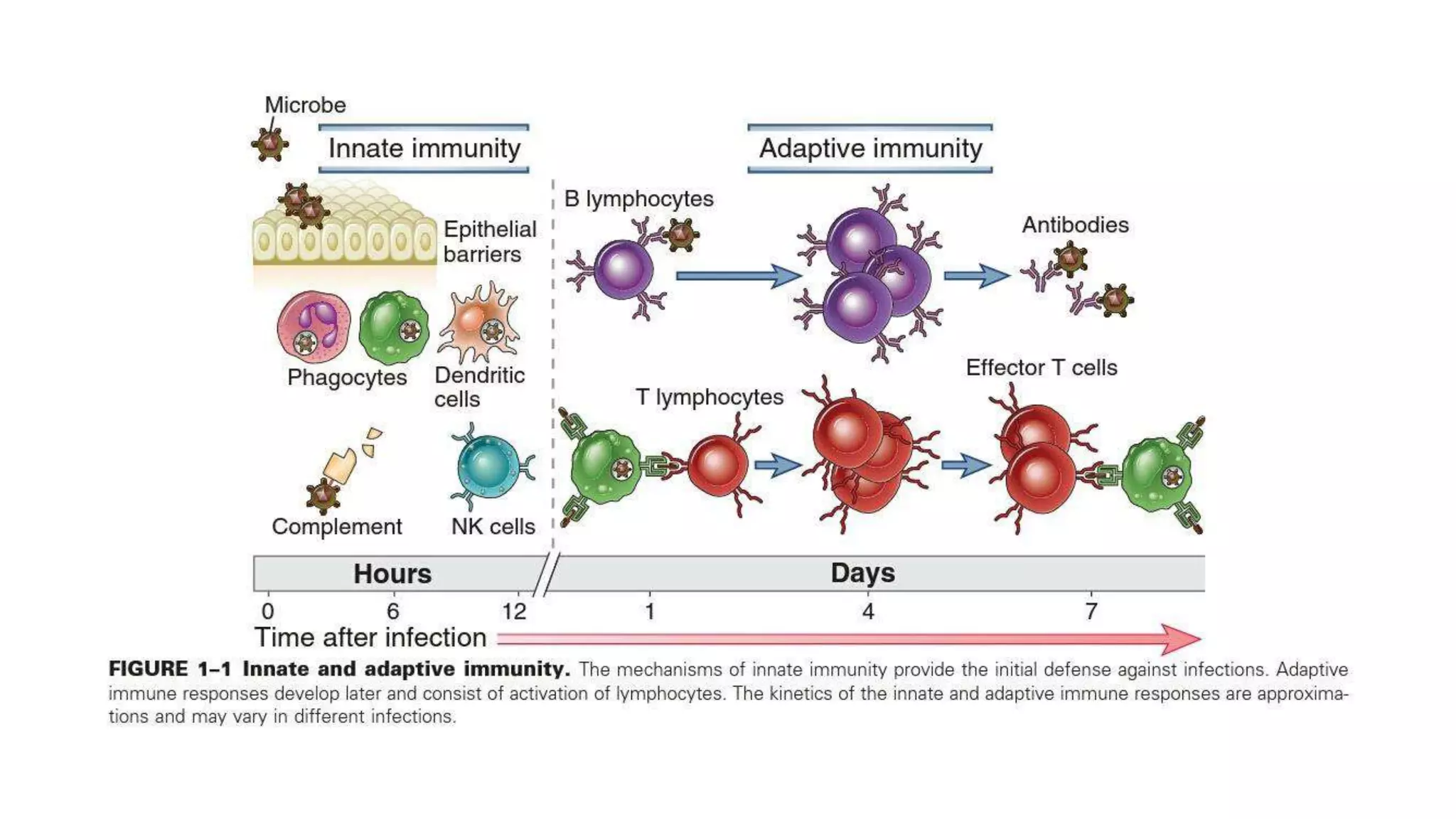
The document provides a comprehensive overview of the host immune response to pathogens, detailing the roles of both the innate and adaptive immune systems. It highlights the mechanisms involved in immunity, such as inflammation, phagocytosis, and antibody synthesis, as well as the types of immunity including natural and acquired. Additionally, it covers the functions of various immune cells, including phagocytes and lymphocytes, and the significance of major histocompatibility complex (MHC) in distinguishing self from non-self.


