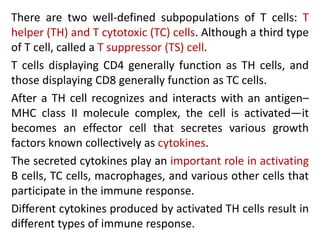
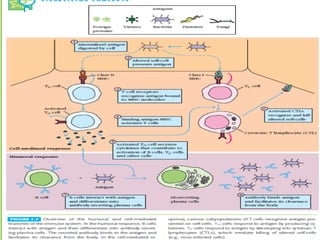
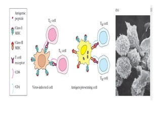
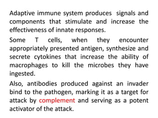
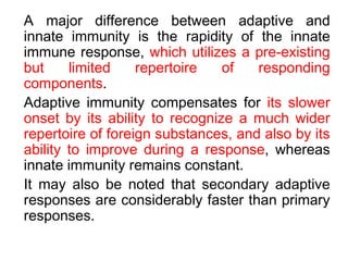
ADAPTIVE IMMUNITY
Dr Sitaram Swain
Adaptive immunity is capable of recognizing and selectively
eliminating specific foreign microorganisms and molecules.
Unlike innate immune responses, adaptive immune responses
are not the same in all members of a species but are reactions
to specific antigenic challenges.
Adaptive immunity displays four characteristic attributes:
• Antigenic specificity
• Diversity
• Immunologic memory
• Self/non-self recognition
The antigenic specificity of the immune system
permits it to distinguish subtle differences among
antigens.
Antibodies can distinguish between two protein
molecules that differ in only a single amino acid.
The immune system is capable of generating
tremendous diversity in its recognition molecules,
allowing it to recognize billions of unique structures
on foreign antigens.
Once the immune system has recognized and
responded to an antigen, it exhibits immunologic
memory; that is, a second encounter with the same
antigen induces heightened state of immune reactivity.
Because of this attribute, the immune system can
confer life-long immunity to many infectious agents
after an initial encounter.
Finally, the immune system normally responds
only to foreign antigens, indicating that it is
capable of self/non-self recognition.
The ability of the immune system to distinguish self
from non self and respond only to non self
molecules is essential for the outcome of an
inappropriate response to self molecules can be
fatal.
The Adaptive Immune System Requires Cooperation
Between Lymphocytes and Antigen-Presenting Cells
An effective immune response involves two major
groups of
cells: T lymphocytes and antigen-presenting cells.
Lymphocytes are one of many types of white
blood cells produced in the bone marrow by the
process of hematopoiesis .
Lymphocytes leave the bone marrow, circulate in
the blood and lymphatic systems, and reside in
various lymphoid organs.
Because they produce and display antigen binding cell-
surface receptors, lymphocytes mediate the defining
immunologic attributes of specificity, diversity, memory,
and self/non-self recognition.
The two major populations of lymphocytes—
B lymphocytes (B cells) and T lymphocytes (T cells)
B LYMPHOCYTES
B lymphocytes mature within the bone marrow; when they
leave it, each expresses a unique antigen-binding receptor
on its membrane. This antigen-binding or B-cell receptor is
a membrane-bound antibody molecule.
When a naive B cell first encounters the antigen that
matches its membrane bound antibody, the binding of the
antigen to the antibody causes the cell to divide rapidly; its
progeny differentiate into memory B cells and effector B
cells called plasma cells.
Memory B cells have a longer life span than naive cells, and
they express the same membrane-bound antibody as their
parent B cell.
Plasma cells produce the antibody in a form that can be
secreted and have little or no membrane-bound antibody.
Although plasma cells live for only a few days, they secrete
enormous amounts of antibody during this time.
It has been estimated that a single plasma cell can secrete
more than 2000 molecules of antibody per second. Secreted
antibodies are the major effector molecules of humoral
immunity.
T LYMPHOCYTES
T lymphocytes also arise in the bone marrow. T cells migrate to
the thymus gland to mature. During its maturation within the
thymus, the T cell comes to express a unique antigen-binding
molecule, called the T-cell receptor, on its membrane.
TCR can recognize only antigen that is bound to cell-
membrane proteins called major histocompatibility complex
(MHC) molecules. MHC molecules that function in this
recognition event, which is termed “antigen presentation,” are
polymorphic glycoproteins found on cell membranes .
There are two major types of MHC molecules: Class I
MHC molecules, which are expressed by nearly all
nucleated cells of vertebrate species, consist of a heavy
chain linked to a small invariant protein called 2-
microglobulin.
Class II MHC molecules, which consist of an alpha and a
beta glycoprotein chain, are expressed only by antigen-
presenting cells.
When a naive T cell encounters antigen combined with a
MHC molecule on cell, T cell proliferates and
differentiates into memory T cells and various effector
cells.
There are two well-defined subpopulations of T cells: T
helper (TH) and T cytotoxic (TC) cells. Although a third type
of T cell, called a T suppressor (TS) cell.
T cells displaying CD4 generally function as TH cells, and
those displaying CD8 generally function as TC cells.
After a TH cell recognizes and interacts with an antigen–
MHC class II molecule complex, the cell is activated—it
becomes an effector cell that secretes various growth
factors known collectively as cytokines.
The secreted cytokines play an important role in activating
B cells, TC cells, macrophages, and various other cells that
participate in the immune response.
Different cytokines produced by activated TH cells result in
different types of immune response.
Under the influence of TH-derived cytokines, a TC cell that
recognizes an antigen–MHC class I molecule complex
proliferates and differentiates into an effector cell called a
cytotoxic T lymphocyte (CTL). In contrast to the TC cell,
the CTL generally does not secrete many cytokines and
instead exhibits cell-killing or cytotoxic activity.
The CTL has a vital function in monitoring the cells of the
body and eliminating any that display antigen, such as
virus-infected cells, tumor cells, and cells of a foreign
tissue graft.
Cells that display foreign antigen complexed with a class I
MHC molecule are called altered self-cells; these are
targets of CTLs.
ANTIGEN-PRESENTING CELLS
Activation of both the humoral and cell-mediated branches of the
immune system requires cytokines produced by TH cells. It is
essential that activation of TH cells themselves be carefully
regulated, because an inappropriate T-cell response to self-
components can have fatal autoimmune consequences.
To ensure carefully regulated activation of TH cells, they can
recognize only antigen that is displayed together with class MHC II
molecules on the surface of antigen-presenting cells (APCs). These
specialized cells, which include macrophages, B lymphocytes, and
dendritic cells, are distinguished by two properties:
(1) they express class II MHC molecules on their membranes, and
(2) they are able to deliver a co-stimulatory signal that is
necessary for TH-cell activation.
Antigen-presenting cells first internalize antigen, either by
phagocytosis or by endocytosis, and then display a part of
that antigen on their membrane bound to a class II MHC
molecule.
The TH cell recognizes and interacts with the antigen–
class II MHC molecule complex on the membrane of the
antigen-presenting cell.
An additional co-stimulatory signal is then produced by
antigen-presenting cell, leading to activation of TH cell.
Humoral Immunity But Not Cellular
Immunity Is Transferred with Antibody
Immune responses can be divided into humoral and cell-
mediated responses. The humoral branch of the immune
system is at work in the interaction of B cells with antigen and
their subsequent proliferation and differentiation into antibody-
secreting plasma cells . Antibody functions as the effector of
the humoral response by binding to antigen and neutralizing it
or facilitating its elimination.
When an antigen is coated with antibody, it can be eliminated
in several ways. For example, antibody can cross-link several
antigens, forming clusters that are more readily ingested by
phagocytic cells. Binding of antibody to antigen on a
microorganism can also activate the complement system,
resulting in lysis of the foreign organism. Antibody can also
neutralize toxins or viral particles by coating them, which
prevents them from binding to host cells.
Effector T cells generated in response to antigen are
responsible for cell-mediated immunity.
Both activated TH cells and cytotoxic T lymphocytes
(CTLs) serve as effector cells in cell-mediated immune
reactions.
Cytokines secreted by TH cells can activate various
phagocytic cells, enabling them to phagocytose and kill
microorganisms more effectively. This type of cell-
mediated immune response is especially important in
ridding the host of bacteria and protozoa contained by
infected host cells.
CTLs participate in cell-mediated immune reactions by
killing altered self-cells; they play an important role in
the killing of virusinfected cells and tumor cells.
Each branch of the immune system is therefore uniquely suited
to recognize antigen. The humoral branch (B cells) recognizes
an enormous variety of epitopes: those displayed on the
surfaces of bacteria or viral particles, as well as those displayed
on soluble proteins, glycoproteins, polysaccharides, or
lipopolysaccharides that have been released from invading
pathogens.
The cell-mediated branch (T cells) recognizes protein epitopes
displayed together with MHC molecules on self-cells, including
altered self-cells also virus-infected self-cells &cancerous cells.
Thus, four related but distinct cell-membrane molecules are
responsible for antigen recognition by the immune system:
Membrane-bound antibodies on B cells
T-cell receptors
Class I MHC molecules
Class II MHC molecules
Antigen Is Recognized Differently by B and T
Lymphocytes
Antigens, which are generally very large and complex, are
not recognized in their entirety by lymphocytes. Instead,
both B and T lymphocytes recognize discrete sites on the
antigen called antigenic determinants, or epitopes.
Epitopes are the immunologically active regions on a
complex antigen, the regions that actually bind to B-cell or
T-cell receptors.
Although B cells can recognize an epitope alone, T cells
can recognize an epitope only when it is associated with an
MHC molecule on the surface of a self-cell (either an
antigen- presenting cell or an altered self-cell).
B and T Lymphocytes Utilize Similar Mechanisms
To Generate Diversity in Antigen Receptors
The antigenic specificity of each B cell is determined by the
membrane-bound antigen-binding receptor (i.e., antibody)
expressed by the cell. As a B cell matures in the bone marrow, its
specificity is created by random rearrangements of a series of gene
segments that encode the antibody molecule .As a result of this
process, each mature B cell possesses a single functional gene
encoding the antibody heavy chain and a single functional gene
encoding the antibody light chain; the cell therefore synthesizes and
displays antibody with one specificity on its membrane.
All antibody molecules on a given B lymphocyte have identical
specificity, giving each B lymphocyte, and the clone of daughter cells
to which it gives rise, a distinct specificity for a single epitope on an
antigen. The mature B lymphocyte is to be antigenically committed.
The random gene rearrangements during B-cell
maturation in the bone marrow generate an enormous
number of different antigenic specificities.
The resulting B-cell population, which consists of
individual B cells each expressing a unique antibody,
is estimated to exhibit collectively more than 1010
different antigenic specificities.
The enormous diversity in the mature B-cell population
is later reduced by a selection process in the bone
marrow that eliminates any B cells with membrane-
bound antibody that recognizes self components.
The selection process helps to ensure that self-
reactive antibodies (auto-antibodies) are not produced.
The attributes of specificity and diversity also characterize
the antigen-binding T-cell receptor (TCR) on T cells. As in B-
cell and T-cell maturation includes random rearrangements
of a series of gene segments that encode the cell’s antigen-
binding receptor .
Each T lymphocyte cell expresses about 105 receptors, and
all of the receptors on the cell and its clonal progeny have
identical specificity for antigen.
The random rearrangement of the TCR genes is capable of
generating on the order of 109 unique antigenic specificities.
This enormous potential diversity is later diminished through
a selection process in the thymus that eliminates any T cell
with self-reactive receptors and ensures that only T cells with
receptors capable of recognizing antigen associated with
MHC molecules will be able to mature .
The Major Histocompatibility Molecules
Bind Antigenic Peptides
MHC is a large genetic complex with multiple loci. The MHC loci encode
two major classes of membrane-bound glycoproteins: class I and class II
MHC molecules.
TH cells generally recognize antigen combined with class II molecules,
whereasTC cells generally recognize antigen combined with class I
molecules .
MHC molecules function as antigen-recognition molecules, but they do
not possess the fine specificity for antigen characteristic of antibodies and
T-cell receptors. Rather, each MHC molecule can bind to a spectrum of
antigenic peptides derived from the intracellular degradation of antigen
molecules.
In both class I and class II MHC molecules the distal
regions (farthest from the membrane) of different alleles
display wide variation in their amino acid sequences.
These variable regions form a cleft within which the
antigenic peptide sits and is presented to T lymphocytes .
Different allelic forms of confer different structures on the
antigen-binding cleft with different specificity. Thus the
ability to present an antigen to T lymphocytes is
influenced by the particular set of alleles that an individual
inherits.
Complex Antigens Are Degraded (Processed) and Displayed
(Presented) with MHC Molecules on the Cell Surface
In order for a foreign protein antigen to be recognized by a
T cell, it must be degraded into small antigenic peptides
that form complexes with class I or class II MHC molecules.
This conversion of proteins into MHC-associated peptide
fragments is called antigen processing and presentation.
Processed and presented antigen together with class I
MHC or class II MHC molecules appears to be determined
by the route that the antigen takes to enter a cell.
Exogenous antigen is produced outside of the host
cell and enters the cell by endocytosis or
phagocytosis.
Antigen presenting cells (macrophages, dendritic
cells, and B cells) degrade ingested exogenous
antigen into peptide fragments within the endocytic
processing pathway.
Class II MHC molecules are expressed within the
endocytic processing pathway and that peptides
produced by degradation of antigen and bind to
the cleft within the class II MHC molecules.
The MHC molecules bearing the peptide are then
exported to the cell surface.
Since expression of class II MHC molecules is limited to
antigen- presenting cells, presentation of exogenous
peptide– class II MHC complexes is limited to these cells. T
cells displaying CD4 recognize antigen combined with
class II MHC molecules and thus are said to be class II
MHC restricted.
These cells generally function as T helper cells.
Endogenous antigen is produced within the host cell itself.
Two common examples are viral proteins synthesized
within virus-infected host cells and unique proteins
synthesized by cancerous cells.
Endogenous antigens are degraded into peptide fragments
that bind to class I MHC molecules within the endoplasmic
reticulum. The peptide–class I MHC complex is then
transported to the cell membrane. Since all nucleated cells
express class I MHC molecules, all cells producing
endogenous antigen use this route to process the antigen.
T cells displaying CD8 recognize antigen associated with
class I MHC molecules and thus are said to be class I MHC
restricted. These cytotoxic T cells attack and kill cells
displaying the antigen–MHC class I complexes for which
their receptors are specific.
Antigen Selection of Lymphocytes
Causes Clonal Expansion
A mature immuno-competent animal contains a large number
of antigen-reactive clones of T and B lymphocytes; the
antigenic specificity of each of these clones is determined by
the specificity of the antigen-binding receptor on the
membrane of the clone’s lymphocytes.
As noted above, the specificity of each T and B lymphocyte is
determined before its contact with antigen by random gene
rearrangements during maturation in the thymus or bone
marrow.
The role of antigen becomes critical when it interacts
with and activates mature, antigenically committed T and
B lymphocytes, bringing about expansion of the
population of cells with a given antigenic specificity. In
this process of clonal selection, an antigen binds to a
particular T or B cell and stimulates it to divide repeatedly
into a clone of cells with the same antigenic specificity as
the original parent cell.
Clonal selection provides a framework for understanding
the specificity and self/nonself recognition that is
characteristic of adaptive immunity. Specificity is shown
because only lymphocytes whose receptors are specific
for a given epitope on an antigen will be clonally
expanded and thus mobilized for an immune response.
Self/nonself discrimination is accomplished by the
elimination, during development, of lymphocytes bearing
self-reactive receptors or by the functional suppression of
these cells in adults.
Immunologic memory also is a consequence of clonal
selection.
During clonal selection, the number of lymphocytes
specific for a given antigen is greatly amplified. Many of
these lymphocytes, referred to as memory cells, appear
to have a longer life span than the naive lymphocytes
from which they arise.
The initial encounter of a naive immunocompetent
lymphocyte with an antigen induces a primary response;
a later contact of the host with antigen will induce a more
rapid and heightened secondary response.
The amplified population of memory cells accounts
for the rapidity and intensity that distinguishes a
secondary response from the primary response.
The primary response has a lag of approximately 5–
7 days before antibody levels start to rise. This lag is
the time required for activation of naive B cells by
antigen and TH cells and for the subsequent
proliferation and differentiation of the activated B
cells into plasma cells.
Antibody levels peak in the primary response at
about day 14 and then begin to drop off as the
plasma cells begin to die.
In the secondary response, the lag is much shorter (only 1–2
days), antibody levels are much higher, and they are sustained
for much longer.
The secondary response reflects the activity of the clonally
expanded population of memory B cells.
These memory cells respond to the antigen more rapidly than
naive B cells; in addition, because there are many more memory
cells than there were naive B cells for the primary response,
more plasma cells are generated in secondary response, and
antibody levels are consequently 100- to 1000-fold higher.
In the cell-mediated branch of the immune system, the
recognition of an antigen-MHC complex by a specific mature T
lymphocyte induces clonal proliferation into various T cells with
effector functions (TH cells & CTLs) and into memory T cells.
When skin from strain C mice is grafted onto strain A mice, a
primary response develops and all the grafts are rejected in
about 10–14 days. If these same mice are again grafted with
strain C skin, it is rejected much more vigorously and rapidly
than the first grafts. However, if animals previously engrafted
with strain C skin are next given skin from an unrelated strain,
strain B, the response to strain B is typical of the primary
response and is rejected in 10–14 days.
That is, graft rejection is a specific immune response. The
same mice that showed a secondary response to graft C
will show a primary response to graft B.
The increased speed of rejection of graft C reflects the
presence of a clonally expanded population of memory TH
and TC cells specific for the antigens of the foreign graft.
This expanded memory population generates more effector
cells, resulting in faster graft rejection.
The Innate and Adaptive Immune Systems
Collaborate, Increasing the Efficiency of Immune Responsiveness
It is important to appreciate that adaptive and innate immunity do
not operate independently—they function as a highly interactive
and cooperative system, producing a combined response more
effective than either branch could produce by itself. Certain
immune components play important roles in both types of
immunity.
An example of cooperation is seen in the encounter between
macrophages and microbes. Interactions between receptors on
macrophages and microbial components generate soluble proteins
that stimulate and direct adaptive immune responses, facilitating
the participation of the adaptive immune system in the elimination
of the pathogen.
Stimulated macrophages also secrete cytokines that can direct
adaptive immune responses against particular intracellular
pathogens.
Adaptive immune system produces signals and
components that stimulate and increase the
effectiveness of innate responses.
Some T cells, when they encounter
appropriately presented antigen, synthesize and
secrete cytokines that increase the ability of
macrophages to kill the microbes they have
ingested.
Also, antibodies produced against an invader
bind to the pathogen, marking it as a target for
attack by complement and serving as a potent
activator of the attack.
A major difference between adaptive and
innate immunity is the rapidity of the innate
immune response, which utilizes a pre-existing
but limited repertoire of responding
components.
Adaptive immunity compensates for its slower
onset by its ability to recognize a much wider
repertoire of foreign substances, and also by its
ability to improve during a response, whereas
innate immunity remains constant.
It may also be noted that secondary adaptive
responses are considerably faster than primary
responses.
Adaptive immunity.pdf

Adaptive immunity.pdf

  • 1.
  • 2.
    Adaptive immunity iscapable of recognizing and selectively eliminating specific foreign microorganisms and molecules. Unlike innate immune responses, adaptive immune responses are not the same in all members of a species but are reactions to specific antigenic challenges. Adaptive immunity displays four characteristic attributes: • Antigenic specificity • Diversity • Immunologic memory • Self/non-self recognition
  • 3.
    The antigenic specificityof the immune system permits it to distinguish subtle differences among antigens. Antibodies can distinguish between two protein molecules that differ in only a single amino acid. The immune system is capable of generating tremendous diversity in its recognition molecules, allowing it to recognize billions of unique structures on foreign antigens.
  • 4.
    Once the immunesystem has recognized and responded to an antigen, it exhibits immunologic memory; that is, a second encounter with the same antigen induces heightened state of immune reactivity. Because of this attribute, the immune system can confer life-long immunity to many infectious agents after an initial encounter.
  • 5.
    Finally, the immunesystem normally responds only to foreign antigens, indicating that it is capable of self/non-self recognition. The ability of the immune system to distinguish self from non self and respond only to non self molecules is essential for the outcome of an inappropriate response to self molecules can be fatal.
  • 6.
    The Adaptive ImmuneSystem Requires Cooperation Between Lymphocytes and Antigen-Presenting Cells An effective immune response involves two major groups of cells: T lymphocytes and antigen-presenting cells. Lymphocytes are one of many types of white blood cells produced in the bone marrow by the process of hematopoiesis . Lymphocytes leave the bone marrow, circulate in the blood and lymphatic systems, and reside in various lymphoid organs.
  • 7.
    Because they produceand display antigen binding cell- surface receptors, lymphocytes mediate the defining immunologic attributes of specificity, diversity, memory, and self/non-self recognition. The two major populations of lymphocytes— B lymphocytes (B cells) and T lymphocytes (T cells)
  • 8.
    B LYMPHOCYTES B lymphocytesmature within the bone marrow; when they leave it, each expresses a unique antigen-binding receptor on its membrane. This antigen-binding or B-cell receptor is a membrane-bound antibody molecule. When a naive B cell first encounters the antigen that matches its membrane bound antibody, the binding of the antigen to the antibody causes the cell to divide rapidly; its progeny differentiate into memory B cells and effector B cells called plasma cells.
  • 9.
    Memory B cellshave a longer life span than naive cells, and they express the same membrane-bound antibody as their parent B cell. Plasma cells produce the antibody in a form that can be secreted and have little or no membrane-bound antibody. Although plasma cells live for only a few days, they secrete enormous amounts of antibody during this time. It has been estimated that a single plasma cell can secrete more than 2000 molecules of antibody per second. Secreted antibodies are the major effector molecules of humoral immunity.
  • 11.
    T LYMPHOCYTES T lymphocytesalso arise in the bone marrow. T cells migrate to the thymus gland to mature. During its maturation within the thymus, the T cell comes to express a unique antigen-binding molecule, called the T-cell receptor, on its membrane. TCR can recognize only antigen that is bound to cell- membrane proteins called major histocompatibility complex (MHC) molecules. MHC molecules that function in this recognition event, which is termed “antigen presentation,” are polymorphic glycoproteins found on cell membranes .
  • 12.
    There are twomajor types of MHC molecules: Class I MHC molecules, which are expressed by nearly all nucleated cells of vertebrate species, consist of a heavy chain linked to a small invariant protein called 2- microglobulin. Class II MHC molecules, which consist of an alpha and a beta glycoprotein chain, are expressed only by antigen- presenting cells. When a naive T cell encounters antigen combined with a MHC molecule on cell, T cell proliferates and differentiates into memory T cells and various effector cells.
  • 13.
    There are twowell-defined subpopulations of T cells: T helper (TH) and T cytotoxic (TC) cells. Although a third type of T cell, called a T suppressor (TS) cell. T cells displaying CD4 generally function as TH cells, and those displaying CD8 generally function as TC cells. After a TH cell recognizes and interacts with an antigen– MHC class II molecule complex, the cell is activated—it becomes an effector cell that secretes various growth factors known collectively as cytokines. The secreted cytokines play an important role in activating B cells, TC cells, macrophages, and various other cells that participate in the immune response. Different cytokines produced by activated TH cells result in different types of immune response.
  • 14.
    Under the influenceof TH-derived cytokines, a TC cell that recognizes an antigen–MHC class I molecule complex proliferates and differentiates into an effector cell called a cytotoxic T lymphocyte (CTL). In contrast to the TC cell, the CTL generally does not secrete many cytokines and instead exhibits cell-killing or cytotoxic activity. The CTL has a vital function in monitoring the cells of the body and eliminating any that display antigen, such as virus-infected cells, tumor cells, and cells of a foreign tissue graft. Cells that display foreign antigen complexed with a class I MHC molecule are called altered self-cells; these are targets of CTLs.
  • 15.
    ANTIGEN-PRESENTING CELLS Activation ofboth the humoral and cell-mediated branches of the immune system requires cytokines produced by TH cells. It is essential that activation of TH cells themselves be carefully regulated, because an inappropriate T-cell response to self- components can have fatal autoimmune consequences. To ensure carefully regulated activation of TH cells, they can recognize only antigen that is displayed together with class MHC II molecules on the surface of antigen-presenting cells (APCs). These specialized cells, which include macrophages, B lymphocytes, and dendritic cells, are distinguished by two properties: (1) they express class II MHC molecules on their membranes, and (2) they are able to deliver a co-stimulatory signal that is necessary for TH-cell activation.
  • 16.
    Antigen-presenting cells firstinternalize antigen, either by phagocytosis or by endocytosis, and then display a part of that antigen on their membrane bound to a class II MHC molecule. The TH cell recognizes and interacts with the antigen– class II MHC molecule complex on the membrane of the antigen-presenting cell. An additional co-stimulatory signal is then produced by antigen-presenting cell, leading to activation of TH cell.
  • 18.
    Humoral Immunity ButNot Cellular Immunity Is Transferred with Antibody Immune responses can be divided into humoral and cell- mediated responses. The humoral branch of the immune system is at work in the interaction of B cells with antigen and their subsequent proliferation and differentiation into antibody- secreting plasma cells . Antibody functions as the effector of the humoral response by binding to antigen and neutralizing it or facilitating its elimination. When an antigen is coated with antibody, it can be eliminated in several ways. For example, antibody can cross-link several antigens, forming clusters that are more readily ingested by phagocytic cells. Binding of antibody to antigen on a microorganism can also activate the complement system, resulting in lysis of the foreign organism. Antibody can also neutralize toxins or viral particles by coating them, which prevents them from binding to host cells.
  • 19.
    Effector T cellsgenerated in response to antigen are responsible for cell-mediated immunity. Both activated TH cells and cytotoxic T lymphocytes (CTLs) serve as effector cells in cell-mediated immune reactions. Cytokines secreted by TH cells can activate various phagocytic cells, enabling them to phagocytose and kill microorganisms more effectively. This type of cell- mediated immune response is especially important in ridding the host of bacteria and protozoa contained by infected host cells. CTLs participate in cell-mediated immune reactions by killing altered self-cells; they play an important role in the killing of virusinfected cells and tumor cells.
  • 20.
    Each branch ofthe immune system is therefore uniquely suited to recognize antigen. The humoral branch (B cells) recognizes an enormous variety of epitopes: those displayed on the surfaces of bacteria or viral particles, as well as those displayed on soluble proteins, glycoproteins, polysaccharides, or lipopolysaccharides that have been released from invading pathogens. The cell-mediated branch (T cells) recognizes protein epitopes displayed together with MHC molecules on self-cells, including altered self-cells also virus-infected self-cells &cancerous cells. Thus, four related but distinct cell-membrane molecules are responsible for antigen recognition by the immune system: Membrane-bound antibodies on B cells T-cell receptors Class I MHC molecules Class II MHC molecules
  • 21.
    Antigen Is RecognizedDifferently by B and T Lymphocytes Antigens, which are generally very large and complex, are not recognized in their entirety by lymphocytes. Instead, both B and T lymphocytes recognize discrete sites on the antigen called antigenic determinants, or epitopes. Epitopes are the immunologically active regions on a complex antigen, the regions that actually bind to B-cell or T-cell receptors. Although B cells can recognize an epitope alone, T cells can recognize an epitope only when it is associated with an MHC molecule on the surface of a self-cell (either an antigen- presenting cell or an altered self-cell).
  • 22.
    B and TLymphocytes Utilize Similar Mechanisms To Generate Diversity in Antigen Receptors The antigenic specificity of each B cell is determined by the membrane-bound antigen-binding receptor (i.e., antibody) expressed by the cell. As a B cell matures in the bone marrow, its specificity is created by random rearrangements of a series of gene segments that encode the antibody molecule .As a result of this process, each mature B cell possesses a single functional gene encoding the antibody heavy chain and a single functional gene encoding the antibody light chain; the cell therefore synthesizes and displays antibody with one specificity on its membrane. All antibody molecules on a given B lymphocyte have identical specificity, giving each B lymphocyte, and the clone of daughter cells to which it gives rise, a distinct specificity for a single epitope on an antigen. The mature B lymphocyte is to be antigenically committed.
  • 23.
    The random generearrangements during B-cell maturation in the bone marrow generate an enormous number of different antigenic specificities. The resulting B-cell population, which consists of individual B cells each expressing a unique antibody, is estimated to exhibit collectively more than 1010 different antigenic specificities. The enormous diversity in the mature B-cell population is later reduced by a selection process in the bone marrow that eliminates any B cells with membrane- bound antibody that recognizes self components. The selection process helps to ensure that self- reactive antibodies (auto-antibodies) are not produced.
  • 24.
    The attributes ofspecificity and diversity also characterize the antigen-binding T-cell receptor (TCR) on T cells. As in B- cell and T-cell maturation includes random rearrangements of a series of gene segments that encode the cell’s antigen- binding receptor . Each T lymphocyte cell expresses about 105 receptors, and all of the receptors on the cell and its clonal progeny have identical specificity for antigen. The random rearrangement of the TCR genes is capable of generating on the order of 109 unique antigenic specificities. This enormous potential diversity is later diminished through a selection process in the thymus that eliminates any T cell with self-reactive receptors and ensures that only T cells with receptors capable of recognizing antigen associated with MHC molecules will be able to mature .
  • 25.
    The Major HistocompatibilityMolecules Bind Antigenic Peptides MHC is a large genetic complex with multiple loci. The MHC loci encode two major classes of membrane-bound glycoproteins: class I and class II MHC molecules. TH cells generally recognize antigen combined with class II molecules, whereasTC cells generally recognize antigen combined with class I molecules . MHC molecules function as antigen-recognition molecules, but they do not possess the fine specificity for antigen characteristic of antibodies and T-cell receptors. Rather, each MHC molecule can bind to a spectrum of antigenic peptides derived from the intracellular degradation of antigen molecules.
  • 26.
    In both classI and class II MHC molecules the distal regions (farthest from the membrane) of different alleles display wide variation in their amino acid sequences. These variable regions form a cleft within which the antigenic peptide sits and is presented to T lymphocytes . Different allelic forms of confer different structures on the antigen-binding cleft with different specificity. Thus the ability to present an antigen to T lymphocytes is influenced by the particular set of alleles that an individual inherits.
  • 28.
    Complex Antigens AreDegraded (Processed) and Displayed (Presented) with MHC Molecules on the Cell Surface In order for a foreign protein antigen to be recognized by a T cell, it must be degraded into small antigenic peptides that form complexes with class I or class II MHC molecules. This conversion of proteins into MHC-associated peptide fragments is called antigen processing and presentation. Processed and presented antigen together with class I MHC or class II MHC molecules appears to be determined by the route that the antigen takes to enter a cell.
  • 29.
    Exogenous antigen isproduced outside of the host cell and enters the cell by endocytosis or phagocytosis. Antigen presenting cells (macrophages, dendritic cells, and B cells) degrade ingested exogenous antigen into peptide fragments within the endocytic processing pathway. Class II MHC molecules are expressed within the endocytic processing pathway and that peptides produced by degradation of antigen and bind to the cleft within the class II MHC molecules. The MHC molecules bearing the peptide are then exported to the cell surface.
  • 30.
    Since expression ofclass II MHC molecules is limited to antigen- presenting cells, presentation of exogenous peptide– class II MHC complexes is limited to these cells. T cells displaying CD4 recognize antigen combined with class II MHC molecules and thus are said to be class II MHC restricted. These cells generally function as T helper cells. Endogenous antigen is produced within the host cell itself. Two common examples are viral proteins synthesized within virus-infected host cells and unique proteins synthesized by cancerous cells.
  • 31.
    Endogenous antigens aredegraded into peptide fragments that bind to class I MHC molecules within the endoplasmic reticulum. The peptide–class I MHC complex is then transported to the cell membrane. Since all nucleated cells express class I MHC molecules, all cells producing endogenous antigen use this route to process the antigen. T cells displaying CD8 recognize antigen associated with class I MHC molecules and thus are said to be class I MHC restricted. These cytotoxic T cells attack and kill cells displaying the antigen–MHC class I complexes for which their receptors are specific.
  • 33.
    Antigen Selection ofLymphocytes Causes Clonal Expansion A mature immuno-competent animal contains a large number of antigen-reactive clones of T and B lymphocytes; the antigenic specificity of each of these clones is determined by the specificity of the antigen-binding receptor on the membrane of the clone’s lymphocytes. As noted above, the specificity of each T and B lymphocyte is determined before its contact with antigen by random gene rearrangements during maturation in the thymus or bone marrow.
  • 35.
    The role ofantigen becomes critical when it interacts with and activates mature, antigenically committed T and B lymphocytes, bringing about expansion of the population of cells with a given antigenic specificity. In this process of clonal selection, an antigen binds to a particular T or B cell and stimulates it to divide repeatedly into a clone of cells with the same antigenic specificity as the original parent cell. Clonal selection provides a framework for understanding the specificity and self/nonself recognition that is characteristic of adaptive immunity. Specificity is shown because only lymphocytes whose receptors are specific for a given epitope on an antigen will be clonally expanded and thus mobilized for an immune response.
  • 36.
    Self/nonself discrimination isaccomplished by the elimination, during development, of lymphocytes bearing self-reactive receptors or by the functional suppression of these cells in adults. Immunologic memory also is a consequence of clonal selection. During clonal selection, the number of lymphocytes specific for a given antigen is greatly amplified. Many of these lymphocytes, referred to as memory cells, appear to have a longer life span than the naive lymphocytes from which they arise. The initial encounter of a naive immunocompetent lymphocyte with an antigen induces a primary response; a later contact of the host with antigen will induce a more rapid and heightened secondary response.
  • 37.
    The amplified populationof memory cells accounts for the rapidity and intensity that distinguishes a secondary response from the primary response. The primary response has a lag of approximately 5– 7 days before antibody levels start to rise. This lag is the time required for activation of naive B cells by antigen and TH cells and for the subsequent proliferation and differentiation of the activated B cells into plasma cells. Antibody levels peak in the primary response at about day 14 and then begin to drop off as the plasma cells begin to die.
  • 38.
    In the secondaryresponse, the lag is much shorter (only 1–2 days), antibody levels are much higher, and they are sustained for much longer. The secondary response reflects the activity of the clonally expanded population of memory B cells. These memory cells respond to the antigen more rapidly than naive B cells; in addition, because there are many more memory cells than there were naive B cells for the primary response, more plasma cells are generated in secondary response, and antibody levels are consequently 100- to 1000-fold higher.
  • 39.
    In the cell-mediatedbranch of the immune system, the recognition of an antigen-MHC complex by a specific mature T lymphocyte induces clonal proliferation into various T cells with effector functions (TH cells & CTLs) and into memory T cells. When skin from strain C mice is grafted onto strain A mice, a primary response develops and all the grafts are rejected in about 10–14 days. If these same mice are again grafted with strain C skin, it is rejected much more vigorously and rapidly than the first grafts. However, if animals previously engrafted with strain C skin are next given skin from an unrelated strain, strain B, the response to strain B is typical of the primary response and is rejected in 10–14 days.
  • 40.
    That is, graftrejection is a specific immune response. The same mice that showed a secondary response to graft C will show a primary response to graft B. The increased speed of rejection of graft C reflects the presence of a clonally expanded population of memory TH and TC cells specific for the antigens of the foreign graft. This expanded memory population generates more effector cells, resulting in faster graft rejection.
  • 43.
    The Innate andAdaptive Immune Systems Collaborate, Increasing the Efficiency of Immune Responsiveness It is important to appreciate that adaptive and innate immunity do not operate independently—they function as a highly interactive and cooperative system, producing a combined response more effective than either branch could produce by itself. Certain immune components play important roles in both types of immunity. An example of cooperation is seen in the encounter between macrophages and microbes. Interactions between receptors on macrophages and microbial components generate soluble proteins that stimulate and direct adaptive immune responses, facilitating the participation of the adaptive immune system in the elimination of the pathogen. Stimulated macrophages also secrete cytokines that can direct adaptive immune responses against particular intracellular pathogens.
  • 44.
    Adaptive immune systemproduces signals and components that stimulate and increase the effectiveness of innate responses. Some T cells, when they encounter appropriately presented antigen, synthesize and secrete cytokines that increase the ability of macrophages to kill the microbes they have ingested. Also, antibodies produced against an invader bind to the pathogen, marking it as a target for attack by complement and serving as a potent activator of the attack.
  • 45.
    A major differencebetween adaptive and innate immunity is the rapidity of the innate immune response, which utilizes a pre-existing but limited repertoire of responding components. Adaptive immunity compensates for its slower onset by its ability to recognize a much wider repertoire of foreign substances, and also by its ability to improve during a response, whereas innate immunity remains constant. It may also be noted that secondary adaptive responses are considerably faster than primary responses.