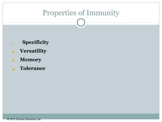
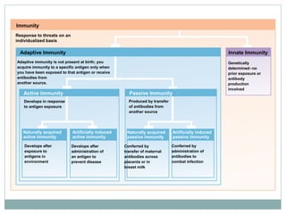
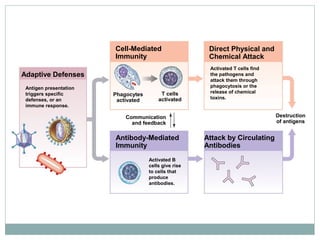
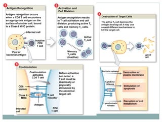
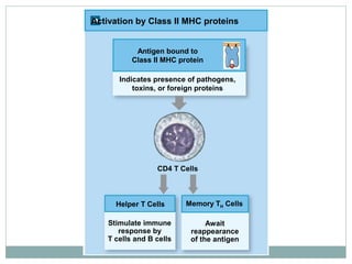
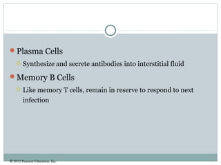
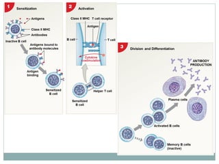
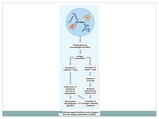
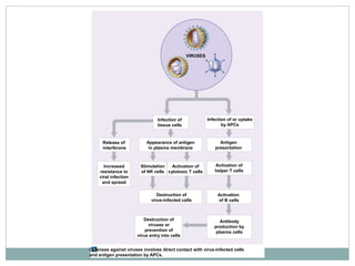
HS 345: HUMAN PHYSIOLOGY
WILMINGTON COLLEGE
LISA REGULA MEYER
Cellular Immunity
© 2012 Pearson Education, Inc.
Properties of Immunity
1. Specificity
2. Versatility
3. Memory
4. Tolerance
Immunity
Response to threats on an
individualized basis
Adaptive Immunity
Active Immunity Passive Immunity
Adaptive immunity is not present at birth; you
acquire immunity to a specific antigen only when
you have been exposed to that antigen or receive
antibodies from
another source.
Develops in response
to antigen exposure
Develops after
exposure to
antigens in
environment
Develops after
administration of
an antigen to
prevent disease
Conferred by
transfer of maternal
antibodies across
placenta or in
breast milk
Conferred by
administration of
antibodies to
combat infection
Naturally acquired
active immunity
Artificially induced
active immunity
Naturally acquired
passive immunity
Artificially induced
passive immunity
Genetically
determined−no
prior exposure or
antibody
production
involved
Innate Immunity
Produced by transfer
of antibodies from
another source
Adaptive Defenses
Cell-Mediated
Immunity
Direct Physical and
Chemical Attack
Antibody-Mediated
Immunity
Attack by Circulating
Antibodies
Destruction
of antigens
Phagocytes
activated
T cells
activated
Communication
and feedback
Antigen presentation
triggers specific
defenses, or an
immune response.
Activated B
cells give rise
to cells that
produce
antibodies.
Activated T cells find
the pathogens and
attack them through
phagocytosis or the
release of chemical
toxins.
© 2012 Pearson Education, Inc.
Major Components of Cellular Immunity
Cytokines
MHC proteins
Lymphocytes
© 2012 Pearson Education, Inc.
Cytokines
 Chemical messengers released by tissue cells
 To coordinate local activities
 To act as hormones to affect whole body
Functions of Cytokines
1. Stimulate T cell divisions
 Produce memory TH cells
 Accelerate cytotoxic T cell maturation
1. Attract and stimulate macrophages
2. Attract and stimulate activity of cytotoxic T cells
3. Promote activation of B cells
© 2012 Pearson Education, Inc.
Types of Cytokines
Interleukins
Lymphokines
Monokines
Chemokines
Interferons
© 2012 Pearson Education, Inc.
MHC Proteins
 The membrane glycoproteins that bind to antigens
 Genetically coded in chromosome 6
 The major histocompatibility complex (MHC)
 Differs among individuals
Antigen Presentation
T cells only recognize antigens that are bound to
glycoproteins in plasma membranes
© 2012 Pearson Education, Inc.
Class I MHC
 Found in membranes of all nucleated cells
 Pick up small peptides in cell and carry them to the surface
 T cells ignore normal peptides
 Abnormal peptides or viral proteins activate T cells to destroy
cell
© 2012 Pearson Education, Inc.
Class II MHC
 Found in membranes of antigen-presenting cells (APCs)
 Found in lymphocytes
 Inserted in plasma membrane to stimulate T cells
© 2012 Pearson Education, Inc.
Lymphocytes
Lymphoid Stem Cells
 Group 1
 Remains in bone marrow and develop with help of stromal cells
 Produces B cells and natural killer cells
 Group 2
 Migrates to thymus
 Produces T cells in environment isolated by blood–thymus
barrier
© 2012 Pearson Education, Inc.
T Cells make up 80% of circulating lymphocytes
Cytotoxic
 Attack cells infected by viruses
 Produce cell-mediated immunity
Memory
 Formed in response to foreign substance
 Remain in body to give “immunity”
Helper*
 Stimulate function of T cells and B cells
Suppressor*
 Inhibit function of T cells and B cells
*Regulatory T Cells control sensitivity of immune response
© 2012 Pearson Education, Inc.
Overview
Antigen presentation
Antigen recognition
T Cell activation
 Antigen binding
 Co-stimulation
T Cell proliferation
T Cell differentiation
© 2012 Pearson Education, Inc.
Cell-Mediated Immune Response
Antigen Presentation
 First step in immune response
 Extracted antigens are “presented” to lymphocytes
 Or attached to dendritic cells to stimulate lymphocytes
 Can be phagocytic or non-phagocytic antigen-presenting
cells
© 2012 Pearson Education, Inc.
 Phagocytic APCs
• Free and fixed
macrophages
 In connective tissues
• Kupffer cells
 Of the liver
• Microglia
 In the CNS
Non-phagocytic APCs
 Langerhans cells
 In the skin
 Dendritic cells
 In lymph nodes and
spleen
© 2012 Pearson Education, Inc.
Antigen Recognition
 Inactive T cell receptors
 Recognize Class I or Class II MHC proteins
 Recognize a specific foreign antigen
 Double recognition
 Binding occurs when MHC protein matches antigen
© 2012 Pearson Education, Inc.
CD Markers
• CD8 Markers
 Found on cytotoxic T
cells and suppressor T
cells
 Respond to antigens on
Class I MHC proteins
• CD4 Markers
 Found on helper T cells
 Respond to antigens on
Class II MHC proteins
© 2012 Pearson Education, Inc.
Co-Stimulation
 For T cell to be activated, it
must be costimulated
 By binding to stimulating
cell at second site
 Which confirms the first
signal
© 2012 Pearson Education, Inc.
Proliferation and Differentiation
After activation, activated T cells enlarge and
multiply
Newly expanded population of T cells differentiate
into
 Cytotoxic T cells
 Memory T cells
 Regulatory T cells
 Suppressor T cells
 Helper T cells
© 2012 Pearson Education, Inc.
Activation of CD8 T Cells
Activation of CD8 T Cells
 Activated by exposure to antigens on MHC proteins
 One responds quickly
 Producing cytotoxic T cells and memory T cells
 The other responds slowly
 Producing suppressor T cells
© 2012 Pearson Education, Inc.
Cytotoxic T Cells
 Seek out and immediately
destroy target cells
1. Release perforin
 To destroy antigenic plasma
membrane
1. Secrete poisonous lymphotoxin
 To destroy target cell
1. Activate genes in target cell
 That cause cell to die
Antigen Recognition Activation and
Cell Division
Infected cell
Inactive
CD8
T cell
Viral or
bacterial antigen
Antigen recognition occurs
when a CD8 T cell encounters
an appropriate antigen on the
surface of another cell, bound
to a Class I MHC protein.
Antigen recognition results
in T cell activation and cell
division, producing active TC
cells and memory TC cells.
Active
TC cell
Memory
TC cells
(inactive)
Infected
cell
CD8
protein
Class I
MHC
T cell
receptor
CD8
T cell
Antigen
Costimulation
activates
CD8 T cell
Before activation
can occur, a
T cell must be
chemically or
physically
stimulated by
the abnormal
target cell.
Costimulation
© 2012 Pearson Education, Inc.
Activation of CD4 T Cells
 Active helper T cells (TH cells)
 Secrete cytokines
 Memory helper (TH) cells
 Remain in reserve
Insert 21_18 here
Activation by Class I MHC proteins
Antigen bound to
Class I MHC protein
Indicates that the cell is infected
or otherwise abnormal
CD8 T Cells
Cytotoxic T Cells Memory TC Cells
Attack and destroy
infected and
abnormal cells
displaying antigen
Await
reappearance
of the antigen
Control or moderate
immune response by
T cells and B cells
Suppressor T Cells
Activation by Class II MHC proteins
Helper T Cells
Stimulate immune
response by
T cells and B cells
Await
reappearance
of the antigen
Memory TH Cells
CD4 T Cells
Indicates presence of pathogens,
toxins, or foreign proteins
Antigen bound to
Class II MHC protein
© 2012 Pearson Education, Inc.
B Cell Sensitization
 Corresponding antigens in interstitial fluids bind to B
cell receptors
 B cell prepares for activation
 Preparation process is sensitization
 During sensitization, antigens are:
 Taken into the B cell
 Processed
 Reappear on surface, bound to Class II MHC protein
© 2012 Pearson Education, Inc.
Helper T Cells
 Sensitized B cell is prepared for activation but needs helper T
cell activated by same antigen
B Cell Activation
 Helper T cell binds to MHC complex
 Secretes cytokines that promote B cell activation and division
© 2012 Pearson Education, Inc.
B Cell Division
 Activated B cell divides into:
 Plasma cells
 Memory B cells
© 2012 Pearson Education, Inc.
Plasma Cells
 Synthesize and secrete antibodies into interstitial fluid
Memory B Cells
 Like memory T cells, remain in reserve to respond to next
infection
© 2012 Pearson Education, Inc.
Pathologies of Cellular Immunity
HIV/AIDS
DiGeorge Syndrome
Wiskott-Aldrich syndrome
© 2012 Pearson Education, Inc.
Combines Responses to Viral Infection
 Similar to bacterial infection
 But cytotoxic T cells and NK cells are activated by contact
with virus-infected cells
BACTERIA
Phagocytosis by
macrophages and APCs
Antigen
presentation
Activation of
cytotoxic T cells
Activation of
helper T cells
Activation
of B cells
Antibody
production by
plasma cells
Destruction of
bacteria by
cell lysis or
phagocytosis
Opsonization
and phagocyte
attraction
Formation of
antigen−antibody
complexes
Defenses against bacteria involve phagocy-
tosis and antigen presentation by APCs.
Release of
interferons
Infection of
tissue cells
Appearance of antigen
in plasma membrane
Infection of or uptake
by APCs
VIRUSES
Antigen
presentation
Activation of
helper T cells
Activation
of B cells
Antibody
production by
plasma cells
Destruction of
viruses or
prevention of
virus entry into cells
Increased
resistance to
viral infection
and spread
Stimulation
of NK cells
Activation of
cytotoxic T cells
Destruction of
virus-infected cells
Defenses against viruses involves direct contact with virus-infected cells
and antigen presentation by APCs.

Wc hum phys 26 feb

  • 1.
    HS 345: HUMANPHYSIOLOGY WILMINGTON COLLEGE LISA REGULA MEYER Cellular Immunity
  • 2.
    © 2012 PearsonEducation, Inc. Properties of Immunity 1. Specificity 2. Versatility 3. Memory 4. Tolerance
  • 3.
    Immunity Response to threatson an individualized basis Adaptive Immunity Active Immunity Passive Immunity Adaptive immunity is not present at birth; you acquire immunity to a specific antigen only when you have been exposed to that antigen or receive antibodies from another source. Develops in response to antigen exposure Develops after exposure to antigens in environment Develops after administration of an antigen to prevent disease Conferred by transfer of maternal antibodies across placenta or in breast milk Conferred by administration of antibodies to combat infection Naturally acquired active immunity Artificially induced active immunity Naturally acquired passive immunity Artificially induced passive immunity Genetically determined−no prior exposure or antibody production involved Innate Immunity Produced by transfer of antibodies from another source
  • 4.
    Adaptive Defenses Cell-Mediated Immunity Direct Physicaland Chemical Attack Antibody-Mediated Immunity Attack by Circulating Antibodies Destruction of antigens Phagocytes activated T cells activated Communication and feedback Antigen presentation triggers specific defenses, or an immune response. Activated B cells give rise to cells that produce antibodies. Activated T cells find the pathogens and attack them through phagocytosis or the release of chemical toxins.
  • 5.
    © 2012 PearsonEducation, Inc. Major Components of Cellular Immunity Cytokines MHC proteins Lymphocytes
  • 6.
    © 2012 PearsonEducation, Inc. Cytokines  Chemical messengers released by tissue cells  To coordinate local activities  To act as hormones to affect whole body Functions of Cytokines 1. Stimulate T cell divisions  Produce memory TH cells  Accelerate cytotoxic T cell maturation 1. Attract and stimulate macrophages 2. Attract and stimulate activity of cytotoxic T cells 3. Promote activation of B cells
  • 7.
    © 2012 PearsonEducation, Inc. Types of Cytokines Interleukins Lymphokines Monokines Chemokines Interferons
  • 8.
    © 2012 PearsonEducation, Inc. MHC Proteins  The membrane glycoproteins that bind to antigens  Genetically coded in chromosome 6  The major histocompatibility complex (MHC)  Differs among individuals Antigen Presentation T cells only recognize antigens that are bound to glycoproteins in plasma membranes
  • 9.
    © 2012 PearsonEducation, Inc. Class I MHC  Found in membranes of all nucleated cells  Pick up small peptides in cell and carry them to the surface  T cells ignore normal peptides  Abnormal peptides or viral proteins activate T cells to destroy cell
  • 10.
    © 2012 PearsonEducation, Inc. Class II MHC  Found in membranes of antigen-presenting cells (APCs)  Found in lymphocytes  Inserted in plasma membrane to stimulate T cells
  • 11.
    © 2012 PearsonEducation, Inc. Lymphocytes Lymphoid Stem Cells  Group 1  Remains in bone marrow and develop with help of stromal cells  Produces B cells and natural killer cells  Group 2  Migrates to thymus  Produces T cells in environment isolated by blood–thymus barrier
  • 13.
    © 2012 PearsonEducation, Inc. T Cells make up 80% of circulating lymphocytes Cytotoxic  Attack cells infected by viruses  Produce cell-mediated immunity Memory  Formed in response to foreign substance  Remain in body to give “immunity” Helper*  Stimulate function of T cells and B cells Suppressor*  Inhibit function of T cells and B cells *Regulatory T Cells control sensitivity of immune response
  • 15.
    © 2012 PearsonEducation, Inc. Overview Antigen presentation Antigen recognition T Cell activation  Antigen binding  Co-stimulation T Cell proliferation T Cell differentiation
  • 16.
    © 2012 PearsonEducation, Inc. Cell-Mediated Immune Response Antigen Presentation  First step in immune response  Extracted antigens are “presented” to lymphocytes  Or attached to dendritic cells to stimulate lymphocytes  Can be phagocytic or non-phagocytic antigen-presenting cells
  • 17.
    © 2012 PearsonEducation, Inc.  Phagocytic APCs • Free and fixed macrophages  In connective tissues • Kupffer cells  Of the liver • Microglia  In the CNS Non-phagocytic APCs  Langerhans cells  In the skin  Dendritic cells  In lymph nodes and spleen
  • 18.
    © 2012 PearsonEducation, Inc. Antigen Recognition  Inactive T cell receptors  Recognize Class I or Class II MHC proteins  Recognize a specific foreign antigen  Double recognition  Binding occurs when MHC protein matches antigen
  • 19.
    © 2012 PearsonEducation, Inc.
  • 20.
    © 2012 PearsonEducation, Inc. CD Markers • CD8 Markers  Found on cytotoxic T cells and suppressor T cells  Respond to antigens on Class I MHC proteins • CD4 Markers  Found on helper T cells  Respond to antigens on Class II MHC proteins
  • 21.
    © 2012 PearsonEducation, Inc. Co-Stimulation  For T cell to be activated, it must be costimulated  By binding to stimulating cell at second site  Which confirms the first signal
  • 22.
    © 2012 PearsonEducation, Inc. Proliferation and Differentiation After activation, activated T cells enlarge and multiply Newly expanded population of T cells differentiate into  Cytotoxic T cells  Memory T cells  Regulatory T cells  Suppressor T cells  Helper T cells
  • 23.
    © 2012 PearsonEducation, Inc. Activation of CD8 T Cells Activation of CD8 T Cells  Activated by exposure to antigens on MHC proteins  One responds quickly  Producing cytotoxic T cells and memory T cells  The other responds slowly  Producing suppressor T cells
  • 24.
    © 2012 PearsonEducation, Inc. Cytotoxic T Cells  Seek out and immediately destroy target cells 1. Release perforin  To destroy antigenic plasma membrane 1. Secrete poisonous lymphotoxin  To destroy target cell 1. Activate genes in target cell  That cause cell to die
  • 25.
    Antigen Recognition Activationand Cell Division Infected cell Inactive CD8 T cell Viral or bacterial antigen Antigen recognition occurs when a CD8 T cell encounters an appropriate antigen on the surface of another cell, bound to a Class I MHC protein. Antigen recognition results in T cell activation and cell division, producing active TC cells and memory TC cells. Active TC cell Memory TC cells (inactive) Infected cell CD8 protein Class I MHC T cell receptor CD8 T cell Antigen Costimulation activates CD8 T cell Before activation can occur, a T cell must be chemically or physically stimulated by the abnormal target cell. Costimulation
  • 26.
    © 2012 PearsonEducation, Inc. Activation of CD4 T Cells  Active helper T cells (TH cells)  Secrete cytokines  Memory helper (TH) cells  Remain in reserve Insert 21_18 here
  • 28.
    Activation by ClassI MHC proteins Antigen bound to Class I MHC protein Indicates that the cell is infected or otherwise abnormal CD8 T Cells Cytotoxic T Cells Memory TC Cells Attack and destroy infected and abnormal cells displaying antigen Await reappearance of the antigen Control or moderate immune response by T cells and B cells Suppressor T Cells
  • 29.
    Activation by ClassII MHC proteins Helper T Cells Stimulate immune response by T cells and B cells Await reappearance of the antigen Memory TH Cells CD4 T Cells Indicates presence of pathogens, toxins, or foreign proteins Antigen bound to Class II MHC protein
  • 30.
    © 2012 PearsonEducation, Inc. B Cell Sensitization  Corresponding antigens in interstitial fluids bind to B cell receptors  B cell prepares for activation  Preparation process is sensitization  During sensitization, antigens are:  Taken into the B cell  Processed  Reappear on surface, bound to Class II MHC protein
  • 31.
    © 2012 PearsonEducation, Inc. Helper T Cells  Sensitized B cell is prepared for activation but needs helper T cell activated by same antigen B Cell Activation  Helper T cell binds to MHC complex  Secretes cytokines that promote B cell activation and division
  • 32.
    © 2012 PearsonEducation, Inc. B Cell Division  Activated B cell divides into:  Plasma cells  Memory B cells
  • 33.
    © 2012 PearsonEducation, Inc. Plasma Cells  Synthesize and secrete antibodies into interstitial fluid Memory B Cells  Like memory T cells, remain in reserve to respond to next infection
  • 35.
    © 2012 PearsonEducation, Inc. Pathologies of Cellular Immunity HIV/AIDS DiGeorge Syndrome Wiskott-Aldrich syndrome
  • 36.
    © 2012 PearsonEducation, Inc. Combines Responses to Viral Infection  Similar to bacterial infection  But cytotoxic T cells and NK cells are activated by contact with virus-infected cells
  • 37.
    BACTERIA Phagocytosis by macrophages andAPCs Antigen presentation Activation of cytotoxic T cells Activation of helper T cells Activation of B cells Antibody production by plasma cells Destruction of bacteria by cell lysis or phagocytosis Opsonization and phagocyte attraction Formation of antigen−antibody complexes Defenses against bacteria involve phagocy- tosis and antigen presentation by APCs.
  • 38.
    Release of interferons Infection of tissuecells Appearance of antigen in plasma membrane Infection of or uptake by APCs VIRUSES Antigen presentation Activation of helper T cells Activation of B cells Antibody production by plasma cells Destruction of viruses or prevention of virus entry into cells Increased resistance to viral infection and spread Stimulation of NK cells Activation of cytotoxic T cells Destruction of virus-infected cells Defenses against viruses involves direct contact with virus-infected cells and antigen presentation by APCs.

Editor's Notes

  • #12 Make up 20–30% of circulating leukocytes Most are stored, not circulating Types T cells Thymus-dependent B cells Bone marrow–derived NK cells Natural killer cells
  • #13 T Cells and B Cells Migrate throughout the body To defend peripheral tissues Retaining their ability to divide Is essential to immune system function Differentiation B cells differentiate With exposure to hormone called cytokine (interleukin-7) T cells differentiate With exposure to several thymic hormones
  • #14 Other T Cells Inflammatory T cells Suppressor/inducer T cells
  • #15 T Cells Provide cell-mediated immunity Defend against abnormal cells and pathogens inside cells B Cells Provide antibody-mediated immunity Defend against antigens and pathogens in body fluids
  • #17 Phagocytic APCs Free and fixed macrophages In connective tissues Kupffer cells Of the liver Microglia In the CNS Non-phagocytic APCs Langerhans cells In the skin Dendritic cells In lymph nodes and spleen
  • #21 CD8 or CD4 Markers Bind to CD3 receptor complex Prepare cell for activation CD Markers Also called cluster of differentiation markers In T cell membranes Molecular mechanism of antigen recognition More than 70 types Designated by an identifying number CD3 Receptor Complex Found in all T cells
  • #36 HIV/AIDS- attacks helper Ts, break humoral/cellular boordination, depresses immune function overall, secondary DiGeorge Syndrome- Cellular only, primary, T cells, genetic (chrom 22 deletion), teratology of fallot Wiskott-Aldrich syndrome- primary, B&T cells affected, cellular and humoral, primarily males, genetic (WAS gene X chromosome), reduced clotting