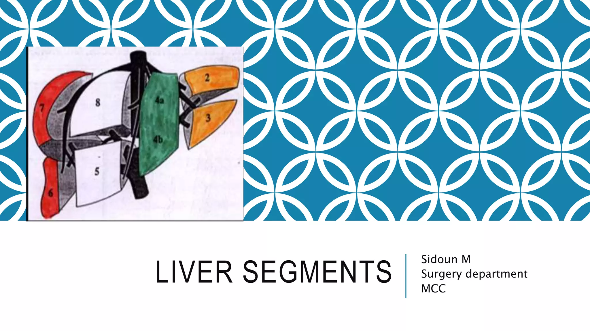
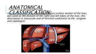
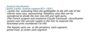
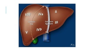
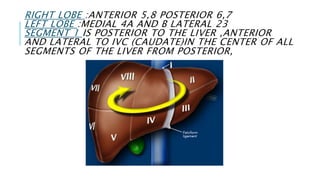
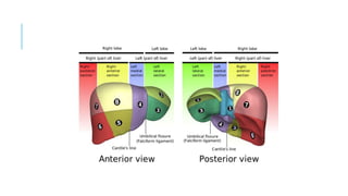
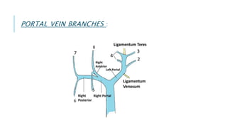
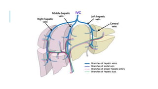
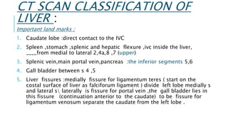
The document discusses anatomical and surgical classifications of liver segments, highlighting limitations in using the falciform ligament for division. It details the Cantlie line and Couinaud's classification, emphasizing vascular supply for segment differentiation, and explains the role of hepatic veins in segment drainage. Important landmarks for CT scan classification of the liver are also described, including the relationship of the caudate lobe, gall bladder, and liver fissures.
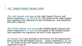


![ONFH[AVN HIP] -TRIPLE REGIME -A NOVAL SURGICAL CONCEPT .pptx](https://cdn.slidesharecdn.com/ss_thumbnails/onfhavnhip2026koaconcalicutdrgokuldevdrmashraf-260210064517-213ec005-thumbnail.jpg?width=640&height=640&fit=bounds)


