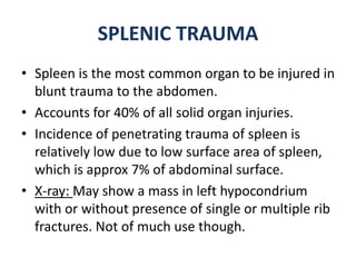
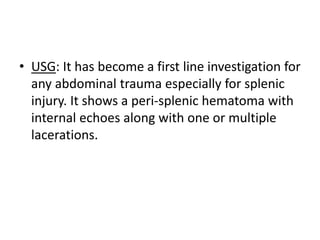
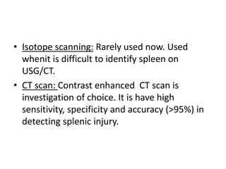
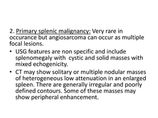
SPLEEN
-By Ankit Beniwal
ANATOMY
• Spleen is the largest organ of lymphatic system in
human body.
• Origin:The spleen develops in the cephalic part of
dorsal mesogastrium (from its left layer; during
the sixth week of intrauterine life) into a number
of nodules that fuse and form a lobulated spleen.
• Location: Lies along the axis of 10th rib.
• Size: normally varies from 7 to 12 cm.
• Formula to assess the size according to age is:
• Weight: 150 to 200grams normally
• Blood supply and venous drainage:
Arterial supply: Splenic artery which is tortous and is
the largest branch of celiac trunk. Divides into multiple
branches just before it enters the hilum of spleen.
Venous drainage: Splenic vein which originates from
splenic hilum joins superior mesentric vein to form
portal vein behind the neck of pancreas.
• Ligaments:
1. Gastro-splenic .(spleen enlarges in it’s direction)
2. Spleno-renal.
3. Spleno-phrenic.
4. Spleno-colic.
• Nerve supply: Sympathetic fibers of celiac plexus
ANATOMICAL VARIATION
1. Accessory spleen or splenunculi:
• Not uncommon and found in 10-30% of the population.
• Mostly lie along the spleen itself(most common site), tail of
pancreas, along splenic artery, at hilum of spleen or in
omental ligament.
• They may enlarge after splencetomy.
• X-ray: It may appear as mass indenting stomach.
• USG: Appears a solid mass which is isoechoic to the spleen.
• CT scan: They have density and enhancing characteristics
similar to the rest of the spleen
• Splenic scintigraphy: It is the investigation of choice to locate
normal functioning splenic tissue. (Explained further on)
Splenunculi abutting the tail of
pancreas
2. Polysplenia:
• They are multiple splenic masses seen along
normal spleen or without a normal appearing
spleen.
• They may vary from 2 to 16 in number.
• Present on patient’s left side but may be present
bilaterally.
• Associated with other anomalies too, eg:
Congenital heart disease, gut malrotation, IVC
anomalies, portal anomalies etc.
3. Splenosis:
• When a normal spleen is auto-transplanted or
moved away from it’s usual location due to
any trauma to abdomen.
4. Asplenia: Two types-
(i) Anatomical asplenia: Absence of spleen
congenitally or iatrogenic.
(ii) Functional asplenia: A spleen is seen but it is
non functional.
• Splenic fossa appears empty in anatomical
asplenia.
• In case of functional asplenia normal functional
tissue can not be traced on splenic scintigrapghy.
SPLENIC TRAUMA
• Spleen is the most common organ to be injured in
blunt trauma to the abdomen.
• Accounts for 40% of all solid organ injuries.
• Incidence of penetrating trauma of spleen is
relatively low due to low surface area of spleen,
which is approx 7% of abdominal surface.
• X-ray: May show a mass in left hypocondrium
with or without presence of single or multiple rib
fractures. Not of much use though.
• USG: It has become a first line investigation for
any abdominal trauma especially for splenic
injury. It shows a peri-splenic hematoma with
internal echoes along with one or multiple
lacerations.
• Isotope scanning: Rarely used now. Used
whenit is difficult to identify spleen on
USG/CT.
• CT scan: Contrast enhanced CT scan is
investigation of choice. It is have high
sensitivity, specificity and accuracy (>95%) in
detecting splenic injury.
CT grading of splenic rupture.
• In it splenic lacerations appears as linear low
attenuation defects that contrast well with
high attenuation vascular spleen.
• Intra splenic hematomas appear as more
diffuse hypo-attenuating region with irregular
margins with splenic swelling.
Grade IV Splenic rupture
Grade III splenic rupture
• Uniform enhancement occurs approximately 50-60
seconds after contrast administration, so imaging
during early arterial phase is useful in detection of
active extravasation and for pseudo-aneurysms.
• Thing to remember when suspecting splenic injury is
that during early phase of enhancement the cord like
geographical enhancement of pulp may look like intra-
splenic lacerations. So to rule these out a delayed
supplementary confirmatory images should be taken.
• Scanning upto pelvis should be done because
sometimes hematoma can only be seen as
hemoperitonium in pelvis.
• Angiography: It may be used in
hemodynamically stable patients with splenic
injury diagnosed on CT to detect whether
active extravasation is present or not. So that
cases who are hemodymanically stable with
active extravasation can be treated with bed
rest or embolization of splenic artery distal to
dorsal pancreatic artery.
Splenomegaly
• X-ray: May show a lump in splenic region with
raised diaphragm.
• USG: Enlarged spleen is seen extending
downwards, anteriorly and to the right,
displacing stomach forwards and medially
with left diaphragm seen pushed upwards.
• CT scan: Shows full extent of spleen.
Massive splenomegaly in Gaucher’s
disease
Splenic masses
1. Splenic abscess: They are rarely seen in
spleen. May be solitary or multiple.
• X-ray: Gas transradiancy is seen i.e. a fluid
level is seen within the splenic mass in erect
radiograph. It is rarely seen but is enough to
make a doagnosis.
• USG: Appearance shows a transition with
time. Early stage shows an ill defined mass
with decreased reflectivity, which may
develop fluid components, debris and
septations. In late stage capsule may develop
and gas within abscess will be seen as focal
areas of high reflectivity with acoustic
shadowing.
• TB also causes splenic abscesses, in miliary TB
lesions may or may not be identifiable on USG
depending on their size.
• Fungal abscess is seen in immunocompromised
patients. Usually appears as central area of
increased refelctivity which is surrounded by area
of decreased reflectivity . If necrosis occurs
centrally, the center shows decreased reflectivity
and shows ‘Wheel within wheel’ appearance.
Miliary TB
• CT scan: They appear similar to infarcts and
hematomas i.e. centrally low density lesions
(20-40 HU ) within splenic parenchyma. .
Minimal peripheral contrast enhancement
may be present when a capsule has developed
Splenic abscess CT
2. Primary splenic malignancy: Very rare in
occurance but angiosarcoma can occur as multiple
focal lesions.
• USG features are non specific and include
splenomegaly with cystic and solid masses with
mixed echogenicity.
• CT may show solitary or multiple nodular masses
of heterogeneous low attenuation in an enlarged
spleen. There are generally irregular and poorly
defined contours. Some of these masses may
show peripheral enhancement.
3. Metastases: Of all metastases 7% is seen in spleen.
Occurs genereally in presence of wide spread metastases
elsewhere. Lymphomatous involvement of spleen is
relatively common where there is splenic enlargement
without an identifiable focal abnormality.
These lesions are usually necrotic and are seen as low
density lesions on CT. They show ring enhancement on
contrast study.
On USG they most commonly appear as a hypoechoic
lesion, although can be iso- or hyperechoic.
Metastases on CT
4. Splenic cyst: Two types- (i) True cyst: Those containing cellular
lining. Eg Hydatid cyst with multiloculated internal structures,
epidermoid cyst.
(ii) False cyst: Those without cellular lining. They are formed due
to previous splenic infarction, infection or trauma.
• USG: Well defined anechoic structure with well defined
rounded margin and posterior acoustic enhancement is seen.
• CT: Typically shows a hypo attenuating relatively well defined
intrasplenic lesion. The wall is thin and has a demarcation to
splenic parenchyma. There is no rim or internal enhancement.
Wall calcification may be present.
Hydatid cyst of Spleen
Splenic infarcts
• Most common cause of infarcts is embolism from
cardio-vascular disease followed by thrombosis from
hematological diseases, eg sickle cell disease, DIC, etc
• USG: They appear as hyporeflective to areflective in
acute phase and appear wedge shaped in peripheral
location. Chronic infarcts appear as area of increased
reflectivity with atrophy of spleen due to scarring.
Multiple and repeated infarcts leads to shrunken spleen
known as Auto-splenectomy, seen in Sickle cell disease.
Splenic infarcts on USG shows no
blood supply in infarcted region.
• CT scan and Radionuclide scan: Typically seen
as localized wedge shaped defects in splenic
parenchyma.
In the chronic phase, infarcts may disappear
completely, but more commonly, they may
reveal progressive volume loss caused by fibrotic
contraction of the infarct.
Massive Splenic infarct on CT
Splenic Calcification
• Calcification in or adjacent to the spleen is common,
especially after age of 50. Calcific plaque in splenic
artery are frequent chance findings on abdominal X-
rays.
• Splenic vein calcification is uncommon but may follow
portal pyaemia or splencetomy.
• Splenic TB granulomas may produce single or multiple
well defined calcification in the parenchyma on X-ray.
• Splenic cyst may show curvilinear or oval calcification.
• A complete infarcted spleen appears as small curved
calcified structure.
SPLENIC CALCIFICATION (TB)
MRI in SPLEEN
• Anatomy and internal structures are well shown on CT
and USG, but MRI is useful in minority of cases.
(i) Portal hypertension: spleen is enlarged with no
change in it’s signal intensity apart from the
occasional findings of siderotic nodules (Gamna-
Gandy bodies) which produce focal areas of low
intensity signals.
(ii) In Transfusion haemosiderosis, the spleen shows
generalized loss of signal in both T1 and T2.
(iii) Cysts: unifirm low T1 signal, high T2 signals and no
enhancement with gadolinium.
• (iv) Haemangioma: Clear well defined margins
with lobulated shape. Uniform low T1 signal and
uniform T2 signal. Dynamic imaging after
gadolinium shows dense peripheral
discontinuous nodular blush, which begins during
arterial phase of liver perfusion and continues
centripetally for several minutes, so that images
taken after 10 minutes often show diffuse
hyperintensity of the lesion. Large haemangioma
may show unenhanced central core even on
delayed imaging.
• (v) Abscess: Rim enhancement on gadolinium
is seen in abscess larger than 1.5cm in size.
But it is also a feature of metastases with
necrotic centers.
Splenic Scintigraphy
• Very rarely used or needed now a days.
• Physiology: Reticluoendothelial cells of spleen takes up
intravenously injected colloids (99mTc) of size 30-
1000nm. In normal cases about 15% of the injected
colloidal acitivity reaches spleen, but in patients with
cirrhosis or hypersplenism a much greater propoprtion
of injected particles will reach spleen and will be
trapped there due to increased blood to the spleen.
• A more specific imaging is done by injecting autologus
RBC’s which have been incubated at 50° C and labelled
with technetium 99. They are called Heat damaged
RBCs.
• It is first choice of technique when localization
of site of functioning splenic tissue in the
absence of a normal spleen on CT or USG, i.e.
in cases of polysplenia/asplenia syndrome.
• Autologous platelets labelled with indium-111
is used to demonstrate rate of clearance of
circulating platelets in patients with Idiopathic
thrombocytopenic purpura, who may be
benefited with splenectomy.
Portal Venography
• Methods: (i) Late phase superior mesentric
angiography.
(ii) Trans-splenic approach. (Discussed below)
(iii) Paraumbilical vein catheterization.
(iv) Transjugular transhepatic approach.
• Indications: (i) To demonstrate prior to operation
the anatomy of the portal system in patients with
portal hypertension.
(ii) To check the patency of a portosystemic
anastomosis.
• Technique for Trans-splenic approach:
 With patient in supine, spleen is identified on USG. Access point must be as low as
possible in the mid axillary line, usually at the level of 10th or 11th intercostal space.
 The region is anaesthetized.
 Patient is asked to hold the breath in mid-inspiration and the needle is then
inserted inwards and upwards into the spleen. Blood will flow back if needle is in
right place. The patient is then asked to breath as shallowly as possible to avoid
trauma to the spleen.
 The small test dose of contrast media (LOCM) can be injected to ensure correct
siting of the cannula.
 When cannula is in right position the splenic pulp pressure can be measured with
manometer (10-15 cm H₂O)
 A hand injection of 50ml of contrast medium is made over 5seconds and recorded
by rapid serial radiography/digital subtraction angiography. Cannula should be
removed as soon as possible after injection to minimize trauma to spleen.
 Occasionaly portal vein will fail to opacify , owing to major portosystemic
collaterals causing reversed floe in portal vein. The final arbiter of portal vein
patency is direct mesentric venography performed at operation. The maximum
width of normal portal vein is said to be 2cm.
THANK YOU

Spleen Radiology

  • 1.
  • 2.
    ANATOMY • Spleen isthe largest organ of lymphatic system in human body. • Origin:The spleen develops in the cephalic part of dorsal mesogastrium (from its left layer; during the sixth week of intrauterine life) into a number of nodules that fuse and form a lobulated spleen. • Location: Lies along the axis of 10th rib. • Size: normally varies from 7 to 12 cm. • Formula to assess the size according to age is: • Weight: 150 to 200grams normally
  • 3.
    • Blood supplyand venous drainage: Arterial supply: Splenic artery which is tortous and is the largest branch of celiac trunk. Divides into multiple branches just before it enters the hilum of spleen. Venous drainage: Splenic vein which originates from splenic hilum joins superior mesentric vein to form portal vein behind the neck of pancreas. • Ligaments: 1. Gastro-splenic .(spleen enlarges in it’s direction) 2. Spleno-renal. 3. Spleno-phrenic. 4. Spleno-colic. • Nerve supply: Sympathetic fibers of celiac plexus
  • 5.
    ANATOMICAL VARIATION 1. Accessoryspleen or splenunculi: • Not uncommon and found in 10-30% of the population. • Mostly lie along the spleen itself(most common site), tail of pancreas, along splenic artery, at hilum of spleen or in omental ligament. • They may enlarge after splencetomy. • X-ray: It may appear as mass indenting stomach. • USG: Appears a solid mass which is isoechoic to the spleen. • CT scan: They have density and enhancing characteristics similar to the rest of the spleen • Splenic scintigraphy: It is the investigation of choice to locate normal functioning splenic tissue. (Explained further on)
  • 6.
    Splenunculi abutting thetail of pancreas
  • 7.
    2. Polysplenia: • Theyare multiple splenic masses seen along normal spleen or without a normal appearing spleen. • They may vary from 2 to 16 in number. • Present on patient’s left side but may be present bilaterally. • Associated with other anomalies too, eg: Congenital heart disease, gut malrotation, IVC anomalies, portal anomalies etc.
  • 9.
    3. Splenosis: • Whena normal spleen is auto-transplanted or moved away from it’s usual location due to any trauma to abdomen.
  • 11.
    4. Asplenia: Twotypes- (i) Anatomical asplenia: Absence of spleen congenitally or iatrogenic. (ii) Functional asplenia: A spleen is seen but it is non functional. • Splenic fossa appears empty in anatomical asplenia. • In case of functional asplenia normal functional tissue can not be traced on splenic scintigrapghy.
  • 12.
    SPLENIC TRAUMA • Spleenis the most common organ to be injured in blunt trauma to the abdomen. • Accounts for 40% of all solid organ injuries. • Incidence of penetrating trauma of spleen is relatively low due to low surface area of spleen, which is approx 7% of abdominal surface. • X-ray: May show a mass in left hypocondrium with or without presence of single or multiple rib fractures. Not of much use though.
  • 13.
    • USG: Ithas become a first line investigation for any abdominal trauma especially for splenic injury. It shows a peri-splenic hematoma with internal echoes along with one or multiple lacerations.
  • 14.
    • Isotope scanning:Rarely used now. Used whenit is difficult to identify spleen on USG/CT. • CT scan: Contrast enhanced CT scan is investigation of choice. It is have high sensitivity, specificity and accuracy (>95%) in detecting splenic injury.
  • 15.
    CT grading ofsplenic rupture.
  • 16.
    • In itsplenic lacerations appears as linear low attenuation defects that contrast well with high attenuation vascular spleen. • Intra splenic hematomas appear as more diffuse hypo-attenuating region with irregular margins with splenic swelling.
  • 17.
  • 18.
  • 19.
    • Uniform enhancementoccurs approximately 50-60 seconds after contrast administration, so imaging during early arterial phase is useful in detection of active extravasation and for pseudo-aneurysms. • Thing to remember when suspecting splenic injury is that during early phase of enhancement the cord like geographical enhancement of pulp may look like intra- splenic lacerations. So to rule these out a delayed supplementary confirmatory images should be taken. • Scanning upto pelvis should be done because sometimes hematoma can only be seen as hemoperitonium in pelvis.
  • 20.
    • Angiography: Itmay be used in hemodynamically stable patients with splenic injury diagnosed on CT to detect whether active extravasation is present or not. So that cases who are hemodymanically stable with active extravasation can be treated with bed rest or embolization of splenic artery distal to dorsal pancreatic artery.
  • 22.
  • 23.
    • X-ray: Mayshow a lump in splenic region with raised diaphragm. • USG: Enlarged spleen is seen extending downwards, anteriorly and to the right, displacing stomach forwards and medially with left diaphragm seen pushed upwards. • CT scan: Shows full extent of spleen.
  • 24.
    Massive splenomegaly inGaucher’s disease
  • 25.
    Splenic masses 1. Splenicabscess: They are rarely seen in spleen. May be solitary or multiple. • X-ray: Gas transradiancy is seen i.e. a fluid level is seen within the splenic mass in erect radiograph. It is rarely seen but is enough to make a doagnosis.
  • 26.
    • USG: Appearanceshows a transition with time. Early stage shows an ill defined mass with decreased reflectivity, which may develop fluid components, debris and septations. In late stage capsule may develop and gas within abscess will be seen as focal areas of high reflectivity with acoustic shadowing.
  • 28.
    • TB alsocauses splenic abscesses, in miliary TB lesions may or may not be identifiable on USG depending on their size. • Fungal abscess is seen in immunocompromised patients. Usually appears as central area of increased refelctivity which is surrounded by area of decreased reflectivity . If necrosis occurs centrally, the center shows decreased reflectivity and shows ‘Wheel within wheel’ appearance.
  • 29.
  • 30.
    • CT scan:They appear similar to infarcts and hematomas i.e. centrally low density lesions (20-40 HU ) within splenic parenchyma. . Minimal peripheral contrast enhancement may be present when a capsule has developed
  • 31.
  • 32.
    2. Primary splenicmalignancy: Very rare in occurance but angiosarcoma can occur as multiple focal lesions. • USG features are non specific and include splenomegaly with cystic and solid masses with mixed echogenicity. • CT may show solitary or multiple nodular masses of heterogeneous low attenuation in an enlarged spleen. There are generally irregular and poorly defined contours. Some of these masses may show peripheral enhancement.
  • 33.
    3. Metastases: Ofall metastases 7% is seen in spleen. Occurs genereally in presence of wide spread metastases elsewhere. Lymphomatous involvement of spleen is relatively common where there is splenic enlargement without an identifiable focal abnormality. These lesions are usually necrotic and are seen as low density lesions on CT. They show ring enhancement on contrast study. On USG they most commonly appear as a hypoechoic lesion, although can be iso- or hyperechoic.
  • 34.
  • 35.
    4. Splenic cyst:Two types- (i) True cyst: Those containing cellular lining. Eg Hydatid cyst with multiloculated internal structures, epidermoid cyst. (ii) False cyst: Those without cellular lining. They are formed due to previous splenic infarction, infection or trauma. • USG: Well defined anechoic structure with well defined rounded margin and posterior acoustic enhancement is seen. • CT: Typically shows a hypo attenuating relatively well defined intrasplenic lesion. The wall is thin and has a demarcation to splenic parenchyma. There is no rim or internal enhancement. Wall calcification may be present.
  • 36.
  • 37.
    Splenic infarcts • Mostcommon cause of infarcts is embolism from cardio-vascular disease followed by thrombosis from hematological diseases, eg sickle cell disease, DIC, etc • USG: They appear as hyporeflective to areflective in acute phase and appear wedge shaped in peripheral location. Chronic infarcts appear as area of increased reflectivity with atrophy of spleen due to scarring. Multiple and repeated infarcts leads to shrunken spleen known as Auto-splenectomy, seen in Sickle cell disease.
  • 38.
    Splenic infarcts onUSG shows no blood supply in infarcted region.
  • 39.
    • CT scanand Radionuclide scan: Typically seen as localized wedge shaped defects in splenic parenchyma. In the chronic phase, infarcts may disappear completely, but more commonly, they may reveal progressive volume loss caused by fibrotic contraction of the infarct.
  • 40.
  • 41.
    Splenic Calcification • Calcificationin or adjacent to the spleen is common, especially after age of 50. Calcific plaque in splenic artery are frequent chance findings on abdominal X- rays. • Splenic vein calcification is uncommon but may follow portal pyaemia or splencetomy. • Splenic TB granulomas may produce single or multiple well defined calcification in the parenchyma on X-ray. • Splenic cyst may show curvilinear or oval calcification. • A complete infarcted spleen appears as small curved calcified structure.
  • 42.
  • 43.
    MRI in SPLEEN •Anatomy and internal structures are well shown on CT and USG, but MRI is useful in minority of cases. (i) Portal hypertension: spleen is enlarged with no change in it’s signal intensity apart from the occasional findings of siderotic nodules (Gamna- Gandy bodies) which produce focal areas of low intensity signals. (ii) In Transfusion haemosiderosis, the spleen shows generalized loss of signal in both T1 and T2. (iii) Cysts: unifirm low T1 signal, high T2 signals and no enhancement with gadolinium.
  • 44.
    • (iv) Haemangioma:Clear well defined margins with lobulated shape. Uniform low T1 signal and uniform T2 signal. Dynamic imaging after gadolinium shows dense peripheral discontinuous nodular blush, which begins during arterial phase of liver perfusion and continues centripetally for several minutes, so that images taken after 10 minutes often show diffuse hyperintensity of the lesion. Large haemangioma may show unenhanced central core even on delayed imaging.
  • 45.
    • (v) Abscess:Rim enhancement on gadolinium is seen in abscess larger than 1.5cm in size. But it is also a feature of metastases with necrotic centers.
  • 46.
    Splenic Scintigraphy • Veryrarely used or needed now a days. • Physiology: Reticluoendothelial cells of spleen takes up intravenously injected colloids (99mTc) of size 30- 1000nm. In normal cases about 15% of the injected colloidal acitivity reaches spleen, but in patients with cirrhosis or hypersplenism a much greater propoprtion of injected particles will reach spleen and will be trapped there due to increased blood to the spleen. • A more specific imaging is done by injecting autologus RBC’s which have been incubated at 50° C and labelled with technetium 99. They are called Heat damaged RBCs.
  • 47.
    • It isfirst choice of technique when localization of site of functioning splenic tissue in the absence of a normal spleen on CT or USG, i.e. in cases of polysplenia/asplenia syndrome. • Autologous platelets labelled with indium-111 is used to demonstrate rate of clearance of circulating platelets in patients with Idiopathic thrombocytopenic purpura, who may be benefited with splenectomy.
  • 48.
    Portal Venography • Methods:(i) Late phase superior mesentric angiography. (ii) Trans-splenic approach. (Discussed below) (iii) Paraumbilical vein catheterization. (iv) Transjugular transhepatic approach. • Indications: (i) To demonstrate prior to operation the anatomy of the portal system in patients with portal hypertension. (ii) To check the patency of a portosystemic anastomosis.
  • 49.
    • Technique forTrans-splenic approach:  With patient in supine, spleen is identified on USG. Access point must be as low as possible in the mid axillary line, usually at the level of 10th or 11th intercostal space.  The region is anaesthetized.  Patient is asked to hold the breath in mid-inspiration and the needle is then inserted inwards and upwards into the spleen. Blood will flow back if needle is in right place. The patient is then asked to breath as shallowly as possible to avoid trauma to the spleen.  The small test dose of contrast media (LOCM) can be injected to ensure correct siting of the cannula.  When cannula is in right position the splenic pulp pressure can be measured with manometer (10-15 cm H₂O)  A hand injection of 50ml of contrast medium is made over 5seconds and recorded by rapid serial radiography/digital subtraction angiography. Cannula should be removed as soon as possible after injection to minimize trauma to spleen.  Occasionaly portal vein will fail to opacify , owing to major portosystemic collaterals causing reversed floe in portal vein. The final arbiter of portal vein patency is direct mesentric venography performed at operation. The maximum width of normal portal vein is said to be 2cm.
  • 50.