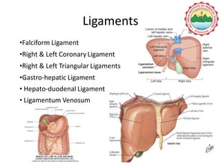
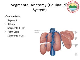
SURGICAL ANATOMY OF THE
LIVER
Dr Ajay Kumar Dhiman
Location of liver
Surfaces of Liver
• Antero-superior surface
• Posterior surface
• Inferior or Visceral surface
Ligaments
•Falciform Ligament
•Right & Left Coronary Ligament
•Right & Left Triangular Ligaments
•Gastro-hepatic Ligament
• Hepato-duodenal Ligament
• Ligamentum Venosum
Lobar Anatomy(American System)
• Right and left lobe determined by Cantlie’s line (portal fissure)
• Cantlie's line runs from the middle of the gallbladder
fossa anteriorly to the inferior vena cava posteriorly
• The MHV lies in the Cantlie’s Line
Segmental Anatomy (Couinaud’s
System)
•Caudate Lobe
Segment I
•Left Lobe
Segments II – IV
• Right Lobe
Segments V-VIII
Bismuth's classification
• Similar to the Couinaud classification
• According to Bismuth 3 hepatic veins divide the liver
into 4 sectors, further divided into segments.
• These sectors are termed portal sectors as each is
supplied by a portal pedicle in the centre.
• The left portal scissura divides the left liver into two sectors:
anterior and posterior. Left anterior sector consists of two
segments: segment IV, which is the quadrate lobe and
segment III, which is anterior part of anatomical left lobe.
These two segments are separated by the left hepatic fissure
or umbilical fissure.
• Left posterior sector consists of only one segment II. It is the
posterior part of left lobe.
Schematic Depiction of the Segemental
Anatomy
Main portal fissure
• Extends from the tip of the GB back to the midpoint of IVC and
contains the middle (main) hepatic vein.
• Separates liver into right and left hemi-livers.
• Segments V and VIII lie to the right and segment IV to the left
of the fissure
Left portal fissure
• Divides left hemi-liver into medial (anterior) and lateral
(posterior) Sectors
• Extends from the mid point of the anterior edge of the liver
between falciform ligament and left triangular ligament to the
point, which marks the confluence of the left and middle
hepatic veins.
• Contains left hepatic vein
• Separates the left anterior and left posterior sectors:
segment III lies anteriorly and segment II posteriorly
Right portal fissure
• Divides right hemi-liver into lateral (posterior) and medial
(anterior) sectors.
• Plane of right fissure is the most variable amongst the main
fissures
• Contains right hepatic vein
• Right fissure marks the thickest point of liver
parenchyma, which is commonly transected during liver
resection.
• Traditionally referred to as segment 1, the caudate lobe has been
divided into 2 segments: 1 and 9.
• Segment 1 refers to the Spiegel lobe, and segment 9 refers to the
paracaval portion and caudate process
• Reidel lobe refers to a ‘‘tongue-like projection of the anterior border
of the right lobe of the liver to the right of the gallbladder’’ below the
costal margin. It is not a true anatomic lobe, but is a normal variant
of segments 5 and 6
Brisbane Classification (2000)
• Liver anatomy is described in first-, second- and third-
order divisions.
• Terminology is based on hepatic artery bile duct
• eight functional zones or segments of the liver.
• As seen from an anterior view, the eight segments are
numbered in a clockwise direction as described by
Couinaud.
• Starting with the proper hepatic artery, the first division is the
right and left hepatic arteries supplying the right and left
hemi-livers or livers, respectively,
• The plane between the two distinct zones of blood supply
extends from the gallbladder bed antero-inferiorly to the
right side of the IVC postero-superiorly and is called the mid-
plane of the liver or Cantlie’s line
• The right and left hepatic arteries then divide into four
second- order sectional arteries each supplying a section of
the liver.
• On the right side, the two sectional arteries supply the right
anterior section and the right posterior section, and the plane
between these sections is the right intersectional plane.
• On the left, the two sectional arteries supply the left
medial section and left lateral section.
• The plane between these sections is the left intersectional
plane and is marked by the round ligament at the umbilical
fissure and the line of attachment of the falciform ligament
to the anterior surface of the liver.
Problems with Brisbane
• Does not address the nomenclature of resections that donot
encompass a complete segment, sometimes referred to as
wedge resections or partial segmentectomies.
• Does not define gray zones between resections; that is, is a
right hepatectomy that includes only a portion of segment 4
classified as a right hepatectomy or a righttrisectionectomy?
Radiological Correlation
How to separate liver segments
on cross sectional imaging
Left lobe: lateral(II/III) vs
medial segment (IVA/B)
Extrapolate a line along the falciform
ligament superiorly to the confluence
of the left and middle hepatic veins at
the IVC (blue line).
Left vs Right lobe: IVA/B vs
V/VIII
Extrapolate a line from the
gallbladder fossa superiorly along the
middle hepatic vein to the IVC (red
line).
Right lobe: anterior (V/VIII) vs
posterior segment (VI/VII)
Extrapolate a line along the right
hepatic vein from the IVC inferiorly to
the lateral liver margin (green line).
Thank you

Ajay liver ppt

  • 1.
    SURGICAL ANATOMY OFTHE LIVER Dr Ajay Kumar Dhiman
  • 2.
  • 3.
    Surfaces of Liver •Antero-superior surface • Posterior surface • Inferior or Visceral surface
  • 4.
    Ligaments •Falciform Ligament •Right &Left Coronary Ligament •Right & Left Triangular Ligaments •Gastro-hepatic Ligament • Hepato-duodenal Ligament • Ligamentum Venosum
  • 5.
    Lobar Anatomy(American System) •Right and left lobe determined by Cantlie’s line (portal fissure) • Cantlie's line runs from the middle of the gallbladder fossa anteriorly to the inferior vena cava posteriorly • The MHV lies in the Cantlie’s Line
  • 7.
    Segmental Anatomy (Couinaud’s System) •CaudateLobe Segment I •Left Lobe Segments II – IV • Right Lobe Segments V-VIII
  • 8.
    Bismuth's classification • Similarto the Couinaud classification • According to Bismuth 3 hepatic veins divide the liver into 4 sectors, further divided into segments. • These sectors are termed portal sectors as each is supplied by a portal pedicle in the centre.
  • 9.
    • The leftportal scissura divides the left liver into two sectors: anterior and posterior. Left anterior sector consists of two segments: segment IV, which is the quadrate lobe and segment III, which is anterior part of anatomical left lobe. These two segments are separated by the left hepatic fissure or umbilical fissure. • Left posterior sector consists of only one segment II. It is the posterior part of left lobe.
  • 11.
    Schematic Depiction ofthe Segemental Anatomy
  • 12.
    Main portal fissure •Extends from the tip of the GB back to the midpoint of IVC and contains the middle (main) hepatic vein. • Separates liver into right and left hemi-livers. • Segments V and VIII lie to the right and segment IV to the left of the fissure
  • 13.
    Left portal fissure •Divides left hemi-liver into medial (anterior) and lateral (posterior) Sectors • Extends from the mid point of the anterior edge of the liver between falciform ligament and left triangular ligament to the point, which marks the confluence of the left and middle hepatic veins. • Contains left hepatic vein • Separates the left anterior and left posterior sectors: segment III lies anteriorly and segment II posteriorly
  • 14.
    Right portal fissure •Divides right hemi-liver into lateral (posterior) and medial (anterior) sectors. • Plane of right fissure is the most variable amongst the main fissures • Contains right hepatic vein • Right fissure marks the thickest point of liver parenchyma, which is commonly transected during liver resection.
  • 15.
    • Traditionally referredto as segment 1, the caudate lobe has been divided into 2 segments: 1 and 9. • Segment 1 refers to the Spiegel lobe, and segment 9 refers to the paracaval portion and caudate process • Reidel lobe refers to a ‘‘tongue-like projection of the anterior border of the right lobe of the liver to the right of the gallbladder’’ below the costal margin. It is not a true anatomic lobe, but is a normal variant of segments 5 and 6
  • 16.
    Brisbane Classification (2000) •Liver anatomy is described in first-, second- and third- order divisions. • Terminology is based on hepatic artery bile duct • eight functional zones or segments of the liver. • As seen from an anterior view, the eight segments are numbered in a clockwise direction as described by Couinaud.
  • 17.
    • Starting withthe proper hepatic artery, the first division is the right and left hepatic arteries supplying the right and left hemi-livers or livers, respectively, • The plane between the two distinct zones of blood supply extends from the gallbladder bed antero-inferiorly to the right side of the IVC postero-superiorly and is called the mid- plane of the liver or Cantlie’s line
  • 18.
    • The rightand left hepatic arteries then divide into four second- order sectional arteries each supplying a section of the liver. • On the right side, the two sectional arteries supply the right anterior section and the right posterior section, and the plane between these sections is the right intersectional plane.
  • 19.
    • On theleft, the two sectional arteries supply the left medial section and left lateral section. • The plane between these sections is the left intersectional plane and is marked by the round ligament at the umbilical fissure and the line of attachment of the falciform ligament to the anterior surface of the liver.
  • 25.
    Problems with Brisbane •Does not address the nomenclature of resections that donot encompass a complete segment, sometimes referred to as wedge resections or partial segmentectomies. • Does not define gray zones between resections; that is, is a right hepatectomy that includes only a portion of segment 4 classified as a right hepatectomy or a righttrisectionectomy?
  • 26.
  • 35.
    How to separateliver segments on cross sectional imaging Left lobe: lateral(II/III) vs medial segment (IVA/B) Extrapolate a line along the falciform ligament superiorly to the confluence of the left and middle hepatic veins at the IVC (blue line). Left vs Right lobe: IVA/B vs V/VIII Extrapolate a line from the gallbladder fossa superiorly along the middle hepatic vein to the IVC (red line). Right lobe: anterior (V/VIII) vs posterior segment (VI/VII) Extrapolate a line along the right hepatic vein from the IVC inferiorly to the lateral liver margin (green line).
  • 43.