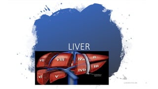
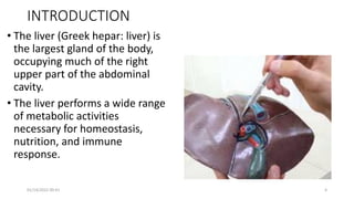
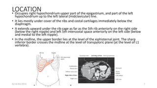
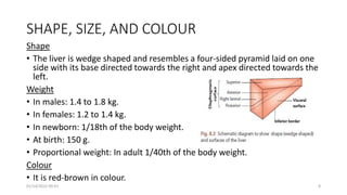
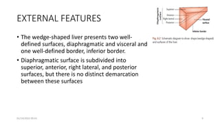
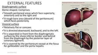
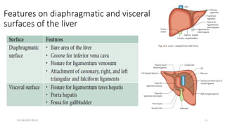
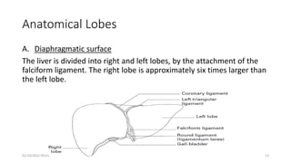
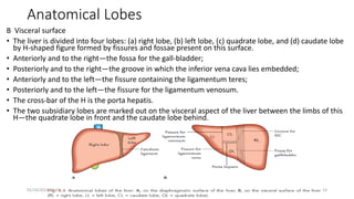
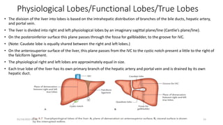
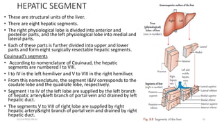
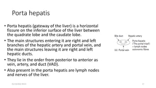
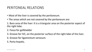
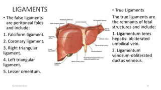
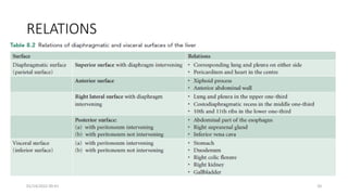
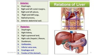
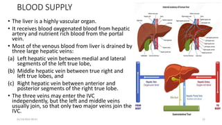
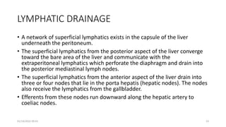
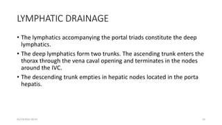
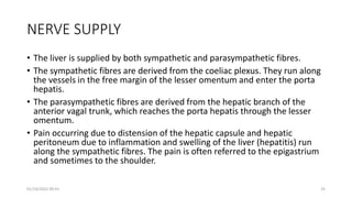
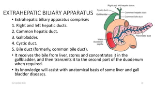
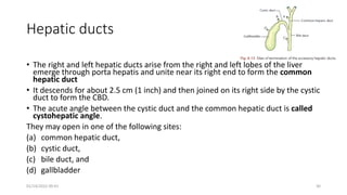
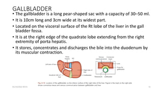
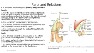
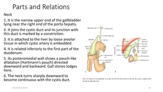
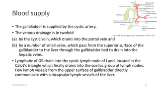
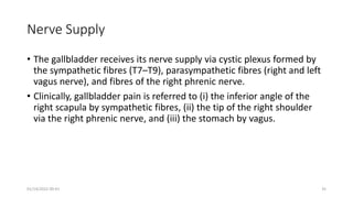
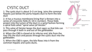
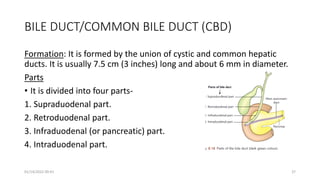
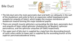
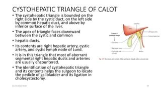
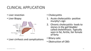
Liver and Extrahepatic
Biliary Apparatus
300 level
MBBS/BDS
OBJECTIVES
• At the end of this lecture, students should be able to
1. Describe the function, location, features and neurovascular supply
of the liver.
2. Write short notes on the classification of the lobes of the liver.
3. Describe hepatic segment and their clinical significance.
4. Write short notes on the portal hepatis.
5. Describe the anatomy of the gall bladder
6. Write short notes on cystic duct, common bile duct and
cystohepatic triangle of Calot
01/14/2022 09:41 2
LIVER
1/14/2022 9:41 AM 3
INTRODUCTION
• The liver (Greek hepar: liver) is
the largest gland of the body,
occupying much of the right
upper part of the abdominal
cavity.
• The liver performs a wide range
of metabolic activities
necessary for homeostasis,
nutrition, and immune
response.
01/14/2022 09:41 4
Functions
i. The liver is a seat of metabolism of carbohydrates, proteins
and fats.
ii. Most serum proteins except the immunoglobulins are
synthesized in liver. Decrease in albumin level in liver disease
may be a cause of edema and ascites.
iii. Blood clotting depends on production of prothrombin and
fibrinogen by the liver. Liver failure results in defective clotting
leading to bleeding tendencies.
iv. Liver is a storage site for glycogen, triglycerides, iron,
copper and fat- soluble vitamins.
01/14/2022 09:41 5
Functions
v. Hepatocytes secrete bile, which is needed for emulsification of fats.
Lack of bile affects digestion of fatty food leading to excretion of fat-
filled light stools (steatorrhea).
vi. Amongst the catabolic functions of the liver, deamination of amino
acids is important. Thus, liver is the site of urea formation. It also
detoxifies hormones and drugs.
vii. Reticuloendothelial cells (Kupffer cells) of the liver kill the
pathogens and other particulate matters reaching the liver through
portal vein from the intestinal tract and from the hepatic artery.
viii. Maintenance of body temperature is also attributed to liver
because it generates lot of heat during the various metabolic reactions.
01/14/2022 09:41 6
LOCATION
• Occupies right hypochondrium upper part of the epigastrium, and part of the left
hypochondrium up to the left lateral (midclavicular) line.
• It lies mostly under cover of the ribs and costal cartilages immediately below the
diaphragm.
• It extends upward under the rib cage as far as the 5th rib anteriorly on the right side
(below the right nipple) and left 5th intercostal space anteriorly on the left side (below
and medial to the left nipple).
• In the midline, the upper border lies at the level of the xiphisternal joint. The sharp
inferior border crosses the midline at the level of transpyloric plane (at the level of L1
vertebra).
01/14/2022 09:41 7
SHAPE, SIZE, AND COLOUR
Shape
• The liver is wedge shaped and resembles a four-sided pyramid laid on one
side with its base directed towards the right and apex directed towards the
left.
Weight
• In males: 1.4 to 1.8 kg.
• In females: 1.2 to 1.4 kg.
• In newborn: 1/18th of the body weight.
• At birth: 150 g.
• Proportional weight: In adult 1/40th of the body weight.
Colour
• It is red-brown in colour.
01/14/2022 09:41 8
EXTERNAL FEATURES
• The wedge-shaped liver presents two well-
defined surfaces, diaphragmatic and visceral and
one well-defined border, inferior border.
• Diaphragmatic surface is subdivided into
superior, anterior, right lateral, and posterior
surfaces, but there is no distinct demarcation
between these surfaces
01/14/2022 09:41 9
EXTERNAL FEATURES
Diaphragmatic surface
Dome-shaped. Comprises
✓Smooth peritoneal areas which face superiorly,
anteriorly and to the right and
✓A rough bare area (devoid of the peritoneum)
which faces posteriorly.
Visceral surface
✓Relatively flat or concave.
✓It is directed downward, backward, and to the left.
✓It is separated in front from the diaphragmatic
surface by the sharp inferior border and behind
from the diaphragm by the posterior layer of
coronary ligament.
✓It is covered by the peritoneum except at the fossa
for gallbladder and the porta hepatis.
01/14/2022 09:41 10
Features on diaphragmatic and visceral
surfaces of the liver
01/14/2022 09:41 11
LOBES OF THE LIVER
• The lobes of the liver are classified into two types:
(a) anatomical lobes and
(b) physiological (functional) lobes.
01/14/2022 09:41 12
Anatomical Lobes
A. Diaphragmatic surface
The liver is divided into right and left lobes, by the attachment of the
falciform ligament. The right lobe is approximately six times larger than
the left lobe.
01/14/2022 09:41 13
Anatomical Lobes
B Visceral surface
• The liver is divided into four lobes: (a) right lobe, (b) left lobe, (c) quadrate lobe, and (d) caudate lobe
by H-shaped figure formed by fissures and fossae present on this surface.
• Anteriorly and to the right—the fossa for the gall-bladder;
• Posteriorly and to the right—the groove in which the inferior vena cava lies embedded;
• Anteriorly and to the left—the fissure containing the ligamentum teres;
• Posteriorly and to the left—the fissure for the ligamentum venosum.
• The cross-bar of the H is the porta hepatis.
• The two subsidiary lobes are marked out on the visceral aspect of the liver between the limbs of this
H—the quadrate lobe in front and the caudate lobe behind.
01/14/2022 09:41 14
Physiological Lobes/Functional Lobes/True Lobes
• The division of the liver into lobes is based on the intrahepatic distribution of branches of the bile ducts, hepatic artery,
and portal vein.
• The liver is divided into right and left physiological lobes by an imaginary sagittal plane/line (Cantlie’s plane/line).
• On the posteroinferior surface this plane passes through the fossa for gallbladder, to the groove for IVC.
• (Note: Caudate lobe is equally shared between the right and left lobes.)
• On the anterosuperior surface of the liver, this plane passes from the IVC to the cystic notch present a little to the right of
the falciform ligament.
• The physiological right and left lobes are approximately equal in size.
• Each true lobe of the liver has its own primary branch of the hepatic artery and portal vein and is drained by its own
hepatic duct.
01/14/2022 09:41 15
HEPATIC SEGMENT
• These are structural units of the liver.
• There are eight hepatic segments.
• The right physiological lobe is divided into anterior and
posterior parts, and the left physiological lobe into medial and
lateral parts.
• Each of these parts is further divided into upper and lower
parts and form eight surgically resectable hepatic segments.
Couinaud’s segments
• According to nomenclature of Couinaud, the hepatic
segments are numbered I to VIII.
• I to IV in the left hemiliver and V to VIII in the right hemiliver.
• From this nomenclature, the segment I&IV corresponds to the
caudate lobe and the quadrate lobe, respectively.
• Segment I to IV of the left lobe are supplied by the left branch
of hepatic artery&left branch of portal vein and drained by left
hepatic duct.
• The segments V to VIII of right lobe are supplied by right
hepatic artery&right branch of portal vein and drained by right
hepatic duct.
01/14/2022 09:41 16
Porta hepatis
• Porta hepatis (gateway of the liver) is a horizontal
fissure on the inferior surface of the liver between
the quadrate lobe and the caudate lobe.
• The main structures entering it are right and left
branches of the hepatic artery and portal vein, and
the main structures leaving it are right and left
hepatic ducts.
• They lie in the order from posterior to anterior as
vein, artery, and duct (VAD).
• Also present in the porta hepatis are lymph nodes
and nerves of the liver.
01/14/2022 09:41 17
PERITONEAL RELATIONS
• Most of the liver is covered by the peritoneum.
• The areas which are not covered by the peritoneum are:
1. Bare area of the liver: It is a triangular area on the posterior aspect of
the right lobe.
2. Fossa for gallbladder
3. Groove for IVC, on the posterior surface of the right lobe of the liver.
4. Groove for ligamentum venosum.
5. Porta hepatis.
01/14/2022 09:41 18
LIGAMENTS
• The false ligaments
are peritoneal folds
and include:
1. Falciform ligament.
2. Coronary ligament.
3. Right triangular
ligament.
4. Left triangular
ligament.
5. Lesser omentum.
• True Ligaments
The true ligaments are
the remnants of fetal
structures and include:
1. Ligamentum teres
hepatis- obliterated
umbilical vein.
2. Ligamentum
venosum-obliterated
ductus venosus.
01/14/2022 09:41 19
RELATIONS
01/14/2022 09:41 20
01/14/2022 09:41 21
BLOOD SUPPLY
• The liver is a highly vascular organ.
• It receives blood oxygenated blood from hepatic
artery and nutrient rich blood from the portal
vein.
• Most of the venous blood from liver is drained by
three large hepatic veins:
(a) Left hepatic vein between medial and lateral
segments of the left true lobe,
(b) Middle hepatic vein between true right and
left true lobes, and
(c) Right hepatic vein between anterior and
posterior segments of the right true lobe.
• The three veins may enter the IVC
independently, but the left and middle veins
usually join, so that only two major veins join the
IVC.
01/14/2022 09:41 22
LYMPHATIC DRAINAGE
• A network of superficial lymphatics exists in the capsule of the liver
underneath the peritoneum.
• The superficial lymphatics from the posterior aspect of the liver converge
toward the bare area of the liver and communicate with the
extraperitoneal lymphatics which perforate the diaphragm and drain into
the posterior mediastinal lymph nodes.
• The superficial lymphatics from the anterior aspect of the liver drain into
three or four nodes that lie in the porta hepatis (hepatic nodes). The nodes
also receive the lymphatics from the gallbladder.
• Efferents from these nodes run downward along the hepatic artery to
coeliac nodes.
01/14/2022 09:41 23
LYMPHATIC DRAINAGE
• The lymphatics accompanying the portal triads constitute the deep
lymphatics.
• The deep lymphatics form two trunks. The ascending trunk enters the
thorax through the vena caval opening and terminates in the nodes
around the IVC.
• The descending trunk empties in hepatic nodes located in the porta
hepatis.
01/14/2022 09:41 24
NERVE SUPPLY
• The liver is supplied by both sympathetic and parasympathetic fibres.
• The sympathetic fibres are derived from the coeliac plexus. They run along
the vessels in the free margin of the lesser omentum and enter the porta
hepatis.
• The parasympathetic fibres are derived from the hepatic branch of the
anterior vagal trunk, which reaches the porta hepatis through the lesser
omentum.
• Pain occurring due to distension of the hepatic capsule and hepatic
peritoneum due to inflammation and swelling of the liver (hepatitis) run
along the sympathetic fibres. The pain is often referred to the epigastrium
and sometimes to the shoulder.
01/14/2022 09:41 25
FACTORS KEEPING THE LIVER IN POSITION
1. Hepatic veins connecting the liver to the IVC.
2. Intra-abdominal pressure maintained by the tone of abdominal
muscles.
3. Peritoneal ligaments connecting the liver to the abdominal walls.
01/14/2022 09:41 26
1/14/2022 9:41 AM 27
EXTRAHEPATIC
BILIARY APPARATUS
1/14/2022 9:41 AM 28
EXTRAHEPATIC BILIARY APPARATUS
• Extrahepatic biliary apparatus comprises
1. Right and left hepatic ducts.
2. Common hepatic duct.
3. Gallbladder.
4. Cystic duct.
5. Bile duct (formerly, common bile duct).
• It receives the bile from liver, stores and concentrates it in the
gallbladder, and then transmits it to the second part of the duodenum
when required.
• Its knowledge will assist with anatomical basis of some liver and gall
bladder diseases.
01/14/2022 09:41 29
Hepatic ducts
• The right and left hepatic ducts arise from the right and left lobes of the liver
emerge through porta hepatis and unite near its right end to form the common
hepatic duct
• It descends for about 2.5 cm (1 inch) and then joined on its right side by the cystic
duct to form the CBD.
• The acute angle between the cystic duct and the common hepatic duct is called
cystohepatic angle.
They may open in one of the following sites:
(a) common hepatic duct,
(b) cystic duct,
(c) bile duct, and
(d) gallbladder
01/14/2022 09:41 30
GALLBLADDER
• The gallbladder is a long pear-shaped sac with a capacity of 30–50 ml.
• It is 10cm long and 3cm wide at its widest part.
• Located on the visceral surface of the Rt lobe of the liver in the gall
bladder fossa.
• It is at the right edge of the quadrate lobe extending from the right
extremity of porta hepatis.
• It stores, concentrates and discharges the bile into the duodenum by
its muscular contraction.
01/14/2022 09:41 31
Parts and Relations
• It is divided into three parts: fundus, body, and neck.
Fundus
1. It is the expanded blind end of the organ. It projects
from the inferior border of the liver and touches the
anterior abdominal wall at the tip of the right 9th costal
cartilage, deep to the point where the right linea
semilunaris meets the costal margin.
2. It is completely surrounded by peritoneum.
3. It is related anteriorly to the anterior abdominal wall
and posteriorly to the transverse colon.
4. It is continuous through the body of the gallbladder
with the narrow neck.
Body
1. It is directed upward, backward, and to the left to
join the neck at the right end of the porta hepatis.
2. Its upper surface is related directly to the liver and is
devoid of the peritoneum.
3. Its undersurface is covered by the peritoneum and is
related to the second part of the duodenum.
01/14/2022 09:41 32
Parts and Relations
Neck
1. It is the narrow upper end of the gallbladder
lying near the right end of the porta hepatis.
2. It joins the cystic duct and its junction with
this duct is marked by a constriction.
3. It is attached to the liver by loose areolar
tissue in which cystic artery is embedded.
4. It is related inferiorly to the first part of the
duodenum.
5. Its posteromedial wall shows a pouch-like
dilatation (Hartmann’s pouch) directed
downward and backward. Gall stones lodges
here.
6. The neck turns sharply downward to
become continuous with the cystic duct.
01/14/2022 09:41 33
Blood supply
• The gallbladder is supplied by the cystic artery.
• The venous drainage is in twofold
(a) by the cystic vein, which drains into the portal vein and
(b) by a number of small veins, which pass from the superior surface of the
gallbladder to the liver through the gallbladder bed to drain into the
hepatic veins.
• Lymphatic of GB drain into the cystic lymph node of Lund, located in the
Calot’s triangle which finally drains into the coeliac group of lymph nodes.
Few lymph vessels from the upper surface of gallbladder directly
communicate with subcapsular lymph vessels of the liver.
01/14/2022 09:41 34
Nerve Supply
• The gallbladder receives its nerve supply via cystic plexus formed by
the sympathetic fibres (T7–T9), parasympathetic fibres (right and left
vagus nerve), and fibres of the right phrenic nerve.
• Clinically, gallbladder pain is referred to (i) the inferior angle of the
right scapula by sympathetic fibres, (ii) the tip of the right shoulder
via the right phrenic nerve, and (iii) the stomach by vagus.
01/14/2022 09:41 35
CYSTIC DUCT
1. The cystic duct is about 3–5 cm long. Joins the common
hepatic just below the porta hepatis duct to form the bile
duct
2. It has a mucous membrane lining that is thrown into a
series of crescentic folds (5–10 in number). These folds
project into the lumen of the duct in a spiral fashion forming
a spiral fold called “spiral valve (of Heister.”
3. This valve of Heister keeps the duct open so that bile can
pass through it both in and out of the gallbladder.
4. When the CBD is closed at its inferior end, bile from the
liver fills the duct and passes through the cystic duct into the
gallbladder.
5. When the CBD is open, the bile flows into it from the
common hepatic and cystic ducts.
01/14/2022 09:41 36
BILE DUCT/COMMON BILE DUCT (CBD)
Formation: It is formed by the union of cystic and common hepatic
ducts. It is usually 7.5 cm (3 inches) long and about 6 mm in diameter.
Parts
• It is divided into four parts-
1. Supraduodenal part.
2. Retroduodenal part.
3. Infraduodenal (or pancreatic) part.
4. Intraduodenal part.
01/14/2022 09:41 37
Bile Duct
• The bile duct joins the main pancreatic duct and both run obliquely in the wall
of the duodenum and unite to form an expansion called hepatopancreatic
ampulla (or ampulla of Vater), which bulges the mucous membrane of
duodenum inward forming the major duodenal papilla.
• There are smooth muscle sphincters surrounding the bile duct, pancreatic duct
and ampulla of Vater called sphincter choledochus (of Boyden), sphincter
pancreaticus, and the sphincter ampullae (of Oddi) respectively.
• The upper part of bile duct is supplied by a twig from the descending branch
of cystic artery while its lower part is supplied by the ascending branch of the
superior pancreaticoduodenal artery.
01/14/2022 09:41 38
CYSTOHEPATIC TRIANGLE OF CALOT
• The cystohepatic triangle is bounded on the
right side by the cystic duct, on the left side
by common hepatic duct, and above by
inferior surface of the liver.
• The apex of triangle faces downward
between the cystic and common
• hepatic ducts.
• Its contents are right hepatic artery, cystic
artery, and cystic lymph node of Lund.
• It is in this triangle that most of aberrant
segmental right hepatic ducts and arteries
are usually encountered.
• The identification of cystohepatic triangle
and its contents helps the surgeon to locate
the pedicle of gallbladder and its ligation in
cholecystectomy.
01/14/2022 09:41 39
CLINICAL APPLICATION
• Liver resection
• Liver Biopsy
• Liver cirrhosis and complications
• Cholecystitis
1. Acute cholecystitis- positive
murphy’s sign
2. Chronic cholecystitis- leads to
stones in the gall bladder
called cholelithiasis. Typically
seen in fat, fertile, fair female
of forty
• Biliary colic
• Obstruction of CBD:
01/14/2022 09:41 40
• Thank you
01/14/2022 09:41 41

Liver and Extrahepatic apparatus.pdf

  • 1.
    Liver and Extrahepatic BiliaryApparatus 300 level MBBS/BDS
  • 2.
    OBJECTIVES • At theend of this lecture, students should be able to 1. Describe the function, location, features and neurovascular supply of the liver. 2. Write short notes on the classification of the lobes of the liver. 3. Describe hepatic segment and their clinical significance. 4. Write short notes on the portal hepatis. 5. Describe the anatomy of the gall bladder 6. Write short notes on cystic duct, common bile duct and cystohepatic triangle of Calot 01/14/2022 09:41 2
  • 3.
  • 4.
    INTRODUCTION • The liver(Greek hepar: liver) is the largest gland of the body, occupying much of the right upper part of the abdominal cavity. • The liver performs a wide range of metabolic activities necessary for homeostasis, nutrition, and immune response. 01/14/2022 09:41 4
  • 5.
    Functions i. The liveris a seat of metabolism of carbohydrates, proteins and fats. ii. Most serum proteins except the immunoglobulins are synthesized in liver. Decrease in albumin level in liver disease may be a cause of edema and ascites. iii. Blood clotting depends on production of prothrombin and fibrinogen by the liver. Liver failure results in defective clotting leading to bleeding tendencies. iv. Liver is a storage site for glycogen, triglycerides, iron, copper and fat- soluble vitamins. 01/14/2022 09:41 5
  • 6.
    Functions v. Hepatocytes secretebile, which is needed for emulsification of fats. Lack of bile affects digestion of fatty food leading to excretion of fat- filled light stools (steatorrhea). vi. Amongst the catabolic functions of the liver, deamination of amino acids is important. Thus, liver is the site of urea formation. It also detoxifies hormones and drugs. vii. Reticuloendothelial cells (Kupffer cells) of the liver kill the pathogens and other particulate matters reaching the liver through portal vein from the intestinal tract and from the hepatic artery. viii. Maintenance of body temperature is also attributed to liver because it generates lot of heat during the various metabolic reactions. 01/14/2022 09:41 6
  • 7.
    LOCATION • Occupies righthypochondrium upper part of the epigastrium, and part of the left hypochondrium up to the left lateral (midclavicular) line. • It lies mostly under cover of the ribs and costal cartilages immediately below the diaphragm. • It extends upward under the rib cage as far as the 5th rib anteriorly on the right side (below the right nipple) and left 5th intercostal space anteriorly on the left side (below and medial to the left nipple). • In the midline, the upper border lies at the level of the xiphisternal joint. The sharp inferior border crosses the midline at the level of transpyloric plane (at the level of L1 vertebra). 01/14/2022 09:41 7
  • 8.
    SHAPE, SIZE, ANDCOLOUR Shape • The liver is wedge shaped and resembles a four-sided pyramid laid on one side with its base directed towards the right and apex directed towards the left. Weight • In males: 1.4 to 1.8 kg. • In females: 1.2 to 1.4 kg. • In newborn: 1/18th of the body weight. • At birth: 150 g. • Proportional weight: In adult 1/40th of the body weight. Colour • It is red-brown in colour. 01/14/2022 09:41 8
  • 9.
    EXTERNAL FEATURES • Thewedge-shaped liver presents two well- defined surfaces, diaphragmatic and visceral and one well-defined border, inferior border. • Diaphragmatic surface is subdivided into superior, anterior, right lateral, and posterior surfaces, but there is no distinct demarcation between these surfaces 01/14/2022 09:41 9
  • 10.
    EXTERNAL FEATURES Diaphragmatic surface Dome-shaped.Comprises ✓Smooth peritoneal areas which face superiorly, anteriorly and to the right and ✓A rough bare area (devoid of the peritoneum) which faces posteriorly. Visceral surface ✓Relatively flat or concave. ✓It is directed downward, backward, and to the left. ✓It is separated in front from the diaphragmatic surface by the sharp inferior border and behind from the diaphragm by the posterior layer of coronary ligament. ✓It is covered by the peritoneum except at the fossa for gallbladder and the porta hepatis. 01/14/2022 09:41 10
  • 11.
    Features on diaphragmaticand visceral surfaces of the liver 01/14/2022 09:41 11
  • 12.
    LOBES OF THELIVER • The lobes of the liver are classified into two types: (a) anatomical lobes and (b) physiological (functional) lobes. 01/14/2022 09:41 12
  • 13.
    Anatomical Lobes A. Diaphragmaticsurface The liver is divided into right and left lobes, by the attachment of the falciform ligament. The right lobe is approximately six times larger than the left lobe. 01/14/2022 09:41 13
  • 14.
    Anatomical Lobes B Visceralsurface • The liver is divided into four lobes: (a) right lobe, (b) left lobe, (c) quadrate lobe, and (d) caudate lobe by H-shaped figure formed by fissures and fossae present on this surface. • Anteriorly and to the right—the fossa for the gall-bladder; • Posteriorly and to the right—the groove in which the inferior vena cava lies embedded; • Anteriorly and to the left—the fissure containing the ligamentum teres; • Posteriorly and to the left—the fissure for the ligamentum venosum. • The cross-bar of the H is the porta hepatis. • The two subsidiary lobes are marked out on the visceral aspect of the liver between the limbs of this H—the quadrate lobe in front and the caudate lobe behind. 01/14/2022 09:41 14
  • 15.
    Physiological Lobes/Functional Lobes/TrueLobes • The division of the liver into lobes is based on the intrahepatic distribution of branches of the bile ducts, hepatic artery, and portal vein. • The liver is divided into right and left physiological lobes by an imaginary sagittal plane/line (Cantlie’s plane/line). • On the posteroinferior surface this plane passes through the fossa for gallbladder, to the groove for IVC. • (Note: Caudate lobe is equally shared between the right and left lobes.) • On the anterosuperior surface of the liver, this plane passes from the IVC to the cystic notch present a little to the right of the falciform ligament. • The physiological right and left lobes are approximately equal in size. • Each true lobe of the liver has its own primary branch of the hepatic artery and portal vein and is drained by its own hepatic duct. 01/14/2022 09:41 15
  • 16.
    HEPATIC SEGMENT • Theseare structural units of the liver. • There are eight hepatic segments. • The right physiological lobe is divided into anterior and posterior parts, and the left physiological lobe into medial and lateral parts. • Each of these parts is further divided into upper and lower parts and form eight surgically resectable hepatic segments. Couinaud’s segments • According to nomenclature of Couinaud, the hepatic segments are numbered I to VIII. • I to IV in the left hemiliver and V to VIII in the right hemiliver. • From this nomenclature, the segment I&IV corresponds to the caudate lobe and the quadrate lobe, respectively. • Segment I to IV of the left lobe are supplied by the left branch of hepatic artery&left branch of portal vein and drained by left hepatic duct. • The segments V to VIII of right lobe are supplied by right hepatic artery&right branch of portal vein and drained by right hepatic duct. 01/14/2022 09:41 16
  • 17.
    Porta hepatis • Portahepatis (gateway of the liver) is a horizontal fissure on the inferior surface of the liver between the quadrate lobe and the caudate lobe. • The main structures entering it are right and left branches of the hepatic artery and portal vein, and the main structures leaving it are right and left hepatic ducts. • They lie in the order from posterior to anterior as vein, artery, and duct (VAD). • Also present in the porta hepatis are lymph nodes and nerves of the liver. 01/14/2022 09:41 17
  • 18.
    PERITONEAL RELATIONS • Mostof the liver is covered by the peritoneum. • The areas which are not covered by the peritoneum are: 1. Bare area of the liver: It is a triangular area on the posterior aspect of the right lobe. 2. Fossa for gallbladder 3. Groove for IVC, on the posterior surface of the right lobe of the liver. 4. Groove for ligamentum venosum. 5. Porta hepatis. 01/14/2022 09:41 18
  • 19.
    LIGAMENTS • The falseligaments are peritoneal folds and include: 1. Falciform ligament. 2. Coronary ligament. 3. Right triangular ligament. 4. Left triangular ligament. 5. Lesser omentum. • True Ligaments The true ligaments are the remnants of fetal structures and include: 1. Ligamentum teres hepatis- obliterated umbilical vein. 2. Ligamentum venosum-obliterated ductus venosus. 01/14/2022 09:41 19
  • 20.
  • 21.
  • 22.
    BLOOD SUPPLY • Theliver is a highly vascular organ. • It receives blood oxygenated blood from hepatic artery and nutrient rich blood from the portal vein. • Most of the venous blood from liver is drained by three large hepatic veins: (a) Left hepatic vein between medial and lateral segments of the left true lobe, (b) Middle hepatic vein between true right and left true lobes, and (c) Right hepatic vein between anterior and posterior segments of the right true lobe. • The three veins may enter the IVC independently, but the left and middle veins usually join, so that only two major veins join the IVC. 01/14/2022 09:41 22
  • 23.
    LYMPHATIC DRAINAGE • Anetwork of superficial lymphatics exists in the capsule of the liver underneath the peritoneum. • The superficial lymphatics from the posterior aspect of the liver converge toward the bare area of the liver and communicate with the extraperitoneal lymphatics which perforate the diaphragm and drain into the posterior mediastinal lymph nodes. • The superficial lymphatics from the anterior aspect of the liver drain into three or four nodes that lie in the porta hepatis (hepatic nodes). The nodes also receive the lymphatics from the gallbladder. • Efferents from these nodes run downward along the hepatic artery to coeliac nodes. 01/14/2022 09:41 23
  • 24.
    LYMPHATIC DRAINAGE • Thelymphatics accompanying the portal triads constitute the deep lymphatics. • The deep lymphatics form two trunks. The ascending trunk enters the thorax through the vena caval opening and terminates in the nodes around the IVC. • The descending trunk empties in hepatic nodes located in the porta hepatis. 01/14/2022 09:41 24
  • 25.
    NERVE SUPPLY • Theliver is supplied by both sympathetic and parasympathetic fibres. • The sympathetic fibres are derived from the coeliac plexus. They run along the vessels in the free margin of the lesser omentum and enter the porta hepatis. • The parasympathetic fibres are derived from the hepatic branch of the anterior vagal trunk, which reaches the porta hepatis through the lesser omentum. • Pain occurring due to distension of the hepatic capsule and hepatic peritoneum due to inflammation and swelling of the liver (hepatitis) run along the sympathetic fibres. The pain is often referred to the epigastrium and sometimes to the shoulder. 01/14/2022 09:41 25
  • 26.
    FACTORS KEEPING THELIVER IN POSITION 1. Hepatic veins connecting the liver to the IVC. 2. Intra-abdominal pressure maintained by the tone of abdominal muscles. 3. Peritoneal ligaments connecting the liver to the abdominal walls. 01/14/2022 09:41 26
  • 27.
  • 28.
  • 29.
    EXTRAHEPATIC BILIARY APPARATUS •Extrahepatic biliary apparatus comprises 1. Right and left hepatic ducts. 2. Common hepatic duct. 3. Gallbladder. 4. Cystic duct. 5. Bile duct (formerly, common bile duct). • It receives the bile from liver, stores and concentrates it in the gallbladder, and then transmits it to the second part of the duodenum when required. • Its knowledge will assist with anatomical basis of some liver and gall bladder diseases. 01/14/2022 09:41 29
  • 30.
    Hepatic ducts • Theright and left hepatic ducts arise from the right and left lobes of the liver emerge through porta hepatis and unite near its right end to form the common hepatic duct • It descends for about 2.5 cm (1 inch) and then joined on its right side by the cystic duct to form the CBD. • The acute angle between the cystic duct and the common hepatic duct is called cystohepatic angle. They may open in one of the following sites: (a) common hepatic duct, (b) cystic duct, (c) bile duct, and (d) gallbladder 01/14/2022 09:41 30
  • 31.
    GALLBLADDER • The gallbladderis a long pear-shaped sac with a capacity of 30–50 ml. • It is 10cm long and 3cm wide at its widest part. • Located on the visceral surface of the Rt lobe of the liver in the gall bladder fossa. • It is at the right edge of the quadrate lobe extending from the right extremity of porta hepatis. • It stores, concentrates and discharges the bile into the duodenum by its muscular contraction. 01/14/2022 09:41 31
  • 32.
    Parts and Relations •It is divided into three parts: fundus, body, and neck. Fundus 1. It is the expanded blind end of the organ. It projects from the inferior border of the liver and touches the anterior abdominal wall at the tip of the right 9th costal cartilage, deep to the point where the right linea semilunaris meets the costal margin. 2. It is completely surrounded by peritoneum. 3. It is related anteriorly to the anterior abdominal wall and posteriorly to the transverse colon. 4. It is continuous through the body of the gallbladder with the narrow neck. Body 1. It is directed upward, backward, and to the left to join the neck at the right end of the porta hepatis. 2. Its upper surface is related directly to the liver and is devoid of the peritoneum. 3. Its undersurface is covered by the peritoneum and is related to the second part of the duodenum. 01/14/2022 09:41 32
  • 33.
    Parts and Relations Neck 1.It is the narrow upper end of the gallbladder lying near the right end of the porta hepatis. 2. It joins the cystic duct and its junction with this duct is marked by a constriction. 3. It is attached to the liver by loose areolar tissue in which cystic artery is embedded. 4. It is related inferiorly to the first part of the duodenum. 5. Its posteromedial wall shows a pouch-like dilatation (Hartmann’s pouch) directed downward and backward. Gall stones lodges here. 6. The neck turns sharply downward to become continuous with the cystic duct. 01/14/2022 09:41 33
  • 34.
    Blood supply • Thegallbladder is supplied by the cystic artery. • The venous drainage is in twofold (a) by the cystic vein, which drains into the portal vein and (b) by a number of small veins, which pass from the superior surface of the gallbladder to the liver through the gallbladder bed to drain into the hepatic veins. • Lymphatic of GB drain into the cystic lymph node of Lund, located in the Calot’s triangle which finally drains into the coeliac group of lymph nodes. Few lymph vessels from the upper surface of gallbladder directly communicate with subcapsular lymph vessels of the liver. 01/14/2022 09:41 34
  • 35.
    Nerve Supply • Thegallbladder receives its nerve supply via cystic plexus formed by the sympathetic fibres (T7–T9), parasympathetic fibres (right and left vagus nerve), and fibres of the right phrenic nerve. • Clinically, gallbladder pain is referred to (i) the inferior angle of the right scapula by sympathetic fibres, (ii) the tip of the right shoulder via the right phrenic nerve, and (iii) the stomach by vagus. 01/14/2022 09:41 35
  • 36.
    CYSTIC DUCT 1. Thecystic duct is about 3–5 cm long. Joins the common hepatic just below the porta hepatis duct to form the bile duct 2. It has a mucous membrane lining that is thrown into a series of crescentic folds (5–10 in number). These folds project into the lumen of the duct in a spiral fashion forming a spiral fold called “spiral valve (of Heister.” 3. This valve of Heister keeps the duct open so that bile can pass through it both in and out of the gallbladder. 4. When the CBD is closed at its inferior end, bile from the liver fills the duct and passes through the cystic duct into the gallbladder. 5. When the CBD is open, the bile flows into it from the common hepatic and cystic ducts. 01/14/2022 09:41 36
  • 37.
    BILE DUCT/COMMON BILEDUCT (CBD) Formation: It is formed by the union of cystic and common hepatic ducts. It is usually 7.5 cm (3 inches) long and about 6 mm in diameter. Parts • It is divided into four parts- 1. Supraduodenal part. 2. Retroduodenal part. 3. Infraduodenal (or pancreatic) part. 4. Intraduodenal part. 01/14/2022 09:41 37
  • 38.
    Bile Duct • Thebile duct joins the main pancreatic duct and both run obliquely in the wall of the duodenum and unite to form an expansion called hepatopancreatic ampulla (or ampulla of Vater), which bulges the mucous membrane of duodenum inward forming the major duodenal papilla. • There are smooth muscle sphincters surrounding the bile duct, pancreatic duct and ampulla of Vater called sphincter choledochus (of Boyden), sphincter pancreaticus, and the sphincter ampullae (of Oddi) respectively. • The upper part of bile duct is supplied by a twig from the descending branch of cystic artery while its lower part is supplied by the ascending branch of the superior pancreaticoduodenal artery. 01/14/2022 09:41 38
  • 39.
    CYSTOHEPATIC TRIANGLE OFCALOT • The cystohepatic triangle is bounded on the right side by the cystic duct, on the left side by common hepatic duct, and above by inferior surface of the liver. • The apex of triangle faces downward between the cystic and common • hepatic ducts. • Its contents are right hepatic artery, cystic artery, and cystic lymph node of Lund. • It is in this triangle that most of aberrant segmental right hepatic ducts and arteries are usually encountered. • The identification of cystohepatic triangle and its contents helps the surgeon to locate the pedicle of gallbladder and its ligation in cholecystectomy. 01/14/2022 09:41 39
  • 40.
    CLINICAL APPLICATION • Liverresection • Liver Biopsy • Liver cirrhosis and complications • Cholecystitis 1. Acute cholecystitis- positive murphy’s sign 2. Chronic cholecystitis- leads to stones in the gall bladder called cholelithiasis. Typically seen in fat, fertile, fair female of forty • Biliary colic • Obstruction of CBD: 01/14/2022 09:41 40
  • 41.