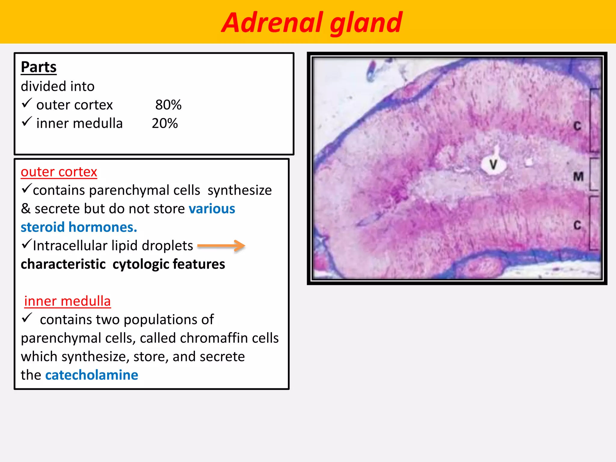
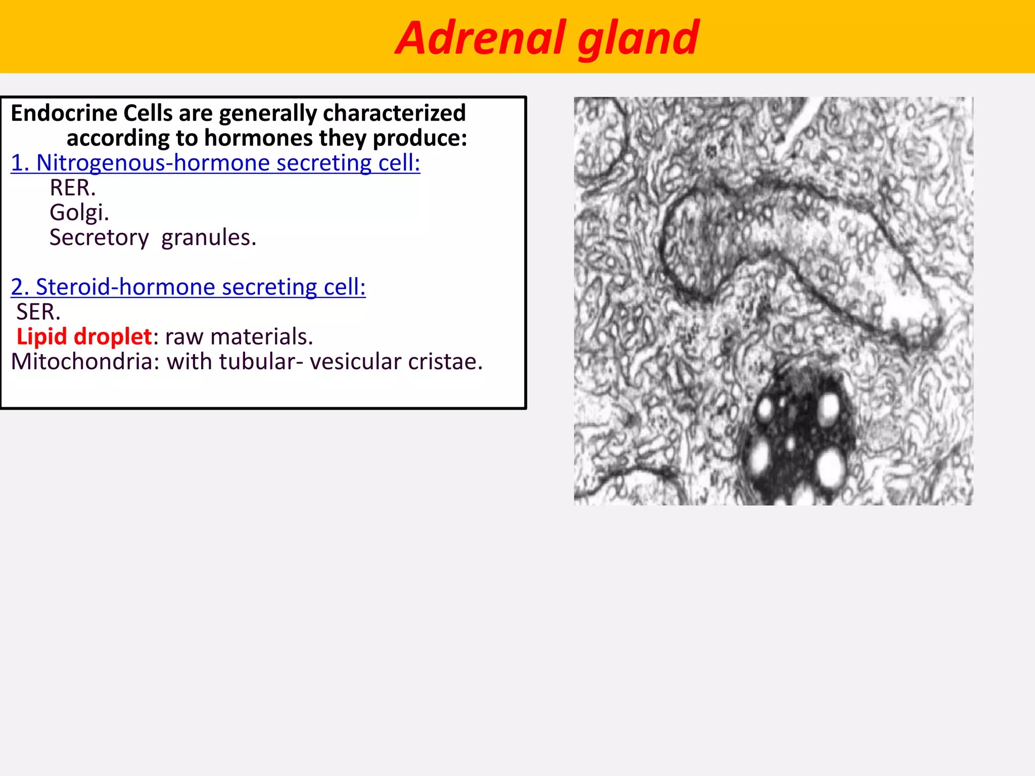
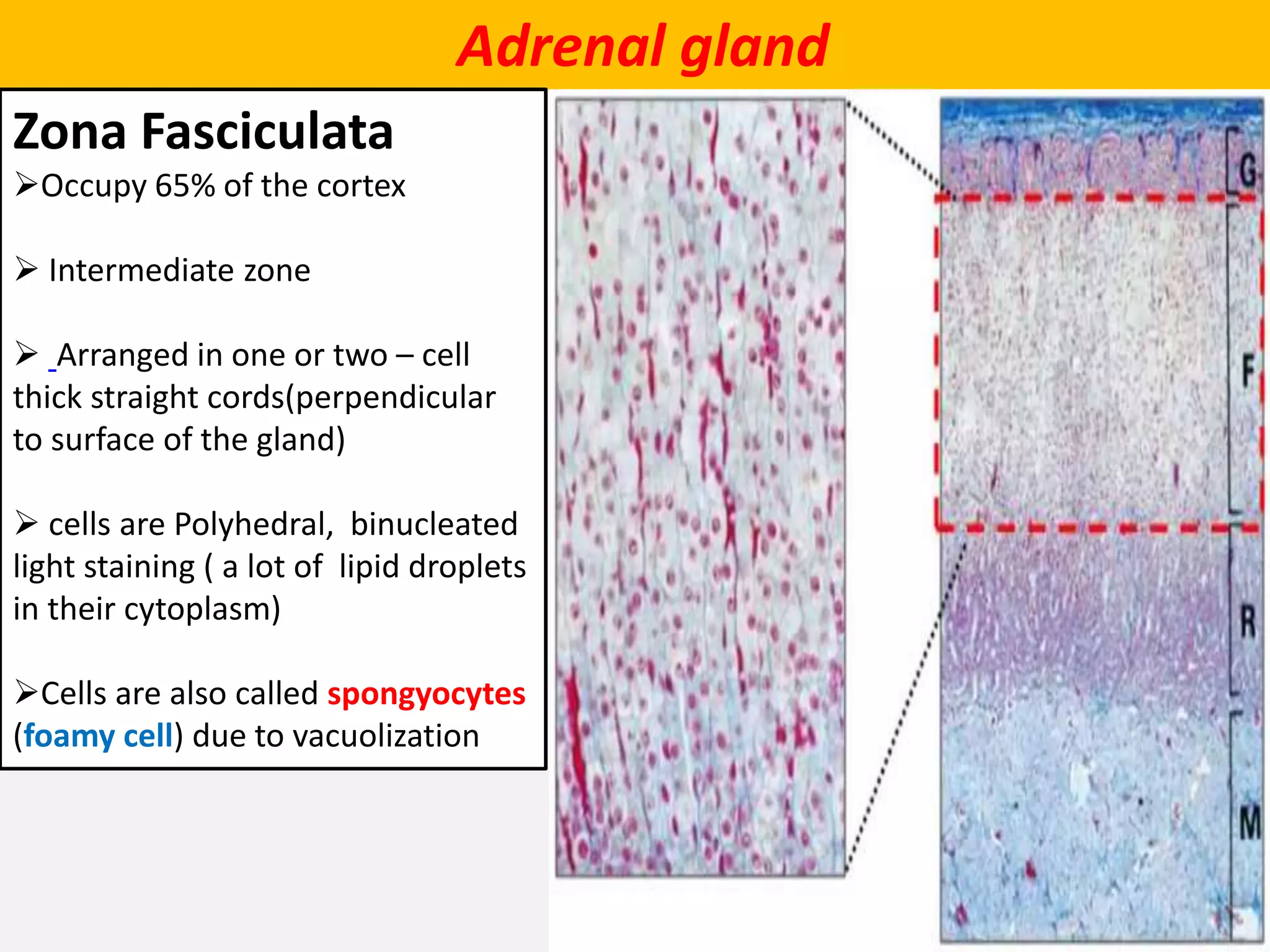
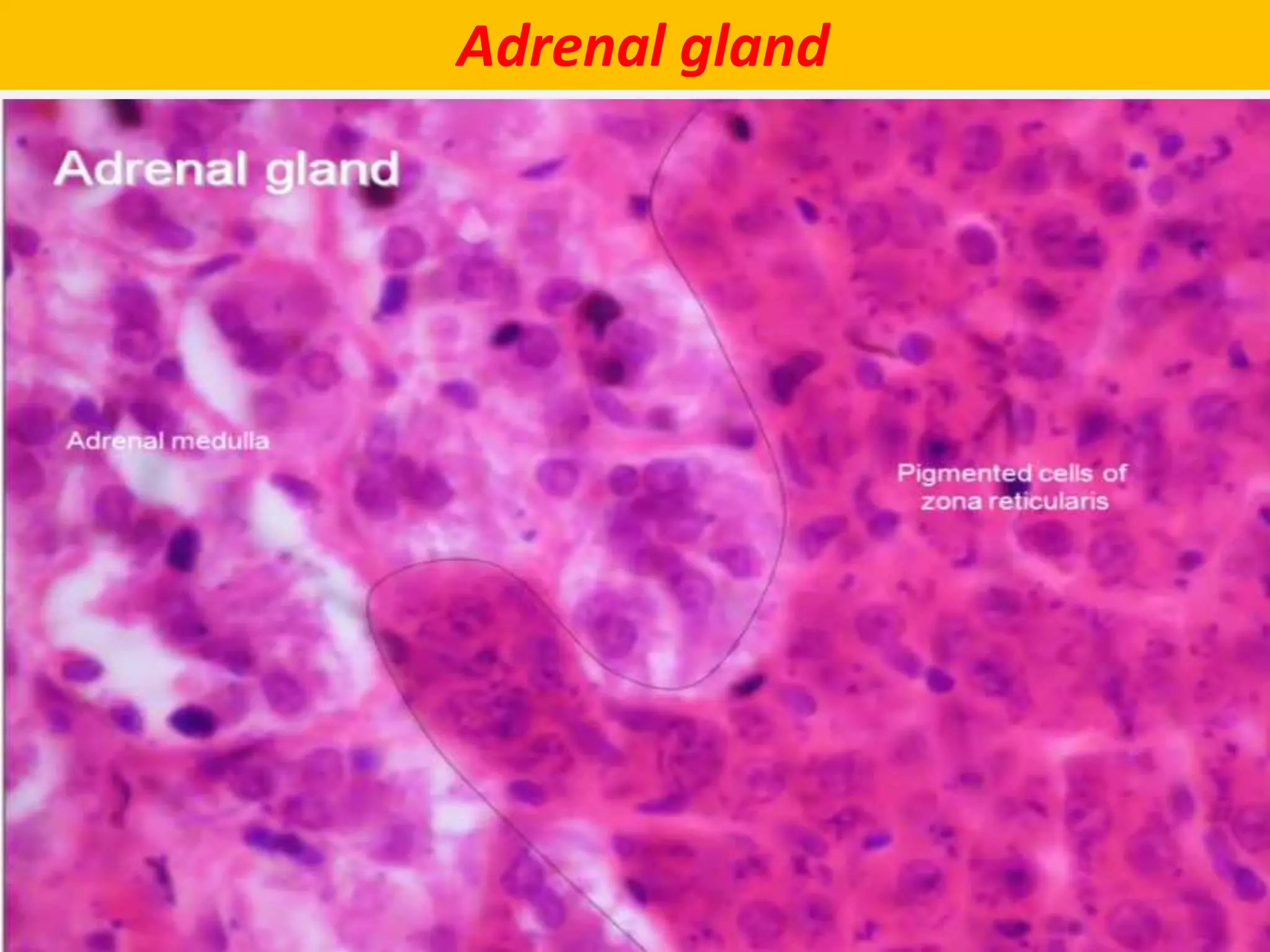
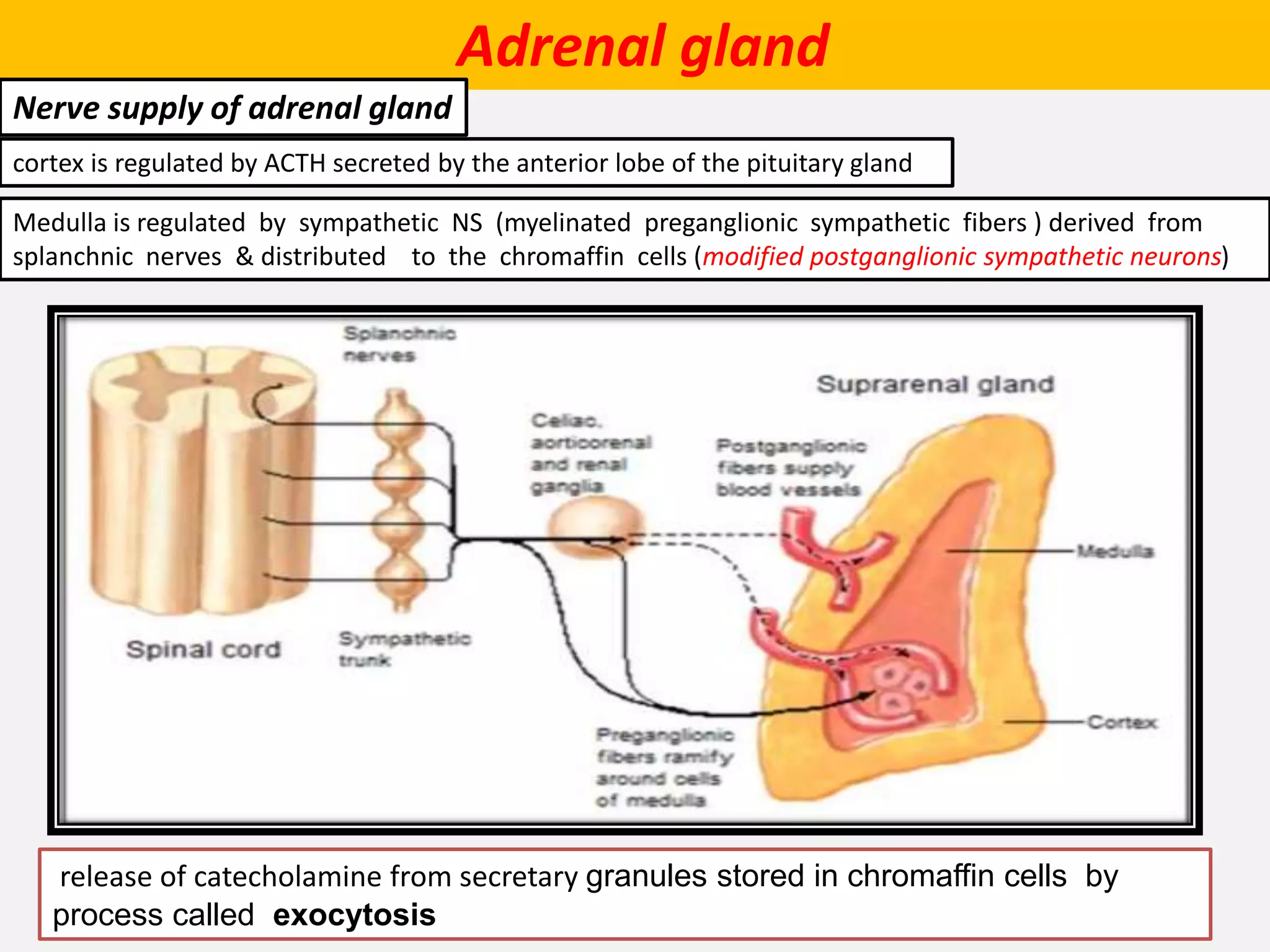
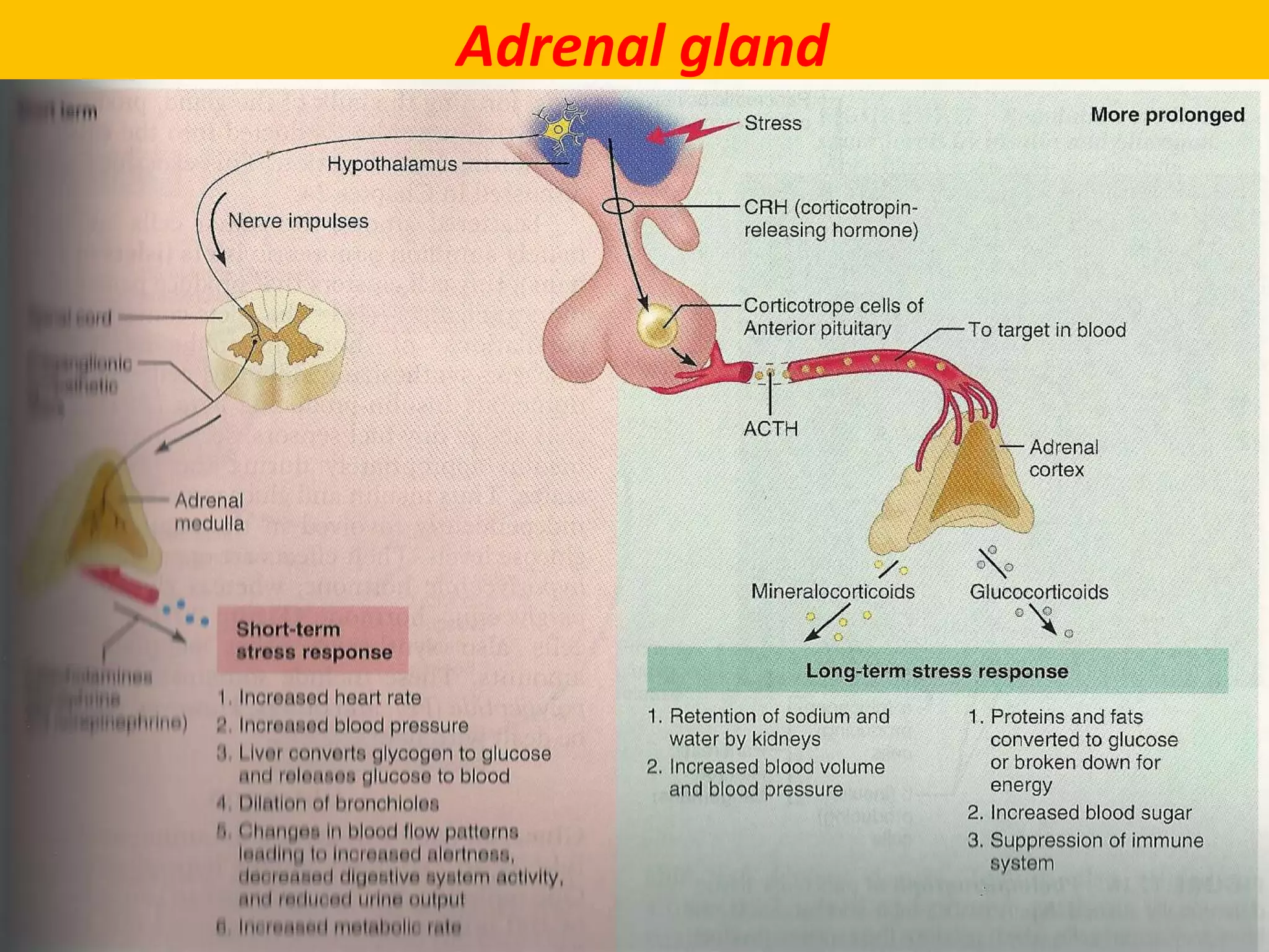
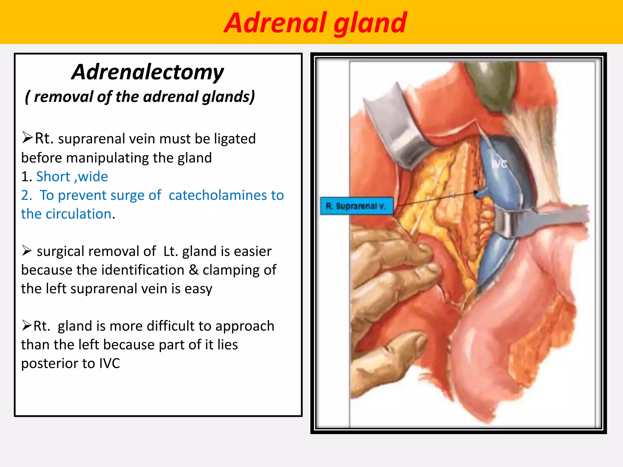
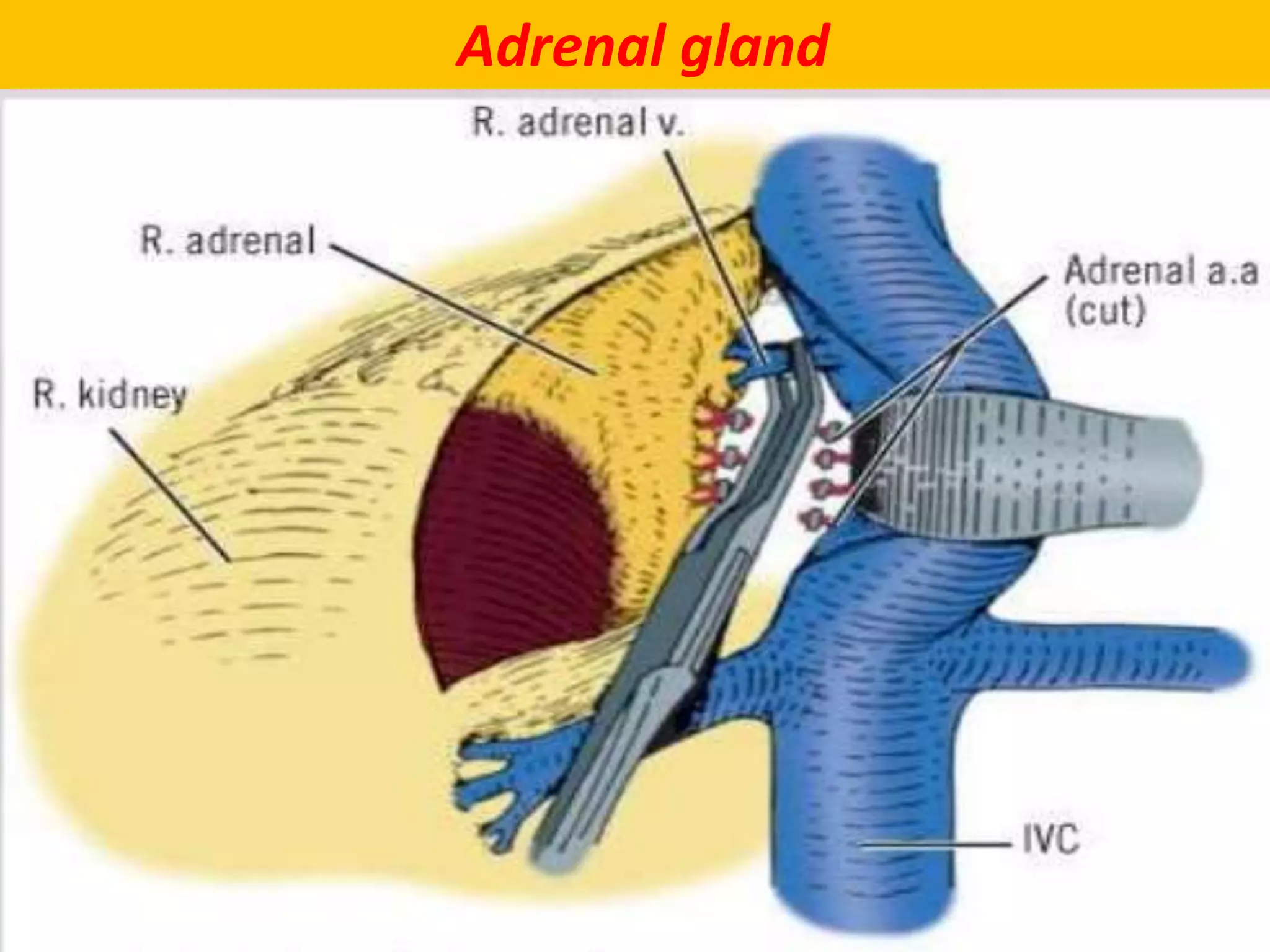
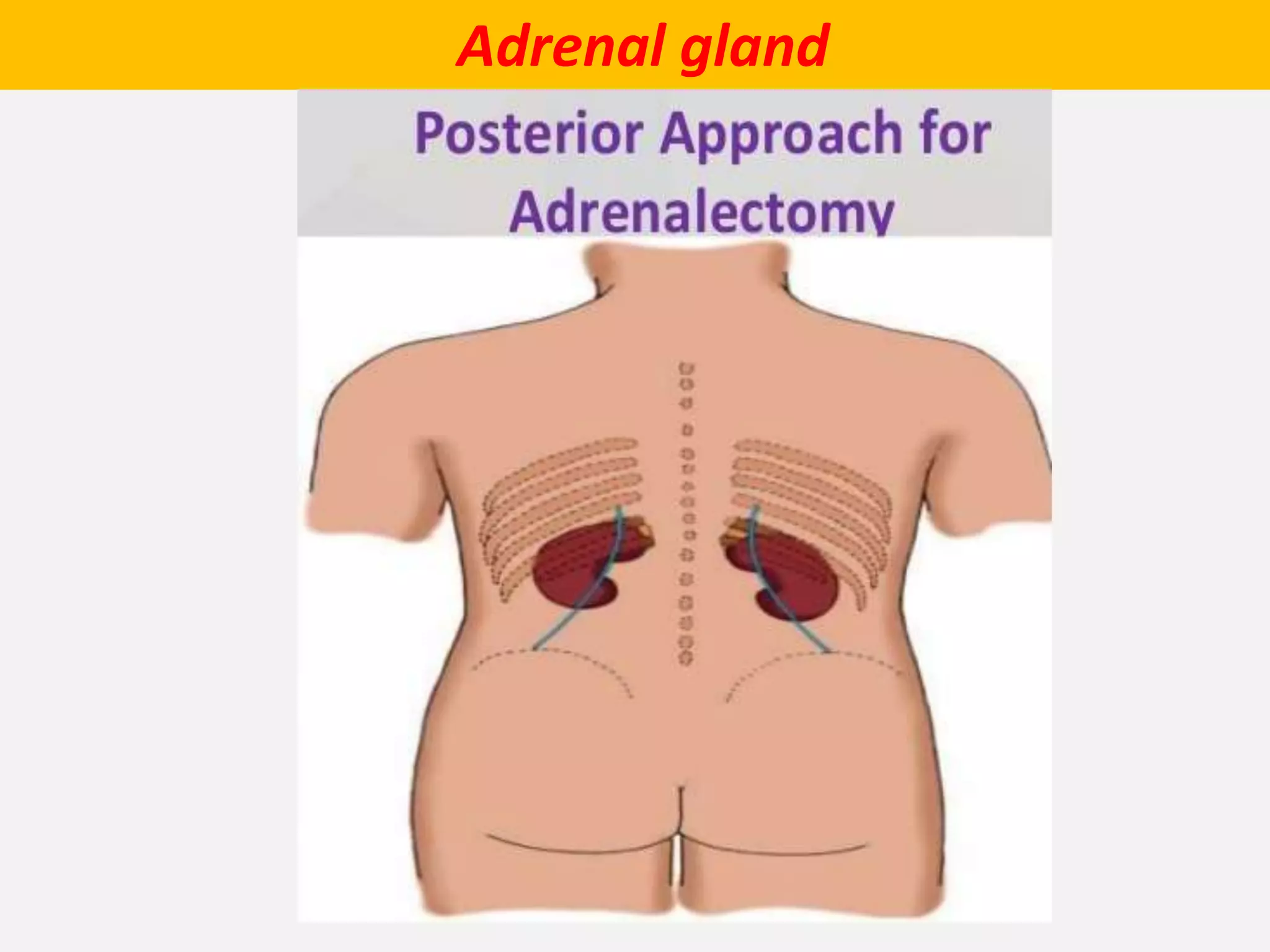
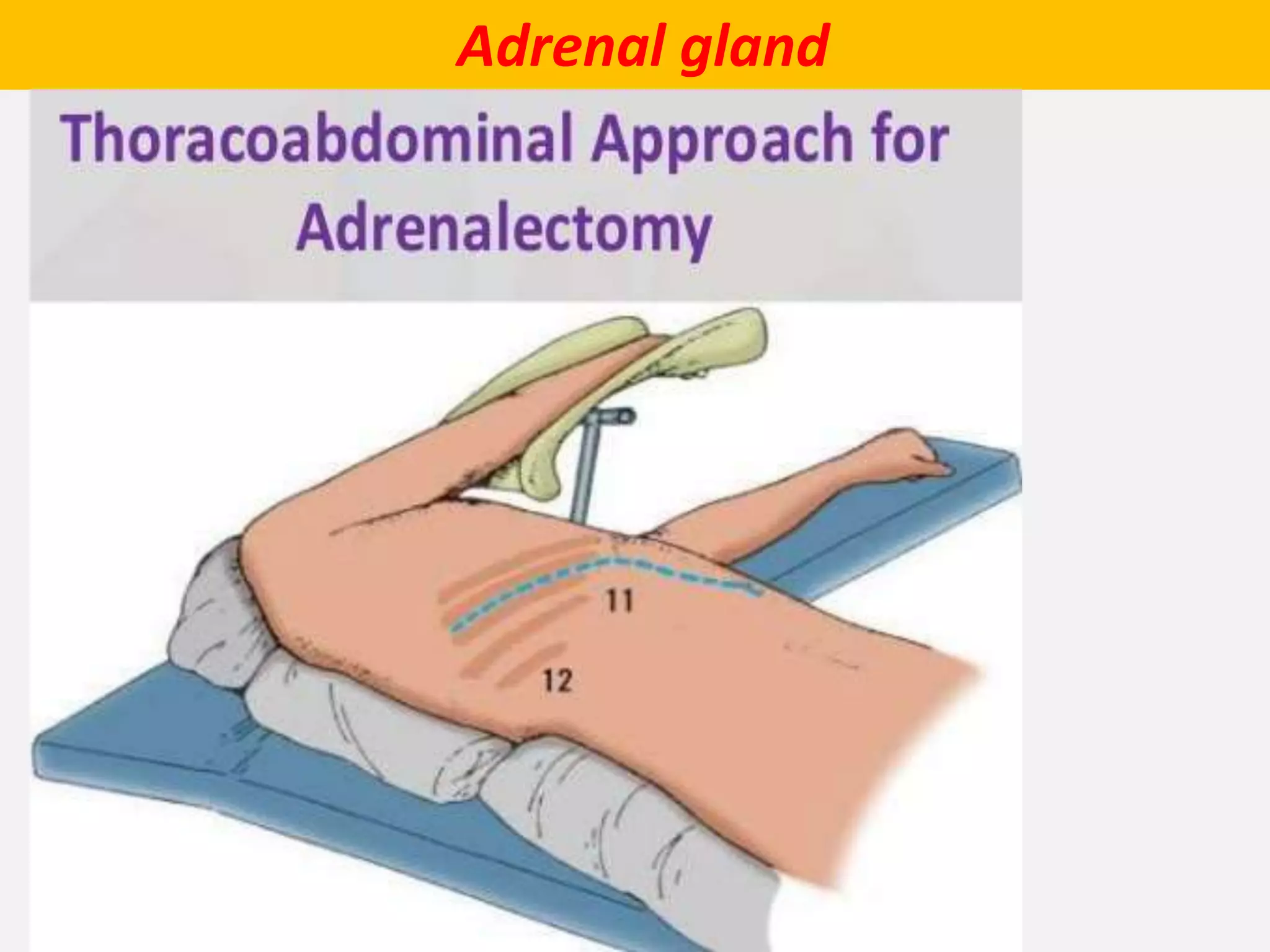
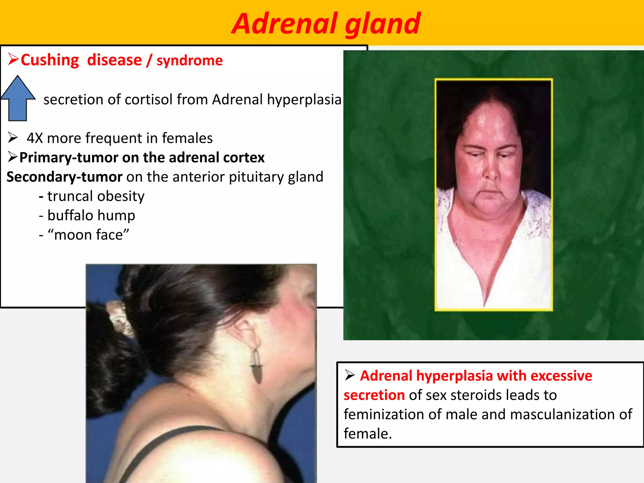
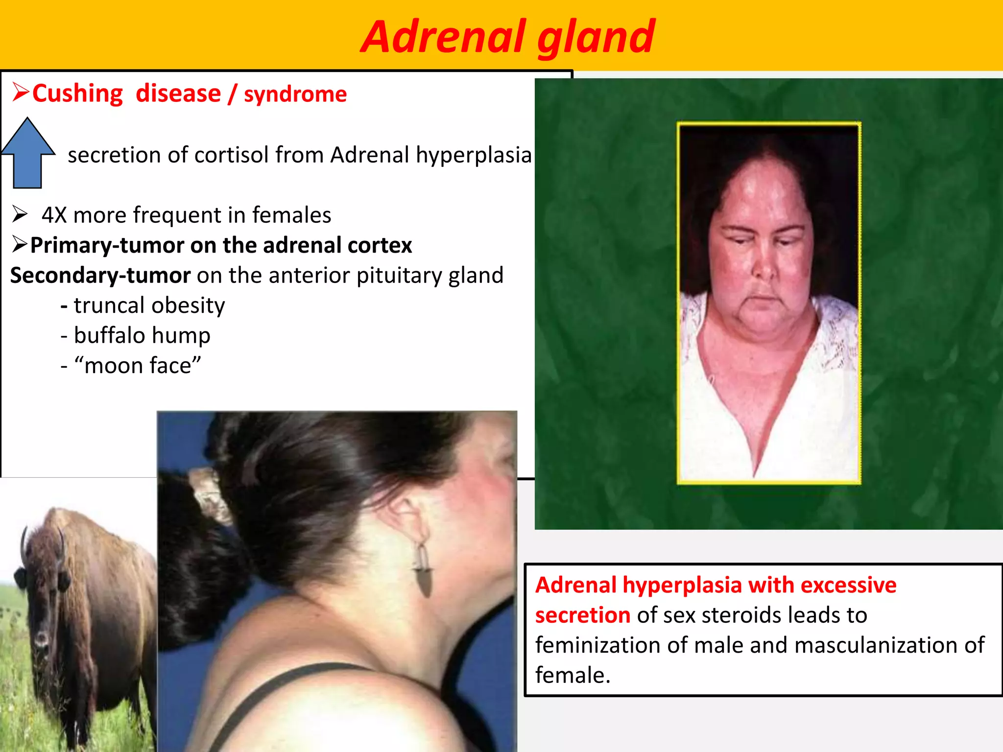
The document describes the anatomy and physiology of the adrenal gland. It discusses the location, shape, size and blood supply of the adrenal glands. It describes the two parts of each adrenal gland - the outer cortex and inner medulla. The cortex contains three zones that secrete different hormones. The medulla contains chromaffin cells that secrete catecholamines. Diseases associated with adrenal gland dysfunction are also mentioned.
Describe the structures, relations, and functions of the adrenal gland.
describe the histological structures and clinical importance of the adrenal gland
Describe the structures, relations, and functions of the adrenal gland.
describe the histological structures and clinical importance of the adrenal gland
location, length, and relation of right an left ureter, raletion of male an female ureter, n physiological site of ureteric constriction, bloo supply an inerve supply of ureter, clinical sinificance of ureter with hysteriectpomy
Anatomy of urinary bladder. surfaces, border of urinary bladder its relation , ligament support, peritoneal relation in male and females, pouches, blood supply of bladder, nerve supply of bladder, true and false ligament of urinary bladder,
It is the review researches based presentation on the topic of "ADRENAL GLAND" in which i describes about anatomical, physiological and pathological aspects of material from different websites and pages from google scholars which i gave references at the end.
The Endocrine System .The endocrine system is made up of glands that make hor...ZionAngelIniovosa
The endocrine system is made up of glands that make hormones. Hormones are the body's chemical messengers. They carry information and instructions from one set of cells to another.
micro teaching on communication m.sc nursing.pdfAnurag Sharma
Microteaching is a unique model of practice teaching. It is a viable instrument for the. desired change in the teaching behavior or the behavior potential which, in specified types of real. classroom situations, tends to facilitate the achievement of specified types of objectives.
Basavarajeeyam is a Sreshta Sangraha grantha (Compiled book ), written by Neelkanta kotturu Basavaraja Virachita. It contains 25 Prakaranas, First 24 Chapters related to Rogas& 25th to Rasadravyas.
New Drug Discovery and Development .....NEHA GUPTA
The "New Drug Discovery and Development" process involves the identification, design, testing, and manufacturing of novel pharmaceutical compounds with the aim of introducing new and improved treatments for various medical conditions. This comprehensive endeavor encompasses various stages, including target identification, preclinical studies, clinical trials, regulatory approval, and post-market surveillance. It involves multidisciplinary collaboration among scientists, researchers, clinicians, regulatory experts, and pharmaceutical companies to bring innovative therapies to market and address unmet medical needs.
Local Advanced Lung Cancer: Artificial Intelligence, Synergetics, Complex Sys...Oleg Kshivets
Overall life span (LS) was 1671.7±1721.6 days and cumulative 5YS reached 62.4%, 10 years – 50.4%, 20 years – 44.6%. 94 LCP lived more than 5 years without cancer (LS=2958.6±1723.6 days), 22 – more than 10 years (LS=5571±1841.8 days). 67 LCP died because of LC (LS=471.9±344 days). AT significantly improved 5YS (68% vs. 53.7%) (P=0.028 by log-rank test). Cox modeling displayed that 5YS of LCP significantly depended on: N0-N12, T3-4, blood cell circuit, cell ratio factors (ratio between cancer cells-CC and blood cells subpopulations), LC cell dynamics, recalcification time, heparin tolerance, prothrombin index, protein, AT, procedure type (P=0.000-0.031). Neural networks, genetic algorithm selection and bootstrap simulation revealed relationships between 5YS and N0-12 (rank=1), thrombocytes/CC (rank=2), segmented neutrophils/CC (3), eosinophils/CC (4), erythrocytes/CC (5), healthy cells/CC (6), lymphocytes/CC (7), stick neutrophils/CC (8), leucocytes/CC (9), monocytes/CC (10). Correct prediction of 5YS was 100% by neural networks computing (error=0.000; area under ROC curve=1.0).
- Video recording of this lecture in English language: https://youtu.be/lK81BzxMqdo
- Video recording of this lecture in Arabic language: https://youtu.be/Ve4P0COk9OI
- Link to download the book free: https://nephrotube.blogspot.com/p/nephrotube-nephrology-books.html
- Link to NephroTube website: www.NephroTube.com
- Link to NephroTube social media accounts: https://nephrotube.blogspot.com/p/join-nephrotube-on-social-media.html
Knee anatomy and clinical tests 2024.pdfvimalpl1234
This includes all relevant anatomy and clinical tests compiled from standard textbooks, Campbell,netter etc..It is comprehensive and best suited for orthopaedicians and orthopaedic residents.
CDSCO and Phamacovigilance {Regulatory body in India}NEHA GUPTA
The Central Drugs Standard Control Organization (CDSCO) is India's national regulatory body for pharmaceuticals and medical devices. Operating under the Directorate General of Health Services, Ministry of Health & Family Welfare, Government of India, the CDSCO is responsible for approving new drugs, conducting clinical trials, setting standards for drugs, controlling the quality of imported drugs, and coordinating the activities of State Drug Control Organizations by providing expert advice.
Pharmacovigilance, on the other hand, is the science and activities related to the detection, assessment, understanding, and prevention of adverse effects or any other drug-related problems. The primary aim of pharmacovigilance is to ensure the safety and efficacy of medicines, thereby protecting public health.
In India, pharmacovigilance activities are monitored by the Pharmacovigilance Programme of India (PvPI), which works closely with CDSCO to collect, analyze, and act upon data regarding adverse drug reactions (ADRs). Together, they play a critical role in ensuring that the benefits of drugs outweigh their risks, maintaining high standards of patient safety, and promoting the rational use of medicines.
The Gram stain is a fundamental technique in microbiology used to classify bacteria based on their cell wall structure. It provides a quick and simple method to distinguish between Gram-positive and Gram-negative bacteria, which have different susceptibilities to antibiotics
Title: Sense of Taste
Presenter: Dr. Faiza, Assistant Professor of Physiology
Qualifications:
MBBS (Best Graduate, AIMC Lahore)
FCPS Physiology
ICMT, CHPE, DHPE (STMU)
MPH (GC University, Faisalabad)
MBA (Virtual University of Pakistan)
Learning Objectives:
Describe the structure and function of taste buds.
Describe the relationship between the taste threshold and taste index of common substances.
Explain the chemical basis and signal transduction of taste perception for each type of primary taste sensation.
Recognize different abnormalities of taste perception and their causes.
Key Topics:
Significance of Taste Sensation:
Differentiation between pleasant and harmful food
Influence on behavior
Selection of food based on metabolic needs
Receptors of Taste:
Taste buds on the tongue
Influence of sense of smell, texture of food, and pain stimulation (e.g., by pepper)
Primary and Secondary Taste Sensations:
Primary taste sensations: Sweet, Sour, Salty, Bitter, Umami
Chemical basis and signal transduction mechanisms for each taste
Taste Threshold and Index:
Taste threshold values for Sweet (sucrose), Salty (NaCl), Sour (HCl), and Bitter (Quinine)
Taste index relationship: Inversely proportional to taste threshold
Taste Blindness:
Inability to taste certain substances, particularly thiourea compounds
Example: Phenylthiocarbamide
Structure and Function of Taste Buds:
Composition: Epithelial cells, Sustentacular/Supporting cells, Taste cells, Basal cells
Features: Taste pores, Taste hairs/microvilli, and Taste nerve fibers
Location of Taste Buds:
Found in papillae of the tongue (Fungiform, Circumvallate, Foliate)
Also present on the palate, tonsillar pillars, epiglottis, and proximal esophagus
Mechanism of Taste Stimulation:
Interaction of taste substances with receptors on microvilli
Signal transduction pathways for Umami, Sweet, Bitter, Sour, and Salty tastes
Taste Sensitivity and Adaptation:
Decrease in sensitivity with age
Rapid adaptation of taste sensation
Role of Saliva in Taste:
Dissolution of tastants to reach receptors
Washing away the stimulus
Taste Preferences and Aversions:
Mechanisms behind taste preference and aversion
Influence of receptors and neural pathways
Impact of Sensory Nerve Damage:
Degeneration of taste buds if the sensory nerve fiber is cut
Abnormalities of Taste Detection:
Conditions: Ageusia, Hypogeusia, Dysgeusia (parageusia)
Causes: Nerve damage, neurological disorders, infections, poor oral hygiene, adverse drug effects, deficiencies, aging, tobacco use, altered neurotransmitter levels
Neurotransmitters and Taste Threshold:
Effects of serotonin (5-HT) and norepinephrine (NE) on taste sensitivity
Supertasters:
25% of the population with heightened sensitivity to taste, especially bitterness
Increased number of fungiform papillae
3. Adrenal gland
LOCATION
paired , retroperitoneal
related to upper pole of kidney
Yellowish in colour
SHAPE AND MEASUREMENTS
Shape: Each gland is flattened A-P
1. RT. : pyramidal in shape
2LT.: crescentric in shape like
Measurements:
Length 50 mm.
Breadth 30 mm.
Thickness 10 mm.
Weight= About 5 g.
At birth the gland is 1/3 of the size of kidney, in adults it is only 1/30th of the
size of kidney
4. Adrenal gland
LOCATION
retroperitoneal
in the epigastric region of abdomen,
anterosuperior to the upper part of
each kidney
yellowish in color
SHAPE AND MEASUREMENTS
Shape: Each gland is flattened A-P
1. RT. : pyramidal in shape
2. LT.: crescentric in shape
Measurements:
Length 50 mm.
Breadth 30 mm.
Thickness 10 mm.
Weight= About 5 g.
At birth the gland is 1/3 of the size of kidney, in adults it is only 1/30th of the
size of kidney
6. Adrenal gland
Sheath
surrounded by two sheaths :
1. Surrounded by fat called perirenal fat.
2. Outer to this, renal fascia encloses
the suprarenal gland together with the
kidney
3. but the gland is separated from the
kidney by a septum.
7. Adrenal gland
ARTERIAL SUPPLY
Each gland is supplied by three arteries
from three different sources :
1. Superior suprarenal artery: A branch of
the inferior phrenic artery.
2. Middle suprarenal artery: A branch of the
abdominal aorta.
3. Inferior suprarenal artery: A branch of the
renal artery.
8. Adrenal gland
VENOUS DRAINAGE
Each gland is drained by only single vein
which emerges from the hilus of the
gland :
1. Right suprarenal vein drains into the
inferior vena cava.
2. Left suprarenal vein drains into the left
renal vein.
9. Adrenal gland
Parts
divided into
outer cortex 80%
inner medulla 20%
outer cortex
contains parenchymal cells synthesize
& secrete but do not store various
steroid hormones.
Intracellular lipid droplets
characteristic cytologic features
inner medulla
contains two populations of
parenchymal cells, called chromaffin cells
which synthesize, store, and secrete
the catecholamine
10. Adrenal gland
Endocrine Cells are generally characterized
according to hormones they produce:
1. Nitrogenous-hormone secreting cell:
RER.
Golgi.
Secretory granules.
2. Steroid-hormone secreting cell:
SER.
Lipid droplet: raw materials.
Mitochondria: with tubular- vesicular cristae.
13. Adrenal gland
Zona glomerulosa
Occupy 15% of the cortex
Immediately beneath the capsule
cells arranged in closely packed,
rounded cluster like glomeruli
cells are smaller , Columnar or
pyramidal with dark stain cytoplasm
14. Adrenal gland
Zona Fasciculata
Occupy 65% of the cortex
Intermediate zone
Arranged in one or two – cell
thick straight cords(perpendicular
to surface of the gland)
cells are Polyhedral, binucleated
light staining ( a lot of lipid droplets
in their cytoplasm)
Cells are also called spongyocytes
(foamy cell) due to vacuolization
15. Adrenal gland
Zona Reticularis
occupy 7% of the cortex
Innermost layer - lies between zona fasciculata and
medulla
Small cells arranged in irregular cords forming
network
Cells are darkly stained, characterize by presence of
lipofuscin pigment granules ( with age)
16. Adrenal gland
Medulla
Lies in the center of the adrenal
gland
Clear line of demarcation
Composed of polyhedral cells
arranged in clusters surrounded
by an extensive capillary network& ,
supported by reticular fiber
network
Composed of :
Chromaffin cells of 2types
A – CELLS
N – CELLS
18. Adrenal gland
Medulla
A – CELLS Characterized by
80%
containing small granules
Store epinephrine
N – CELLS Characterized by
20%
containing large dense granules
Store Norepinephrine
19. Adrenal gland
Medulla
has a dual blood supply that arises
from
(1) medullary arterioles that bypass
the adrenal cortex
(2) capillary sinusoids that perfuse the
adrenal cortex (adrenocortical
sinusoids).
central adrenomedullary vein drains
to IVC(Rt.)
or
Lt. renal V.(Lt.)
20. Adrenal gland
Nerve supply of adrenal gland
cortex is regulated by ACTH secreted by the anterior lobe of the pituitary gland
Medulla is regulated by sympathetic NS (myelinated preganglionic sympathetic fibers ) derived from
splanchnic nerves & distributed to the chromaffin cells (modified postganglionic sympathetic neurons)
release of catecholamine from secretary granules stored in chromaffin cells by
process called exocytosis
22. Adrenal gland
Radiology of Suprarenal Gland
deeply located difficult to visualize on plain X-ray.
CT scan of abdomen inverted Y shape with medial & lateral limbs
23. Adrenal gland
Adrenalectomy
( removal of the adrenal glands)
Rt. suprarenal vein must be ligated
before manipulating the gland
1. Short ,wide
2. To prevent surge of catecholamines to
the circulation.
surgical removal of Lt. gland is easier
because the identification & clamping of
the left suprarenal vein is easy
Rt. gland is more difficult to approach
than the left because part of it lies
posterior to IVC
32. Adrenal gland
Cushing disease / syndrome
secretion of cortisol from Adrenal hyperplasia
4X more frequent in females
Primary-tumor on the adrenal cortex
Secondary-tumor on the anterior pituitary gland
- truncal obesity
- buffalo hump
- “moon face”
Adrenal hyperplasia with excessive
secretion of sex steroids leads to
feminization of male and masculanization of
female.
33. Adrenal gland
Cushing disease / syndrome
secretion of cortisol from Adrenal hyperplasia
4X more frequent in females
Primary-tumor on the adrenal cortex
Secondary-tumor on the anterior pituitary gland
- truncal obesity
- buffalo hump
- “moon face”
Adrenal hyperplasia with excessive
secretion of sex steroids leads to
feminization of male and masculanization of
female.
35. Adrenal gland
Addison’s disease
hypofunction of adrenal cortex
What hormones will you have too little
of???
- glucocorticoids or _______
- mineralocorticoids or _______
- sex steroid or ____________
36. Adrenal gland
Addison’s disease
hypofunction of adrenal cortex
What hormones will you have too little
of???
- glucocorticoids or _______
- mineralocorticoids or _______
- sex steroid or ____________
38. Adrenal gland
Embryologically : adrenal gland
consists of two parts develops from two
different sources at 5th week :
(a) a large outer part called cortex :
is mesodermal in origin and develops
from celomic epithelium
(b) a small inner part called medulla.
develops from neural crest.
39. Adrenal gland
A. Cortex
1. forms from two episodes of mesoderm proliferation that occur between the root of the
dorsal mesentery and the gonad.
first episode forms the inner fetal cortex.
second episode forms the outer adult cortex
2. During the fetal period , the suprarenal glands are very large due to the size of the fetal
cortex( 10–20 times larger than adult adrenal gland).
3. suprarenal glands become smaller as the fetal cortex involutes rapidly during the first 2
weeks after birth and continues to involute during the first year of life.
4. zona glomerulosa and zona fasciculata of the adult cortex are present at birth, but the zona
reticularis is not formed until age 3 years.
B. Medulla
1. forms when neural crest cells aggregate at the medial aspect of the fetal cortex and become
surrounded by the fetal and adult cortex.
2. The neural crest cells differentiate into chromaffin cells
40. Adrenal gland
Congenital anomalies
1. Ectopic adrenal tissue/adrenal gland:
The adrenal tissue or complete adrenal gland may be found fused to kidney deep
in its capsule or in the right lobe of the liver.
2. adrenal hyperplasia
a) Congenital adrenal hyperplasia:
It is most commonly caused by mutation of genes for enzymes involved in adrenocortical
steroid biosynthesis (e.g., 21-hydroxylase deficiency),
b) Adrenogenital syndrome:
It occurs due to congenital hyperplasia of the cells of the adrenal cortex, which secrete
androgen.
C/F differ in male and female :
(a) In male: (adrenogenital syndrome)
It leads to a very early development of secondary sexual characters.
(b) In female: (pseudohermaphroditism)
female child may be mistaken as a male.
44. Neural Crest cells
Derivatives of neural crest:
1. C.T. and bones of the face and skull.
2. ganglia of the cranial nerves.
3. C-cells of the thyroid gland.
4. conotruncal septum of the heart.
5. odontoblast cells.
6. dermis of the face and neck.
7. spinal ganglia.
8. autonomic ganglia.
9. Chromaffin cells (adrenal medulla).
10. schwan cells.
11. glial cells of the brain.
12. melanocytes.
13. arachnoid and pia matter
45. Adrenal gland
Sites of chromaffin tissue :
1. para-aortic bodies
2. Along sympathetic chain near sympathetic ganglia
3. Along sympathetic plexuses
4. Near splanchnic nerves.
chromaffin tissue
1. stain intensely with chromium salts
yellow-brown .
2. Neural crest derivatives
Chromaffin cells can be found in
extrasuprarenal sites at birth, but these sites
normally regress completely by puberty.
In a normal adult, chromaffin cells are found
only in the suprarenal medulla
Pheochromocytoma:
tumor arising from chromaffin cells
usually from extra-adrenal
50. Sympathetic Chain
Extension:
from the base of the skull to the coccyx.
bears a number of ganglia along its length
On either side of V. column (paravertebral ganglion)
Parts
cervical, thoracic, lumbar and sacral
Ganglion
Cervical :3
usually one ganglion less than the number of nerves:
11 thoracic; 4 lumbar; and 4 sacral
2 neurons
(pre & postganglionic )
Preganglionic N : T1- L2 in lat. horn
Communication with spinal nerves
T1-L2 : by grey &white communicants
Above & below this level: only grey communicants
NO white communicants
Why?
51. Fate of preganglionic fibres
preganglionic fibres arise from the lateral horns of spinal segments with ventral root
4 options
Sympathetic ganglion of sympathetic trunk
(paravertebral ganglion
prevertebral ganglion
52. preganglionic fibres arise from the lateral horns of spinal segments with ventral root
preganglionic fibres enter the sympathetic ganglion via white rami communicantes of the
spinal nerve.
Rely (synopsis) in the ganglion
Postganglionic fibres from the ganglion re-enter the spinal nerve via grey rami
communicantes
supply the corresponding dermatome of the upper limb and the body wall.
53. BRANCHES of thoracic part
Visceral & somatic Efferent Fibers
divided into two groups: medial & lateral
Medial branches(visceral)
1) postganglionic fibres
from 1st to 5th ganglia
distributed to the heart, great vessels,
lungs, and esophagus through the
following plexuses:
a) Pulmonary plexus bronchodilatation
b) b) Cardiac plexus. vasoconstriction
c) Aortic plexus. Secretion
d) Esophageal plexus
HR & dilate coronary art.
2) preganglionic fibres
5th to 12th thoracic ganglia
form three splanchnic nerves :
a) Greater splanchnic nerve(5-9th)
b) Lesser splanchnic nerve(10 &11th )
c) Least (lowest) splanchnic nerve(12th (renal)
54. BRANCHES of thoracic part
Lateral branches(somatic)
supply:
• erector pili of hair follicle ( pilomotor)
• Sweat gland (sudomotor)
• Bl. V of the skin ( vasomotor)
dilate Cutaneous Bl. V of limbs and
body wall