To define the hepatobiliary system
To outline the embryological development and congenital anomalies of the hepatobiliary system.
To describe the gross anatomy and histology of the hepatobiliary system.
To outline the clinical anomalies associated with the hepatobiliary system
Composed of the liver and the bile ducts.
Mainly concerned with formation, transport, concentration and secretion of bile.
Bile is produced by the liver and transported by the bile ducts into the small intestines
Liver, biliary system, pancreas and spleeness_online
This document discusses the anatomy of the liver, pancreas, and portal system. It begins with the embryological development of these organs, then describes the anatomy and blood supply of the liver in detail. It also discusses the gallbladder, biliary tree, pancreas, and their related structures and variations. Key functions of the liver are outlined. The document provides a thorough overview of the anatomy relevant to upper gastrointestinal and hepatobiliary surgery.
The liver is the largest gland in the human body, weighing approximately 1500 grams. It performs many important functions like absorbing nutrients from the gastrointestinal tract, storing glycogen, and secreting bile. The liver has two lobes - the right lobe, which is the largest, and the left lobe. It is situated in the right upper quadrant of the abdomen and has two surfaces - the diaphragmatic surface and the visceral surface. The liver receives blood from the hepatic portal vein and hepatic artery and is supported by various ligaments.
This document provides an overview of the liver and biliary system. It describes the anatomy and functions of the liver, including its lobes and surfaces. It also details the structures of the extrahepatic biliary system, including the hepatic ducts, common bile duct, gallbladder, and their relations. Additionally, it discusses the blood supply, lymphatics, and innervation of the liver and biliary structures. Clinical considerations related to the liver and gallbladder are also briefly mentioned.
Development of liver , extrahepatic biliary apparatus , pancreas and spleen.SaachiGupta4
embryology- development and developmental anomalies of the liver, extrahepatic biliary apparatus, pancreas and spleen.
Stages of development of liver, reidel's lobe, annular pancreas.
Alimentary tract embryology
The liver is the largest gland in the body, located in the upper right quadrant of the abdomen under the diaphragm and within the rib cage. It has four lobes - two major lobes and two minor lobes - and ducts that carry bile from the liver to the gallbladder and join the pancreatic duct. The liver is made up of lobules containing hepatocytes that radiate outward from a central vein and produce and secrete bile. Blood flows to the liver through the hepatic portal vein and supplies the liver with nutrients and oxygen. The liver performs many important metabolic functions like synthesizing and breaking down substances, and excreting waste from the bloodstream into bile.
This document provides an overview of the surgical anatomy of the hepatobiliary system. It discusses the liver, gallbladder, and biliary tree in detail. Key points include the blood supply to the liver from the hepatic artery and portal vein, segmentation of the liver, ligaments supporting the liver, variations in arterial and venous anatomy, embryological development of the hepatobiliary system, and congenital anomalies that can occur. The gallbladder, cystic duct, common bile duct, and variations are also described.
The document provides information on the anatomy and physiology of the spleen:
1. The spleen is the largest lymphoid organ located in the left upper quadrant of the abdomen. It has important immunological and filtration functions.
2. The spleen receives its blood supply from the splenic artery and drains into the splenic vein. It has a complex network of trabeculae and sinusoids that allow it to filter blood and remove old red blood cells.
3. The spleen plays roles in immune function and filtration of the blood, removing aged red blood cells, platelets, and pathogens from circulation. Though other organs share these functions, the spleen is a major contributor.
The liver receives its blood supply from both the hepatic portal vein and hepatic arteries. The hepatic portal vein supplies around 75% of the liver's blood flow, carrying nutrient-rich but oxygen-poor blood from the spleen, gastrointestinal tract and other organs. The hepatic arteries supply the remaining blood flow, carrying oxygen-rich but nutrient-poor arterial blood. Both sources provide oxygen, with around half coming from each vessel, and most venous drainage from the liver passes into the three hepatic veins which drain into the inferior vena cava.
This is not a substitute for Books. Let it just help you understand some concepts in liver anatomy.
Continuation of this work will depend on your feedback. Stay Blessed.
The liver is the largest visceral organ located in the upper right quadrant of the abdominal cavity. It performs over 200 essential functions including nutrient storage, breakdown of red blood cells, bile secretion, and synthesis of proteins and cholesterol. The liver receives blood from the hepatic portal vein and hepatic artery and drains into the hepatic veins. It is divided into four lobes and has both a diaphragmatic and visceral surface. The gallbladder stores and concentrates bile produced by the liver. Cirrhosis is a condition where the liver develops scar tissue due to chronic damage.
The document provides an overview of the anatomy of the abdomen, including:
1) It describes the surface anatomy of the abdomen using planes and regions.
2) It details the layers of the anterior abdominal wall including muscles and fascia.
3) It explains the anatomy of the inguinal canal including its walls, openings, contents and Hasselbach's triangle.
4) It briefly outlines the muscles and fascia of the posterior abdominal wall.
The liver is the largest gland in the body located under the right rib cage. It is divided into four lobes and has two surfaces - a diaphragmatic surface and a visceral surface. The porta hepatis contains the hepatic artery, portal vein and hepatic ducts. Blood flows into the liver through the hepatic artery and portal vein and exits through the hepatic veins. The gallbladder stores and concentrates bile produced by the liver. The biliary system consists of the hepatic ducts, cystic duct, common hepatic duct, gallbladder and common bile duct which empties into the duodenum.
This document discusses the development of several organ systems, including:
- The liver, pancreas, spleen, and respiratory system develop from the endoderm and mesoderm. The liver forms from the hepatic bud, while the pancreas forms from dorsal and ventral buds. The spleen forms from mesoderm in the dorsal mesogastrium.
- The respiratory system develops from a median diverticulum of the foregut that divides into right and left lung buds. The larynx and trachea develop from the cranial part of the diverticulum. The lung buds undergo repeated division to form the bronchial tree and lungs.
- The peritoneal, pericardial, and pleural
The liver is the largest abdominal organ and glandular organ of the GI system. It is located in the right upper quadrant of the abdomen and has two main surfaces - the diaphragmatic surface which faces the diaphragm and is convex, and the visceral surface which faces inferiorly and to the left. The visceral surface contains the porta hepatis, a deep groove that contains the hepatic vessels and bile ducts. The liver is divided functionally into left and right lobes and anatomically into right, left, caudate and quadrate lobes.
The document provides an overview of liver anatomy including:
- The liver's position in the right hypochondrium and epigastric region and its weight of 1.5kg on average.
- It has two surfaces: the diaphragmatic surface against the diaphragm and the visceral surface covered in peritoneum except at the gallbladder fossa and porta hepatis.
- The visceral surface is related to other structures like the stomach, duodenum, and right kidney.
- Couinaud described the liver as being divided into 8 segments based on arterial, portal, and biliary drainage.
The document provides information on the gross anatomy and histology of the liver. It begins with describing the liver's location, shape, size, and functions. It then discusses the liver's external features including surfaces and impressions. It also describes the liver's internal structures such as lobes, vascular supply from the hepatic artery and portal vein, lymphatic drainage, and innervation. Finally, it examines the liver's histology including its parenchyma, sinusoids, liver lobules in their classic, portal, and acinar forms, and the perisinusoidal space of Disse.
The document provides an overview of the liver, biliary apparatus, and related diseases. It describes the anatomy and blood supply of the liver and gallbladder. It discusses hepatocellular carcinoma and bile duct cancers, including risk factors, clinical presentation, diagnosis, and treatment. Hepatocellular carcinoma is often associated with liver cirrhosis and viral hepatitis. Gallbladder cancer commonly occurs in the setting of gallstones and has a poor prognosis due to late presentation. Bile duct cancer risk factors include liver flukes and primary sclerosing cholangitis. Obstructive jaundice is a common symptom of bile duct cancers.
The liver develops from the endoderm of the foregut. During the 4th week, the hepatic diverticulum buds off from the foregut and divides into the pars hepatica and pars cystica. The pars hepatica gives rise to the liver parenchyma of hepatocytes and bile ducts. It expands between the layers of the septum transversum mesenchyme. The pars cystica develops into the gallbladder and cystic duct. By week 8, the basic structure of the liver and biliary tree is established.
describes about peritoneal cavity and clinical importance of it. it describes in deatils about lesser sac, greater sac, pouch of Morrison, pouch of Douglas.
1. Respiration includes ventilation, gas exchange, and oxygen utilization. Ventilation is the mechanical process of breathing that moves air in and out of the lungs. Gas exchange occurs through diffusion of oxygen and carbon dioxide between the alveoli and blood.
2. The lungs contain over 300 million alveoli which provide a large surface area for gas exchange. Each alveolus is lined by fluid and surfactant that helps reduce surface tension to facilitate breathing.
3. During inspiration, contraction of the diaphragm and intercostal muscles increases the thoracic cavity volume, lowering pressure and drawing air into the lungs. Expiration is passive as the lungs and chest wall recoil, increasing pressure to push
Anatomy of liver, biliary tract and portal systemTanat Tabtieang
This document provides an overview of the anatomy of the liver, biliary tract, and portal venous system. It describes the liver's location, lobes and segments, surfaces, ligaments, blood supply, and drainage. The gallbladder, biliary tract, and portal venous system are also summarized. Specifically, it discusses the gallbladder's location and parts, as well as the tributaries and anastomoses of the portal venous system. Complications of portal hypertension like esophageal varices are also mentioned. The objectives are to describe the surface anatomy and functional anatomy of the liver and associated structures.
Development of liver, pancreas, spleen and extrahepatic biliary apparatusSaachiGupta4
Embryology- anatomy
Topic: Development of liver, pancreas, spleen, and extrahepatic biliary apparatus.
For M.B.B.S. students. It gives knowledge on the development of the organs mentioned above and their developmental anomalies
The esophagus extends from the pharynx to the stomach. It has 3 parts - cervical, thoracic, and abdominal. The stomach is J-shaped and located in the upper abdomen. It has a fundus, body, and pylorus. Both organs receive their blood supply from branches of the celiac trunk and drain into the portal vein. They are innervated by sympathetic and parasympathetic fibers from the vagus nerves.
anatomy of spleen, surfaces of spleen, peritoneal an visceral relations, blood supply an lymphatic drainage, hilum of spleen , function of spleen, splenomegaly,
The liver is the second largest organ and largest gland in the body. It has a dual blood supply and is divided into lobules that contain hepatocytes, sinusoids, and a central vein. Blood flows into lobules through the portal vein and hepatic artery and out through hepatic veins. Hepatocytes are arranged in plates separated by sinusoids and produce bile that flows into canaliculi and out of the liver. The liver has regenerative abilities and its segmentation allows for resection of parts without damaging remaining tissue.
The document describes the anatomy and segmentation of the liver. It notes that the liver is divided into eight functionally independent segments, with each segment having its own vascular inflow, outflow and biliary drainage. It describes Couinaud's division of the liver into a functional left and right liver divided by Cantlie's line, which runs from the middle of the gallbladder fossa to the inferior vena cava. Because of this segmentation, individual segments can be surgically resected without damaging remaining segments.
The liver and gallbladder develop from the foregut endoderm. An outgrowth called the hepatic diverticulum or liver bud appears at the junction of the foregut and midgut during the third week of development. It penetrates the septum transversum and divides into right and left lobes which receive hematopoietic cells and connective tissue from the septum. Meanwhile, the bile duct and gallbladder form. Initially the bile duct opens into the duodenum but later rotates to open dorsally due to duodenal rotation. The liver performs hematopoiesis and bile production in the fetus. Anomalies can include duplication or absence of the gallbladder and abnormalities of the bile duct
This document provides an overview of lung and pleural anatomy from Dr. Noura El Tahawy. It discusses the structure and layers of the pleura, noting that the pleural cavities are lined by mesothelial membranes and contain the lungs. Each lung remains attached to the mediastinum at the root, which contains the airways, blood vessels, lymphatics and nerves. The document also reviews the surfaces and borders of the lungs, the root and hilum structures, the bronchial tree and segments, vasculature, innervation and lymphatic drainage of the lungs.
The liver, gallbladder, and bile ducts make up the hepatobiliary system. The liver is the largest organ located in the right upper abdomen. It has two surfaces and receives 80% of its blood supply from the portal vein. The gallbladder stores and concentrates bile before it is released into the small intestine. Bile ducts drain bile from the liver and gallbladder and include the right and left hepatic ducts which join to form the common hepatic duct and eventually the common bile duct. Variations can occur in the anatomy of these structures. Ultrasound is useful for evaluating the normal anatomy and identifying any abnormalities.
Mr. N.N., a 56-year-old male, presents with abdominal distention, confusion, and other symptoms after a history of excessive alcohol use for 20 years. He is diagnosed with alcohol-induced liver cirrhosis. The document then proceeds to describe in detail the anatomy of the liver and biliary system, including blood supply, ligaments, histology of the liver and gallbladder, portal vein formation, and clinical cases of cholelithiasis and chronic cholecystitis.
The document provides information on the structure and functions of the liver and pancreas. It discusses the liver's location, lobes, ligaments, vascular and biliary supply. The liver receives blood from the hepatic portal vein and hepatic arteries. It secretes bile into canaliculi between hepatocytes. The bile ducts drain into the right and left hepatic ducts. The pancreas is also mentioned. The peritoneum and its derivatives are briefly introduced.
1.Antomy and physiology of liver by worku.pptxGoldGetnet
The liver has complex anatomy and vasculature. It is divided into 4 lobes and 8 segments based on blood supply. The liver receives dual blood supply from the hepatic artery and portal vein. It has 3 major functions - metabolism, protein synthesis, and bile production. The bile duct drains bile from the liver into the small intestine.
The document provides an overview of sonography of the liver. It discusses liver anatomy, development, lobes and surfaces. It describes the vascular supply including the portal vein and hepatic arteries. Common congenital abnormalities are mentioned such as hepatic cysts, peribiliary cysts, and polycystic liver disease. Imaging findings for these abnormalities on ultrasound are summarized. The document also briefly covers blood supply, nerve supply, lymphatic drainage and Couinaud liver segmentation.
C:\documents and settings\user\desktop\gastrointestinal 0406 liverpdfMBBS IMS MSU
This document provides an overview of the anatomy of the gastrointestinal system, with a focus on the liver, gallbladder, and biliary system. It describes the structure and connections of the liver, including its lobes, ligaments, and vascular supply. It then discusses the gallbladder, its attachment to the liver, blood supply, and structure. Finally, it details the biliary system, including the branching of the hepatic ducts, formation of the common bile duct, and termination of the bile and pancreatic ducts in the duodenum.
Liver is the largest internal organ of the body weighing about 1500g in adults. It occupies the right hypochondrium and extends into the epigastrium and left hypochondrium .
The liver, gallbladder, pancreas, and spleen are described. The liver is the largest gland and has many functions including bile production, carbohydrate and fat metabolism, and vitamin processing. The gallbladder stores and concentrates bile from the liver. The pancreas produces enzymes and hormones to aid digestion. The spleen filters blood and stores blood cells. All four organs have specific locations, blood supply from the hepatic and splenic arteries, and drainage into the portal vein and lymphatics.
The liver is the largest solid organ located in the upper right abdomen. It performs hundreds of vital functions including removing toxins from the blood, maintaining blood sugar levels, and regulating blood clotting. The liver receives 20% of its blood supply from the hepatic artery and 80% from the portal vein. It is divided into four lobes and has five surfaces. The liver plays a crucial role in metabolism and detoxification.
The liver is the largest gland in the body located in the right upper quadrant of the abdomen. It has two surfaces - diaphragmatic and visceral, and plays a key role in metabolism and detoxification. The liver receives blood from the hepatic artery and portal vein and excretes bile into the common hepatic duct. The gallbladder stores and concentrates bile before it is released into the duodenum through the cystic and common bile ducts. The pancreas is a retroperitoneal organ that extends from the duodenum to the spleen and contains both exocrine and endocrine components. Its exocrine secretions include enzymes that aid in digestion while its endocrine islets of Langerhans regulate blood
The liver is the largest gland in the body. It is located in the right hypochondrium and performs many essential functions including bile production, carbohydrate metabolism, detoxification, and vitamin storage. The liver has four lobes - right, left, caudate, and quadrate. It receives blood from the hepatic artery and portal vein and drains into the hepatic veins. The ligaments that connect the liver include the falciform, coronary, and ligamentum teres.
This document discusses liver anatomy, function tests, and imaging. It covers the embryological development of the liver, its lobes and ligament attachments. It describes the dual blood supply, biliary drainage system, and microscopic anatomy. Common liver function tests are outlined including those assessing synthesis, damage, and detoxification. Ultrasound imaging of the liver is also summarized, noting its advantages of being inexpensive and non-invasive but limitations in imaging certain areas.
Liver and extra hepatic biliary apparatus.pptxSundip Charmode
The document provides information about the liver including its shape, weight, position, surfaces, borders, lobes, ligaments, blood supply, lymphatic drainage and clinical significance. It discusses the gallbladder, cystic duct, common hepatic duct, and common bile duct which make up the extrahepatic biliary apparatus. The document describes the anatomy and relations of these structures in detail.
This document provides information on abdominal ultrasound indications and liver anatomy and segmentation. It discusses common reasons for abdominal ultrasound exams, including abdominal pain, jaundice, and liver or gallbladder abnormalities. It then details Couinaud's classification of liver segmentation, which divides the liver into eight functionally independent segments based on vascular supply and drainage. Each segment is examined using ultrasound, with descriptions of imaging views and anatomical landmarks to identify the different segments.
The accessory organs of the digestive system include the salivary glands, liver, gallbladder and pancreas. The liver performs many vital functions including metabolic regulation, hematological regulation and bile production. It regulates nutrient levels, filters toxins, stores vitamins and produces plasma proteins and bile. The liver lobule is the basic functional unit, with hepatocytes arranged in plates around a central vein and sinusoids. Blood enters through the hepatic artery and portal vein and leaves through hepatic veins.
The document provides information about the liver, gallbladder, and spleen. It discusses the formation, location, lobes, blood supply, and clinical notes of the liver. It describes the gallbladder's formation, location, vasculature, and clinical issues like biliary colic and cholecystitis. For the spleen, it notes the location under the left ribs and impressions from other organs on its surface.
The document summarizes CT anatomy of the liver in 3 sentences:
The liver is the largest abdominal organ, surrounded by Glisson's capsule. It has five surfaces and is divided into four sectors by structures forming an "H" on its inferior surface. The liver has eight functionally independent segments based on its vascular inflow, outflow and biliary drainage.
The document discusses the anatomy of the liver. It notes that the liver is the largest gland in the body, located in the right upper quadrant of the abdomen. It has both endocrine and exocrine functions, and performs many metabolic activities related to nutrition, hemostasis, and the immune system. The liver secretes bile and stores glycogen. It receives around 20% of its blood supply from the hepatic artery and 80% from the portal vein. The liver has 8 segments and is drained by the hepatic veins. It is supplied by both the sympathetic and parasympathetic nervous systems. The document outlines the liver's shape, size, blood supply, drainage and clinical applications.
ANATOMY AND PHYSIOLOGY OF LIVER AND GALL BLADDERJyothiK38
The liver lies in the right hypochondriac and epigastric regions. It has four lobes and weighs about 1.5 kg. Its functions include metabolic regulation and bile production. The liver receives blood from the hepatic artery and portal vein and drains into the hepatic veins and inferior vena cava. It is covered by peritoneum and connected to other structures by ligaments. The gallbladder stores and concentrates bile produced by the liver.
The document provides an overview of the anatomy of the liver, gallbladder, and pancreas. It describes the characteristics, lobes, surfaces, ligaments, vasculature including arteries, veins and lymphatics, and imaging appearance of the liver. For the gallbladder, it discusses the anatomy including relations, arterial blood supply, venous and lymphatic drainage, and the cystic duct. Finally, it outlines the parts, location, blood supply including arteries and veins, ducts, lymphatic drainage and imaging of the pancreas.
Similar to Liver & Hepatobiliary System .pptx (20)
Pyramidal, bony cavity facial skeleton
Base anterior, apex posterior
Contains and protects eyeball, muscles, nerves, vessels & most of the lacrimal apparatus
Bones forming orbit lined with periorbita
Forms Fascial sheath of the eyeball
By the end of the lecture, students should be able to:
Describe briefly development of the thyroid & parathyroid glands.
Describe the shape, position, relations and structure of the thyroid gland.
Describe the shape, position, blood supply & lymphatic drainage of the parathyroid glands.
List the blood supply & lymphatic drainage of the thyroid gland.
Describe the most common congenital anomalies of the thyroid gland.
List the nerves endanger with thyroidectomy operation.
Is a multilayered structure with the layers that can be defined by the word itself.
Extends from;
The supraorbital margins anteriorly
To the highest nuchal line posteriorly
Down to the ears & zygomatic arches laterally.
The forehead is common to both the scalp & face.
1. The document discusses the meninges, cerebral spinal fluid, and dural venous sinuses. It describes the three meningeal layers - dura mater, arachnoid mater, and pia mater.
2. It then provides details on the various dural venous sinuses, including their locations, tributaries, and drainage. Key sinuses discussed include the superior sagittal sinus, straight sinus, transverse sinus, sigmoid sinus, and cavernous sinus.
3. The document also covers cerebral spinal fluid, including its composition and functions. The choroid plexus is described as actively secreting CSF in the ventricles.
The document discusses the temporomandibular joint (TMJ) and types of dislocations that can occur. It notes that the masticatory system includes the TMJ and masticatory muscles. There are four types of TMJ dislocations: anterior from contraction of lateral pterygoid muscles, lateral from blows to the jaw when open, posterior which are uncommon, and those caused by fractures involving the mandible neck. Treatment involves reduction maneuvers. The document also lists clinical correlates of TMJ issues like arthritis, developmental disorders, metabolic/neoplastic disorders, and inflammatory/pain dysfunction syndromes.
The region on the lateral surface of the face that comprises the parotid gland & the structures immediately related to it
Largest of the salivary glands
Located subcutaneously, below and in front of the external auditory meatus
Occupies the deep hollow behind the ramus of the mandible
Wedge-shaped when viewed externally, with the base above & the apex behind the angle of the mandible
Part of the body between the head and the thorax
Contains a number of vessels, nerves and structures connecting the head to the trunk and upper limbs
These include the esophagus, trachea, brachial plexus, carotid arteries, jugular veins, vagus and accessory nerves, lymphatics among others
A layer of pseudostratified ciliated columnar epithelial cells that secrete mucus
Found in nose, sinuses, pharynx, larynx and trachea
Mucus can trap contaminants
Cilia move mucus up towards mouth
Has a free tip and attached to forehead by the bridge.
External orifices (nares) bounded laterally by the ala & medially by nasal septum.
Framework above made up of: nasal bones, frontal process of maxilla, nasal part of frontal bone.
Framework below : by plates of hyaline cartilage; upper and lower nasal cartilages, and septal cartilage
The head and neck region of four week human embryo somewhat resemble these regions of a fish embryo of comparable stage
This explains the former use of designation branchial apparatus
Branchial is derived from the Greek word branchia or gill
Located on the side of the head
Extends from the superior temporal lines to the zygomatic arch.
Communicates with the infratemporal fossa deep to the zygomatic arch.
Contains a numbers of structures that include a muscle, nerves, blood vessels
The larynx is a respiratory organ located located within the anterior aspect of the neck.
Anterior to the inferior portion of the pharynx but superior to the trachea, lies below the hyoid bone in the midline at C3-6 vertebra level.
Its primary function is to provide a protective sphincter for air passages.
This document provides an overview of the anatomy of the upper and lower urinary tract. It describes the kidneys, including their location, internal structure consisting of the cortex, medulla and renal sinus. It discusses the vascular segments and blood supply to the kidneys. It also describes the ureters that carry urine from the kidneys to the urinary bladder, and provides details on the anatomy of the urinary bladder in both males and females.
The esophagus is a muscular tube connecting the throat (pharynx) with the stomach. The esophagus is about 8 inches long, and is lined by moist pink tissue called mucosa. The esophagus runs behind the windpipe (trachea) and heart, and in front of the spine. Just before entering the stomach, the esophagus passes through the diaphragm.
This document summarizes the internal female genitalia, including the ovaries, fallopian tubes, uterus, cervix, and upper part of the vagina. It describes the location, structure, blood supply, functions, and common disorders of each organ. The ovaries produce eggs and sex hormones. The fallopian tubes receive eggs from the ovaries, provide a site for fertilization, and transport fertilized eggs to the uterus. The uterus receives and nourishes a fertilized egg. The cervix connects the uterus to the vagina, which acts as a birth canal. Common disorders like ovarian cysts, ovarian cancer, and ectopic pregnancies are also discussed.
At the end of the presentation ,we should be able to describe the:
Location, shape and relations of the right and left adrenal glands.
Blood supply, lymphatic drainage and nerve supply of right and left adrenal glands
Parts of adrenal glands and function of each part.
Development of adrenal gland and common anomalies.
The pericardium is the sac that encloses the heart. It consists of an outer fibrous part known as the fibrous pericardium, and a double layered serous sac known as the serous pericardium.
The pericardium prevents
sudden dilatation of the heart, especially the right chamber, and displacement of the heart and great vessels,
minimizes friction between the heart and surrounding structures, and
prevents the spread of infection or cancer from the lung or pleura.
Major Function:
Makes sperm cells (gametes) and transfer the sperm into the female reproductive system in order to fertilize the female gametes to produce a zygote.
Include:
the testes, the epididymis, the vas deferens, the seminal vesicles, the prostate gland, and the Cowper’s glands.
The testes, (To Testify) the paired, oval-shaped organs that produce sperm and male sex hormones, are located in the scrotum.
They are highly innervated and sensitive to touch and pressure.
The testes produce testosterone, which is responsible for the development of male sexual characteristics and sex drive (libido).
The azygos vein connects the inferior vena cava and the superior vena cava
The thoracic duct is the largest lymph vessel that ultimately drains lymph from all parts of the body into the blood circulation
We shall look at them one at a time
TEST BANK For Community Health Nursing A Canadian Perspective, 5th Edition by...Donc Test
TEST BANK For Community Health Nursing A Canadian Perspective, 5th Edition by Stamler, Verified Chapters 1 - 33, Complete Newest Version Community Health Nursing A Canadian Perspective, 5th Edition by Stamler, Verified Chapters 1 - 33, Complete Newest Version Community Health Nursing A Canadian Perspective, 5th Edition by Stamler Community Health Nursing A Canadian Perspective, 5th Edition TEST BANK by Stamler Test Bank For Community Health Nursing A Canadian Perspective, 5th Edition Pdf Chapters Download Test Bank For Community Health Nursing A Canadian Perspective, 5th Edition Pdf Download Stuvia Test Bank For Community Health Nursing A Canadian Perspective, 5th Edition Study Guide Test Bank For Community Health Nursing A Canadian Perspective, 5th Edition Ebook Download Stuvia Test Bank For Community Health Nursing A Canadian Perspective, 5th Edition Questions and Answers Quizlet Test Bank For Community Health Nursing A Canadian Perspective, 5th Edition Studocu Test Bank For Community Health Nursing A Canadian Perspective, 5th Edition Quizlet Test Bank For Community Health Nursing A Canadian Perspective, 5th Edition Stuvia Community Health Nursing A Canadian Perspective, 5th Edition Pdf Chapters Download Community Health Nursing A Canadian Perspective, 5th Edition Pdf Download Course Hero Community Health Nursing A Canadian Perspective, 5th Edition Answers Quizlet Community Health Nursing A Canadian Perspective, 5th Edition Ebook Download Course hero Community Health Nursing A Canadian Perspective, 5th Edition Questions and Answers Community Health Nursing A Canadian Perspective, 5th Edition Studocu Community Health Nursing A Canadian Perspective, 5th Edition Quizlet Community Health Nursing A Canadian Perspective, 5th Edition Stuvia Community Health Nursing A Canadian Perspective, 5th Edition Test Bank Pdf Chapters Download Community Health Nursing A Canadian Perspective, 5th Edition Test Bank Pdf Download Stuvia Community Health Nursing A Canadian Perspective, 5th Edition Test Bank Study Guide Questions and Answers Community Health Nursing A Canadian Perspective, 5th Edition Test Bank Ebook Download Stuvia Community Health Nursing A Canadian Perspective, 5th Edition Test Bank Questions Quizlet Community Health Nursing A Canadian Perspective, 5th Edition Test Bank Studocu Community Health Nursing A Canadian Perspective, 5th Edition Test Bank Quizlet Community Health Nursing A Canadian Perspective, 5th Edition Test Bank Stuvia
Cell Therapy Expansion and Challenges in Autoimmune DiseaseHealth Advances
There is increasing confidence that cell therapies will soon play a role in the treatment of autoimmune disorders, but the extent of this impact remains to be seen. Early readouts on autologous CAR-Ts in lupus are encouraging, but manufacturing and cost limitations are likely to restrict access to highly refractory patients. Allogeneic CAR-Ts have the potential to broaden access to earlier lines of treatment due to their inherent cost benefits, however they will need to demonstrate comparable or improved efficacy to established modalities.
In addition to infrastructure and capacity constraints, CAR-Ts face a very different risk-benefit dynamic in autoimmune compared to oncology, highlighting the need for tolerable therapies with low adverse event risk. CAR-NK and Treg-based therapies are also being developed in certain autoimmune disorders and may demonstrate favorable safety profiles. Several novel non-cell therapies such as bispecific antibodies, nanobodies, and RNAi drugs, may also offer future alternative competitive solutions with variable value propositions.
Widespread adoption of cell therapies will not only require strong efficacy and safety data, but also adapted pricing and access strategies. At oncology-based price points, CAR-Ts are unlikely to achieve broad market access in autoimmune disorders, with eligible patient populations that are potentially orders of magnitude greater than the number of currently addressable cancer patients. Developers have made strides towards reducing cell therapy COGS while improving manufacturing efficiency, but payors will inevitably restrict access until more sustainable pricing is achieved.
Despite these headwinds, industry leaders and investors remain confident that cell therapies are poised to address significant unmet need in patients suffering from autoimmune disorders. However, the extent of this impact on the treatment landscape remains to be seen, as the industry rapidly approaches an inflection point.
Muktapishti is a traditional Ayurvedic preparation made from Shoditha Mukta (Purified Pearl), is believed to help regulate thyroid function and reduce symptoms of hyperthyroidism due to its cooling and balancing properties. Clinical evidence on its efficacy remains limited, necessitating further research to validate its therapeutic benefits.
One health condition that is becoming more common day by day is diabetes.
According to research conducted by the National Family Health Survey of India, diabetic cases show a projection which might increase to 10.4% by 2030.
8 Surprising Reasons To Meditate 40 Minutes A Day That Can Change Your Life.pptxHolistified Wellness
We’re talking about Vedic Meditation, a form of meditation that has been around for at least 5,000 years. Back then, the people who lived in the Indus Valley, now known as India and Pakistan, practised meditation as a fundamental part of daily life. This knowledge that has given us yoga and Ayurveda, was known as Veda, hence the name Vedic. And though there are some written records, the practice has been passed down verbally from generation to generation.
Does Over-Masturbation Contribute to Chronic Prostatitis.pptxwalterHu5
In some case, your chronic prostatitis may be related to over-masturbation. Generally, natural medicine Diuretic and Anti-inflammatory Pill can help mee get a cure.
- Video recording of this lecture in English language: https://youtu.be/kqbnxVAZs-0
- Video recording of this lecture in Arabic language: https://youtu.be/SINlygW1Mpc
- Link to download the book free: https://nephrotube.blogspot.com/p/nephrotube-nephrology-books.html
- Link to NephroTube website: www.NephroTube.com
- Link to NephroTube social media accounts: https://nephrotube.blogspot.com/p/join-nephrotube-on-social-media.html
Promoting Wellbeing - Applied Social Psychology - Psychology SuperNotesPsychoTech Services
A proprietary approach developed by bringing together the best of learning theories from Psychology, design principles from the world of visualization, and pedagogical methods from over a decade of training experience, that enables you to: Learn better, faster!
Promoting Wellbeing - Applied Social Psychology - Psychology SuperNotes
Liver & Hepatobiliary System .pptx
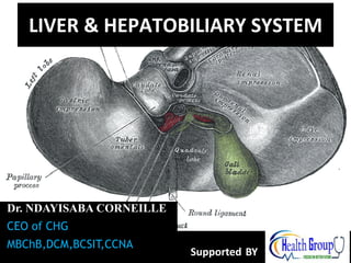
1. LIVER & HEPATOBILIARY SYSTEM
Dr. NDAYISABA CORNEILLE
CEO of CHG
MBChB,DCM,BCSIT,CCNA
Supported BY 1
2. objectives
• To define the hepatobiliary system
• To outline the embryological development
and congenital anomalies of the hepatobiliary
system.
• To describe the gross anatomy and histology
of the hepatobiliary system.
• To outline the clinical anomalies associated
with the hepatobiliary system
2
3. The hepatobiliary system
• Composed of the liver and the bile ducts.
• Mainly concerned with formation, transport,
concentration and secretion of bile.
• Bile is produced by the liver and transported
by the bile ducts into the small intestines
3
5. Embryological development
• Devt. starts during 3rd week of IUL as an
outgrowth of the distal end of the foregut.
• Endodermal in origin.
• The developing liver bud proliferates and
penetrates septum trnsversum. As
proliferation continues, the connecting stalk
forms the bile duct.
• A ventral outgrowth of the bile duct forms the
cystic duct and gall bladder.
5
6. Embryological development
• The liver primordium appears in the middle of the
third week as an outgrowth of the endodermal
epithelium at the distal end of the foregut.
• This outgrowth, the hepatic diverticulum, or liver
bud, consists of rapidly proliferating cells that
penetrate the septum transversum.
• As proliferation continues, the connection between
the hepatic diverticulum and the foregut
(duodenum) narrows, forming the bile duct
6
7. Embryological development
• A small outgrowth of the bile duct develops hence
forming the cystic duct and the gall bladder.
• During further development, epithelial liver cords
intermingle with the vitelline and umbilical veins,
which form hepatic sinusoids.
• Liver cords differentiate into the parenchyma (liver
cells) and form the lining of the biliary ducts
7
8. Embryological development
• Kupfer cells, heamatopoietic cells and
connective cells are mesodermal in origin.
• Formation of blood cells starts during 10th
week of IUL. At this time liver is approx 10% of
body weight.
• In the last 2 mths, this fxn reduces and by
birth it is 5% of total body wt.
• Bile production starts during 12th week of IUL
8
10. Congenital anomalies
• Accessory hepatic ducts.
• Duplication of the gall bladder.
• Extrahepatic biliary atresia: occurs in 1/15000
live births. Can be fatal if not corrected in
15%.
• Intrahepatic biliary duct atresia
• Hypoplasia: rare(1/100,000). Can result from
fetal infections. May be lethal or can run a
benign course.
10
12. Clinical significance of portal and
biliary system
• Portal hypertension: liver cirrhosis leading to
portal systemic anastomoses.
• Gall stones: obstruction of biliary system
causing jaundice.
12
13. Liver
• The liver is the largest gland in the body and has a wide
variety of functions
• Weight: 1/50 of body weight in adult & 1/20 of body
weight in infant
• It is exocrine(bile) & endocrine organ(Albumen ,
prothrombin & fibrinogen)
• Function of the liver
• Secretion of bile & bile salt
• Metabolism of carbohydrate, fat and protein
• Formation of heparin & anticoagulant substances
• Detoxication
• Storage of glycogen and vitamins
• Activation of vita .D 13
15. Surface anatomy of the liver
-The greater part
of the liver is
situated under
cover of the
right costal
margin
- Diaphragm
separates it from
the pleura,
lungs,
pericardium, and
heart. 15
16. Anterior View of the liver
• Right lobe
• Cut edge of the
Falciform ligament
left lobe
• Diverging cut edges
of the superior part
of the coronary
ligament
• Fundus of the gall
bladder
16
17. Surfaces of the liver, their relations &
impressions
• Postero - inferior
surface= visceral
surface
• Superior surface
= Diaphragmatic
surface
• Anterior surface
• Posterior surface
• Right surface
17
18. Posteroinfero surface= visceral surface
Relations
• I.V.C
• the esophagus
• the stomach
• the duodenum
• the right colic flexure
• the right kidney
• Right Suprarenal gland
• the gallbladder.
• Porta hepatic( bile duct,H.A.H.V)
• Fissure for lig. Venoosum & lesser
omentum
• Tubular omentum
• Lig.teres 18
20. Sup. Surface of the liver
• Right & left lobes
• Cut edge of the Falciform
ligament
• The cut edges of the
superior and inferior parts
of the coronary ligament
• The left triangular ligament
• The right triangular ligament
• Bare area of the liver (where
there is no peritoneum
covering the liver
• Groove for the inferior vena
cava and the hepatic veins
• Caudate lobe of the liver
more or less wrapping
around the groove of the
inferior vena cava
• Fundus of gall bladder
• Ligamentum teres
20
21. Relations of Sup . surface of liver
• Diaphragm
• Pleura & lung
• Pericardium &
heart
21
22. Relations of the liver Anteriorly
• Diaphragm
• Right & Left pleura and lung
• Costal cartilage
• Xiphoid process
• Anterior abdominal wall
22
24. Posterior relation of the liver
• Diaphragm
• Right Kidney
• Supra renal gland
• Transverse
colon(hepatic flexure
• Duodenum
• Gall bladder
• I.V.C
• Esophagus
• Fundus of stomach
24
25. Lobes of the liver
• Right Lobe
• Left lobe
• Quadrate lobe
• Caudate lobe
25
26. Separation of the four lobes of the liver:
• Right sagittal fossa -
groove for inferior
vena cava and gall
bladder
• left sagittal fissure -
contains the
Ligamentum
Venoosum and round
ligament of liver
• Transverse fissure (also
porta hepatis) - bile
ducts, portal vein,
hepatic arteries
26
27. Right Lobe
-Largest lobe
- Occupies the right
hypochondrium
- Divided into anterior
and posterior sections
by the right hepatic
vein
- Reidel’s Lobe extend
as far caudally as the
iliac crest
27
28. Left Lobe
–Varied in size
–Lies in the
epigastric and left
hypochondriac
regions
–Divided into lateral
and medial
segments by the
left hepatic vein
28
29. Lobes of the liver…..cont
Right & Left lobe separated by
• Falciform ligament
• Ligamentum Venosum
• Ligamentum teres
29
30. Caudate Lobe
-present in the posterior
surface from the Rt.
Lobe
Two processes
1- c- process
2- papillary process
Relations of caudate lobe
- Inferior the porta
hepatis
- The right the fossa for
the inferior vena cava
- The left the fossa for
the lig.venosum.
30
31. Quadrate lobe
Present on the inferior
surface from the Rt. Lobe
Relation
- Anterior anterior
margin of the liver
- Superior porta hepatis
- Right fossa for the
gallbladder
- Left by the fossa for lig.
teres
31
32. Porta hepatis
-It is the hilum of the liver
-It is found on the
posteroinferior surface
- lies between the caudate
and quadrate lobes
-Lesser omentum attach to its
margin
Contents
- Gallbladder anterior
- Hepatic Artery + nerve+
lymphatic node middle.
- Portal vein posterior
32
34. Peritoneum of the liver
• The liver is covered by
peritoneum (intraperitoneal
organ)except at bare area(it
is origin from septum
transversum)
• Inferior surface covered
with peritoneum of greater
sac except porta hepatis,
Gall Bladder & Lig. teres
fissure
• Right Lateral surface
covered by peritoneum,
related to diaphragm which
separate it from Right
Pleura , lung and the Right
Ribs (6-11) 34
35. 1- The Falciform ligament of liver
2- The Ligamentum teres hepatis
3- The coronary ligament
4- The right triangular ligament
5- The left triangular ligament
6- The Hepatogastric ligament
7- The hepatoduonedenal ligament
8- The Ligamentum Venoosum
The ligaments of the liver
35
38. • Falciform ligament of
liver
– Consists of double
peritoneal layer
– Sickle shape
– Extends from
anterior abdominal
wall (umbilicus) to
liver
– Free border of the
ligament contains
Ligamentum teres
(obliterated
umbilical vein) 38
39. • Coronary ligament
the area between upper
and lower layer of the
coronary ligament is the
bare area of liver which
contract with the
diaphragm;
• Left and right
triangular ligaments
formed by left and right
extremity of coronary
ligament
39
41. The Ligamentum
Venoosum
-Fibrous band that is
the remains of the
ductus venosus
- Is attached to the left
branch of the portal
vein and ascends in a
fissure on the visceral
surface of the liver to
be attached above to
the inferior vena cava
41
42. Bare area.
• This is the area of the liver between the
coronary ligament which is not lined by
visceral peritoneum.
• Ligamentum teres: obliterated umbilical vein.
• Ligamentum venosum: obliterated ductus
venosum which was joining the portal vein to
the inferior venacava hence bypassing the
liver in fetal circulation.
42
45. LIVER Histology
• lobules >> roughly
hexagonal structures
consisting of
hepatocytes. Radiate
outward from a central
vein.
• At each of the six
corners of a lobule is a
portal triad (
p.arteriole,p.venule &
bile duct)
•Between the
hepatocytes are the
liver sinusoids.
45
47. Where do the two blood supplies mix?
• Liver surrounded by a thin capsule at
portahepatic(it is thick)Glisson’s
capsule invests the liver and send septa
into liver subset subdivide the
parenchyma into lobules
47
48. Segmental anatomy of the liver
• Rt .& Lt. lobes anatomically
no morphological
significance. Separation by
ligaments (Falciform, lig.
Venoosum & Lig.teres)
• True morphological and
physiological division by a line
extend from fossa of GD to
fossa of I.V.C each has its
own arterial blood supply,
venous drainage and biliary
drainage
• No anastomosis between
divisions
• 3 major hepatic veins Rt,
Lt & central
• 8 segments based on hepatic
and portal venous segments 48
52. Blood supply of the liver
• Proper hepatic
artery The right
and left hepatic
arteries enter the
porta hepatis.
• The right hepatic
artery usually
gives off the cystic
artery, which runs
to the neck of the
gallbladder.
52
53. Blood Circulation through the Liver
• The blood vessels conveying
blood to the liver are the hepatic
artery (30%) and portal vein
(70%).
• The hepatic artery brings
oxygenated blood to the liver,
and the portal vein brings venous
blood rich in the products of
digestion, which have been
absorbed from the
gastrointestinal tract.
• The arterial and venous blood is
conducted to the central vein of
each liver lobule by the liver
sinusoids.
• The central veins drain into the
right and left hepatic veins, and
these leave the posterior surface
of the liver and open directly into
the inferior vena cava. 53
54. Vein drainage of the liver
• The portal vein divides
into right and left
terminal branches that
enter the porta
hepatis behind the
arteries.
• The hepatic veins
(three or more)
emerge from the
posterior surface of
the liver and drain into
the inferior vena cava.
54
56. Lymphatic drainage of the liver
• Liver produce large amount of lymph~
one third – one half of total body lymph
• Lymph leave the liver and enters several
lymph nod in porta hepatis efferent
vessels pass to celiac nods
• A few vessels pass from the bare area of
the liver through the diaphragm to the
posterior Mediastinal lymph nodes.
56
57. Nerve supply
• Sympathetic hepatic plexus>>> celiac
plexuses thoracic ganglion chain T1-T12
• Parasympathetic vagus nerve( anterior
part)
• Sympathetic and parasympathetic nerves
form the celiac plexus.
• The anterior vagal trunk gives rise to a large
hepatic branch, which passes directly to the
liver
57
58. Endoscopic retrograde cholangiopancreatography
(ERCP)
• It is a technique that combines the use of endoscopy
and fluoroscopy to diagnose and treat certain
problems of the biliary or pancreatic ductal systems.
Through the endoscope, the physician can see the
inside of the stomach and duodenum, and inject dyes
into the ducts in the biliary tree and pancreas so they
can be seen on X-rays.
• ERCP is used primarily to diagnose and treat
conditions of the bile ducts, including gallstones,
inflammatory strictures (scars), leaks (from trauma and
surgery), and cancer.
58
61. Biliary Apparatus :
• It collects bile from the liver, stores in the
gallbladder & transmits to 2nd part of duodenum.
• Gall bladder.
• Cystic duct.
• Right and left hepatic ducts which unite to form
Common Hepatic Duct.
• Common Bile duct formed by the union of cystic
duct and common hepatic duct.
61
65. ANATOMICAL POSITION OF GB
- Epigastric - Right
hypochondrium region
- At the tip of the 9th right
coastal cartilage
- Green muscular organ
- Pear-shaped, hollow
structure
- On inferior surface of liver
- Between quadrate and right
lobes
- Has a short mesentery
- Capacity 40- 60 cc
- Body and neck
Directed toward porta hepatis
65
66. Structure of GB
Fundus
-Ant:ant.abdominal wall
- Post.inf: transverscolon
Body
sup: liver
post.inf: Tranverse colon. End of 1st part
of doudenum , begins of 2nd part of
doudenum
Neck
- Form the cystic duct, 4cm
Hartmann’s Pouch
1. Lies between body and neck of gallbladder
2. A normal variation
3. May obscure cystic duct
4. If very large, may see cystic duct arising from
pouch 66
70. Arterial Supply to the Gallbladder
• Cystic artery
• Right hepatic artery
• Proper hepatic artery
• Common hepatic artery
70
71. Blood supply of GB:
- Cystic artery
branch of Rt. Hepatic
artery
- Cystic vein end in
portal vein
- Small branches (
arteries and veins run
between liver and gall
bladder
Common Hepatic
Artery
Proper Hepatic
Artery
Gastroduodenal
Artery
71
72. Lymphatic drainage of GB
1. Terminate at celiac nodes
2. Cystic node at neck of GB
a. Actually a hepatic node
b. Lies at junction of cystic
& common hepatic ducts
3. Other lymph vessels also drain
into hepatic nodes 72
74. Extra hepatic biliary system
Rt. hepatic duct
+
Lt hepatic duct
↓
Common hepatic duct
+
Cystic duct
↓
Common bile duct
- 4cm
- Descend in free edge of
lesser omentum
- Supra duodenal part
Retro duodenal part
Retro pancreatic part
Common bile duct
74
75. Bile duct……. parts and relations
-3 inc long
-1st part
-Located in right free margin of lesser omentum
- in front of the opening into the lesser sac
(Epiploic opening)
-Rt to hepatic artery and portal vein
- 2nd part
-Behind the 1st part of the duodenum
-Rt to the gastroduodenal artery
-3 rd part
-Posterior surface of the head of the pancreas
-Contact with main pancreatic duct
-Related with IVC, gastroduodenal artery, portal
vein
-End in the half second part of duodenum at
ampulla of Vater
75
80. Blood supply of CBD
Small arteries supplying CBD
a. Arise from cystic artery
b. Posterior branch of superior
pancreaticoduodenal artery
80
81. What is bile?
• Bile composed of water, ions, bile
acids, organic molecules (including
cholesterol, phospholipids,
bilirubin)
• Gallstones are mostly cholesterol
• Acids and salts emulsify fats for
absorption across wall of small
intestines into lacteal lymph
capillaries (review)
• Contains waste products from RBC
breakdown and other metabolic
processing (color of feces from
bilirubin in bile)(review)
• Ions buffer chyme from stomach
(review) 81
82. Cholelithiasis
• GB shows likely sites of
stone
formation/deposition
• Gangrene of
gallbladder is rare
• Stone in C.B.D obstruct
jaundice & pancreatitis
82
83. Gallbladder Diseases
1- Cholelithiasis & Cholecystitis
Cholecystitis = inflammation of GB
Cholelithisi = Stone(s) in GB
2- Obstructive jaundice: liver patterns
3- Gangrene of gall bladder rare
4- Congenital defects
83
84. END
THANKS FOR LISTENING
By
DR NDAYISABA CORNEILLE
MBChB,DCM,BCSIT,CCNA
Contact us:
amentalhealths@gmail.com/
ndayicoll@gmail.com
whatsaps :+256772497591
/+250788958241
84