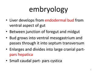
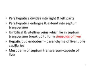
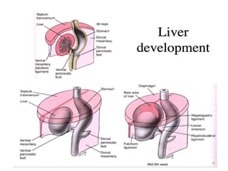
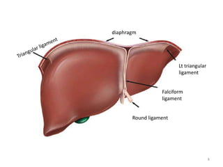
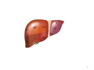
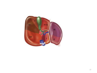
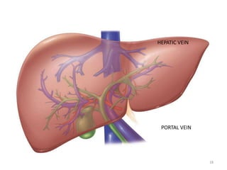
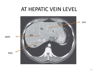
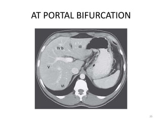
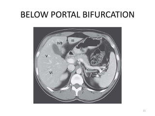
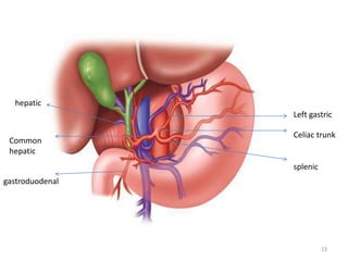
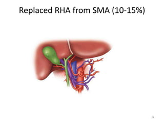
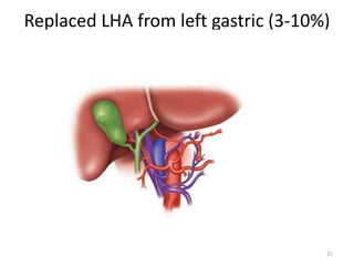
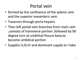
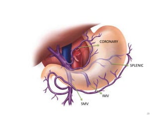
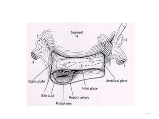
This document discusses liver anatomy, function tests, and imaging. It covers the embryological development of the liver, its lobes and ligament attachments. It describes the dual blood supply, biliary drainage system, and microscopic anatomy. Common liver function tests are outlined including those assessing synthesis, damage, and detoxification. Ultrasound imaging of the liver is also summarized, noting its advantages of being inexpensive and non-invasive but limitations in imaging certain areas.
Surgical Anatomy of the Liver : Ηepatectomies - Dimitris P. KorkolisDimitris P. Korkolis
- The liver is the largest gland in the body and has a wide variety of functions
- Weight: 1/50 of body weight in adult & 1/20 of body weight in infant
- It is exocrine(bile) & endocrine organ(Albumin , prothrombin & fibrinogen)
Function of the liver :
- Secretion of bile & bile salt
- Metabolism of carbohydrate, fat and protein
- Formation of heparin & anticoagulant substances
- Detoxication
- Storage of glycogen and vitamins
- Activation of vita .D
This is not a substitute for Books. Let it just help you understand some concepts in liver anatomy.
Continuation of this work will depend on your feedback. Stay Blessed.
To define the hepatobiliary system
To outline the embryological development and congenital anomalies of the hepatobiliary system.
To describe the gross anatomy and histology of the hepatobiliary system.
To outline the clinical anomalies associated with the hepatobiliary system
Composed of the liver and the bile ducts.
Mainly concerned with formation, transport, concentration and secretion of bile.
Bile is produced by the liver and transported by the bile ducts into the small intestines
Portal Vein and portocaval Anastomosis. Anatomy of portal vein, tributaries, branches and course, formations and relations. Anatomy of portal vein and adjacent structures, their relation to liver and intestine, relation to IVC and Aorta, clinical and applied anatomy for both undergraduates and postgraduates. portal hypertension is an increase in blood pressure, however, rather than being systematic, it's localized to the portal system. Portal hypertension is most commonly caused by liver cirrhosis which in itself can be caused by alcoholism or other liver disease. It can also be caused by blood clots in the portal vein and schistosomiasis amongst other things. This increase in blood pressure can affect areas of anastomosis between the portal vasculature which we just discussed and the caval musculature which are classified as the vessels not relating to the portal system resulting in pressure pushing larger blood volumes into these anastomotic areas. This in turn can cause the vessels to dilate and form varicose veins which can result in potentially fatal hemorrhage. Some of these important porto-caval anastomotic areas are listed below – the first vein being the portal vein and the second vein being the caval vein – the superior rectal and inferior rectal veins, the left gastric and esophageal veins, the colonic veins and the retroperitoneal veins and the para-umbilical and epigastric veins.
In severe cases, the last anastomosis mentioned between the para-umbilical veins which are the small veins that run within the round ligament of the liver and the epigastric veins which are found in the anterior abdominal wall can form large dilations. These dilations can form the clinical presentation caput medusa or the head of the medusa as the dilated veins look like the snakes of the head of the medusa or Gorgon from Greek mythology. In this image on the right, we can only see the beginnings of a presentation of the caput medusa as in a true caput medusa, the veins would be raised and enlarged.
Hello everyone! This is Nicole from Kenhub, and today we're going to talk about the hepatic portal vein.
We are going to discuss the hepatic portal vein and to do so we'll be using this image here which is a ventral view of the portal hepatic vein with the central portion of the liver cut out so we can see the portal vein and other portal vessels. You can also see the aorta just here as well as the inferior vena cava just posterior to the portal hepatic vein. The portal venous system is an important system that has its own unique flow and we'll talk about how this works in tandem with the venous system in the coming slides.
The portal vein is one of the most important vessels in the body.
Its main functions are to direct blood to the liver from the gastrointestinal tract and receive nutrient rich blood from the intestines.
The portal hepatic vein also receives blood from the spleen, the pancreas and the gallbladder which are channels within the vessel.
Operation “Blue Star” is the only event in the history of Independent India where the state went into war with its own people. Even after about 40 years it is not clear if it was culmination of states anger over people of the region, a political game of power or start of dictatorial chapter in the democratic setup.
The people of Punjab felt alienated from main stream due to denial of their just demands during a long democratic struggle since independence. As it happen all over the word, it led to militant struggle with great loss of lives of military, police and civilian personnel. Killing of Indira Gandhi and massacre of innocent Sikhs in Delhi and other India cities was also associated with this movement.
We all have good and bad thoughts from time to time and situation to situation. We are bombarded daily with spiraling thoughts(both negative and positive) creating all-consuming feel , making us difficult to manage with associated suffering. Good thoughts are like our Mob Signal (Positive thought) amidst noise(negative thought) in the atmosphere. Negative thoughts like noise outweigh positive thoughts. These thoughts often create unwanted confusion, trouble, stress and frustration in our mind as well as chaos in our physical world. Negative thoughts are also known as “distorted thinking”.
How to Split Bills in the Odoo 17 POS ModuleCeline George
Bills have a main role in point of sale procedure. It will help to track sales, handling payments and giving receipts to customers. Bill splitting also has an important role in POS. For example, If some friends come together for dinner and if they want to divide the bill then it is possible by POS bill splitting. This slide will show how to split bills in odoo 17 POS.
How to Create Map Views in the Odoo 17 ERPCeline George
The map views are useful for providing a geographical representation of data. They allow users to visualize and analyze the data in a more intuitive manner.
The French Revolution, which began in 1789, was a period of radical social and political upheaval in France. It marked the decline of absolute monarchies, the rise of secular and democratic republics, and the eventual rise of Napoleon Bonaparte. This revolutionary period is crucial in understanding the transition from feudalism to modernity in Europe.
For more information, visit-www.vavaclasses.com
How to Make a Field invisible in Odoo 17Celine George
It is possible to hide or invisible some fields in odoo. Commonly using “invisible” attribute in the field definition to invisible the fields. This slide will show how to make a field invisible in odoo 17.
Read| The latest issue of The Challenger is here! We are thrilled to announce that our school paper has qualified for the NATIONAL SCHOOLS PRESS CONFERENCE (NSPC) 2024. Thank you for your unwavering support and trust. Dive into the stories that made us stand out!
Model Attribute Check Company Auto PropertyCeline George
In Odoo, the multi-company feature allows you to manage multiple companies within a single Odoo database instance. Each company can have its own configurations while still sharing common resources such as products, customers, and suppliers.
Students, digital devices and success - Andreas Schleicher - 27 May 2024..pptxEduSkills OECD
Andreas Schleicher presents at the OECD webinar ‘Digital devices in schools: detrimental distraction or secret to success?’ on 27 May 2024. The presentation was based on findings from PISA 2022 results and the webinar helped launch the PISA in Focus ‘Managing screen time: How to protect and equip students against distraction’ https://www.oecd-ilibrary.org/education/managing-screen-time_7c225af4-en and the OECD Education Policy Perspective ‘Students, digital devices and success’ can be found here - https://oe.cd/il/5yV
2. embryology
• Liver develops from endodermal bud from
ventral aspect of gut
• Between junction of foregut and midgut
• Bud grows into ventral mesogastrium and
passes through it into septum transversum
• Enlarges and divides into large cranial part-
pars hepatica
• Small caudal part- pars cystica
2
3. • Pars hepatica divides into right & left parts
• Pars hepatica enlarges & extend into septum
transversum
• Umbilical & vitelline veins which lie in septum
transversum break up to form sinusoids of liver
• Hepatic bud endoderm- parenchyma of liver , bile
capillaries
• Mesoderm of septum transversum-capsule of
liver
3
5. ANATOMY
• Largest organ in body
• 1500 gm
• In right upper abdomen ,beneath diaphragm
,protected by rib cartilage
• surrounded by a fibrous sheath known as
Glisson’s capsule.
5
6. • Liver is held in place by several ligaments
• ROUND LIGAMENT
– a remnant of obliterated umbilical vein
– enters the left liver hilum at the front edge of the
falciform ligament
• FALCIFORM LIGAMENT
– separates the left lateral and left medial segments
along the umbilical Fissure
– anchors the liver to the anterior abdominal wall
• LEFT & RIGHT TRIANGULAR LIGAMENTS
– ,secure two sides of liver to diapragm
• CORONARY LIGAMENTS .
– Extensions of triangular ligament anteriorly on liver
6
7. • The right coronary ligament also extends from
the right undersurface of the liver to the
peritoneum overlying the right kidney, thereby
anchoring the liver to the right
retroperitoneum.
• Surgical importance -These ligaments (round,
falciform, triangular,and coronary) can be
divided in a bloodless plane to fully mobilize
the liver to facilitate hepatic resection.
7
9. • just to the left of the gallbladder fossa, the liver
attaches via hepatoduodenal and the gastrohepatic
ligaments .
• The HEPATODODENAL LIGAMENT
– is known as the porta hepatis
– contains the common bile duct, the hepatic artery, and
the portal vein.,nerves & lymphatics
– From the right side and deep (dorsal) to the porta hepatis
is the foramen of Winslow, also known as the
epiploic foramen.
– anatomical relationship of thesestructures is for the bile
duct to be within the free edge, the hepatic artery to be
above and medial, and the portal vein to lie posteriorly.
– This passage connects directly to the lesser sac and allows
complete vascular inflow control to the liver when the
hepatoduodenal ligament is clamped using the Pringle
maneuver. 9
11. SEGMENTAL ANATOMY
• Liver is grossly seperated into right & left lobes
by cantlie line
• REX-CANTLIE’S line – plane from gall bladder
fossa anteriorly to IVC posteriorly passing
through middle hepatic vein
• Also known as scissura or portal fissure
11
12. Couinaud functional classification
• Based on hepatic vein & portal vein
liver
Caudate
lobe
Left lobe
II,III,IV
Right lobe
V,VI,VII,VIII
Cantlie line
12
13. • The caudate lobe lies to the left and anterior of the IVC
• contains 3 subsegments:
– the Spiegel lobe,
– the paracaval portion,
– the caudate process
• Blood supply- independently from left & right portal
veins,hepatic arteries
• Drainage- independendtly into IVC
• Biliary drainage into both right & left hepatic duct at
their confluence
13
14. • Left hepatic vein divides left lobe into medial
& lateral sectors
• Left lateral- superior-II
inferior- III
• Left medial- superior -IV a
inferior- IV b
• Supplied by-left hepatic artery ,left portal vein
• Drainage-left hepatic duct,left hepatic vein
(II,III), middle hepatic vein (IV)
14
15. • Right hepatic vein divides right lobe into
anterior & posterior sectors
• Right anterior -inferior- V
• - superior- VIII
• Right posterior-inferior-VI
• - superior-VII
• Supplied by right hepatic artery,right portal
vein,
• Drainage- right hepatic duct,right hepatic
vein(V,VI,VII,VIII), middle hepatic vein(V,VIII)
15
26. Portal vein
• formed by the confluence of the splenic vein
and the superior mesenteric vein.
• Traverses through porta hepatis
• Then left portal vein branches from main vein
,consists of transverse portion ,followed by 90
degree turn at umbilical fissure base,to
become umbilical portion
• Supplies II,III,IV and dominant supply to I lobe
26
27. • Right portal vein branches near to liver
parenchyma,
• Supplies V,VI,VII,VIII lobes
• Other branches- coronary/left gastric vein
• -superior pancreaticoduodenal
vein
27
30. • The portal vein drains the splanchnic blood from
the stomach,pancreas, spleen, small intestine,
and majority of the colon to the liver before
returning to the systemic circulation.
• The portal vein pressure in normal individual is 3
to 5 mmHg.
• The portal vein is valveless,
• in the setting of portal hypertension, the
pressure can be quite high (20 to 30 mmHg).
• This results in decompression of the systemic
circulation through portocaval anastomoses,
most commonly via the coronary (left gastric)
vein, which produces esophageal and gastric
varices with a propensity for major hemorrhage
30
31. Hepatic Veins and Inferior Vena Cava
• venous drain of liver is by 3 hepatic veins
• Right, middle & left hepatic veins & drain into
suprahepatic IVC
• RHV drains V,VI,VII,VIII segments
• MHV drains IV,V,VIII segments
• LHV drains II,III segments
• Segment I / caudate drains directly into IVC
31
32. • In 95 % Persons, LHV , RHV together forms a
common trunk & drain into IVC
• In 20 % people , accessory RHV may be present in
hepatocaval ligament
• This can be source of torrential bleed during right
hepatectomy
• umbilical vein is additional vein that runs under
the falciform ligament, between the left and
middle veins,and empties into the left hepatic
vein
32
33. • Hepatic vein bisects the portal branches in
liver parenchyma
• RHV runs between right anterior & posterior
portal veins
• MHV runs between right anterior & left portal
vein
• LHV runs between branches of left portal vein
supplying II,III segments
33
34. Bile ducts & hepatic ducts
• Intrahepatic bile ducts along with respective
portal vein & hepatic artery is known as portal
pedicle
• the bile duct branches are usually superior to the
portal vein, whereas the hepatic artery branches
run inferiorly
• Left hepatic duct –present at base of umbilical
fissure -drains II,III,IV segments,
• Right hepatic duct formed by anterior & posterior
branches- drains V,VI,VII,VIII segments
34
35. • Caudate lobe has its own biliary drainage both
into left & right hepatic ducts
• Longer left hepatic duct joins with right
hepatic duct to form confluence anterior to
right portal vein
• This forms common hepatic duct- 4mm
diameter
• Below cystic duct ,it is common bile duct-6mm
diameter
35
36. Nerve supply
• Parasympathetic innervation – by vagus
– Left vagus- anterior hepatic branch
– Right vagus- posterior hepatic branch
• Sympathetic innervation is by
– greater thoracic sphlanchnic nerves,
– celiac ganglion
• Source of reffered pain to right shoulder &
scapula is phrenic nerve stimulation due to
glisson capsule stretch or diaphragm irritation
36
37. Lymphatic drainage
• Lymph produced in liver
• Drains via perisinusoidal space of disse &
periportal clefts of mall to
• Cystic duct lymph node,CBD,hepatic
artery,retropancreatic & celiac lymph
nodes(along hepatic arteries)
• Also drains superiorly into cardiophrenic
lymph nodes(along hepatic veins)
37
38. Plate system of liver
• On the ventral surface of liver connective tissue
condenses & forms system of fibrous plates &
sheaths
• They extend into liver along with biliovascular
structures
• consists of a sheath which surrounds the bile duct
and blood vessels (hepatic artery and portal vein)
• continuous with the Glisson’s capsule intra-
hepatically
• the hepatoduodenal ligament extra-hepatically.
38
39. • system also contains a large number of
lymphatics, nerves and a small vascular
network.
• Three plates are found in the hilar area:
– the hilar plate,
– the cystic plate and
– the umbilical plate.
39
42. • upper curved edge of the hilar plate is
dissected free from the undersurface of the
liver to expose the left hepatic duct, the biliary
ductal confluence and the right hepatic duct
when repairing a biliary stricture
42
43. • during cholecystectomy, the cystic plate is left
behind
• severe inflammation and fibrosis—
– , the cystic plate may become short and thick and
– the distance between the cystic plate and right portal
pedicle may be markedly reduced.
– the plane between the gallbladder and the cystic plate
may be obliterated
– may enter the plane behind the cystic plate.
• Continued dissection in this plane will eventually
reach the right portal pedicle and if the sheath of
the pedicle is breached, there is a very high risk
of injury to the right hepatic artery and right
portal vein
43
44. Microscopic anatomy
• Hepatic parenchyma organised into microscopic
functional units – acinus/lobule
• Lobule is formed bya central terminal hepatic
venule surrounded by 4 – 6 portal traids
• In between central venule and portal triad
hepatocytes are arranged in single layer
surrounded by sinusoids
• Blood flows from
• portal triad sinusoids central venule
44
45. • Bile flows from
• Hepatocyte terminal canaliculi bile ducts
portal triad
• Between portal triad & central venule there are 3
zones
• Zone 1 – periportal zone- rich in oxygen,nutrients
• Zone 2-intermediate zone
• Zone 3- perivenular zone-poor in
oxygen,nutrients
• Hence zone 3 more prone to ischemia & zone 1 to
toxic injury
45
47. LIVER FUNCTION TESTS
• SYNTHESIS FUNCTION
• Serum albumin
• Synthesized exclusively by hepatocyte 10 gm/day
• Half life – 20 days,hence not a marker for acute
dysfunction
• Low albumin-
– chronic liver disease
– neprotic syndrome
– protein malnutrition
– chronic infections
47
48. • Acute liver damage
• PT/INR
• All clotting factors except viii –syntesized in hepatocyte
• hAlf life 6hrs to 4 days
• 2,7,9,10 factors – collectively measured by serum
prothrombin time
• Elevated – hepatitis,cirrhosis,obstuctive jaundice, fat
malabsorption
• PT not correctable with parenteral vit k-
hepatitis,DIC,portal vein obstruction
48
49. • Serum enzymes that reflect damage to
hepatocytes
• Aminotransferases- within cytoplasm of
hepatocyte
• Aspartate aminotransferase AST / SGOT
• Alanine aminotransferase ALT / SGPT
• Normal levels < 20 u/L
• Upto 300 - nonspecific
49
50. • These enzymes are elevated in serum in great
amounts when there is damage to liver cell
membrane resulting in increased permeability
• > 1000 u/L - 1) viral hepatitis
• 2) ischemic liver injury
• 3)toxic / drug induced liver injury
• Also a sensitive indicator of transplant rejection
• AST/ALT > 2:1 – Alcohol liver disease
50
51. • Enzymes reflecting cholestasis
• Alkaline phosphatase
• 5 nucleotidase
• Gamma glutamyl transpeptidase
• ALP & 5 nucleotidase found in bile canalicular
membrane of hepatocytes
• GGT in bile duct epithelium & endoplasmi
reticulum of hepatocyte
51
52. • ALP – greater than a four fold rise of normal
value indicates cholestasis,liver
infiltration,amyloidosis,pagets disease
• Also present in bone and placenta
• Cannot differentiate intra or extrahepatic
biliary obstruction
52
53. • Serum bilurubin- produced in reticulo endothelial
system , breakdown product of eme containing protein
• Conjugated in in hepatocyte
• Excreted through bile
• Hepatocellular damage – both direct and indirect
bilurubin level increases
• Impaired excretion – increase in direct & total bilurubin
levels
• NORMAL serum bilurubin < 1mg/dl
• Direct – 30 % of normal
53
54. • DETOXIFICATION FUNCTION
– Liver converts ammonia to urea
– Normal < 60 mmol/L
– Raised levels – hepatic encephalopathy
• Serum ceruloplasmin
– Copper binding protein secreted by liver
– Decreased – wilson disease
• Alpha feto protein
• Pathognomonic for primary liver malignancy
54
56. ULTRASOUND
• Based on pulse- echo principle
• The ultrasound transducer converts electrical
energy to high-frequency sound energy that is
transmitted into tissue.
• waves are transmitted through the tissue, some
are reflected back,
• ultrasound image is produced when the receiver
detects those reflected waves.
• augmented by Doppler flow imaging.
• can detect the presence of blood vessels but also
can determine the direction and velocity of blood
flow 56
57. advantages
• inexpensive,
• widely available,
• no radiation exposure,
disadvantages
• Incomplete imaging ( at dome,beneath ribs,lesion
boundaries)
• Obesity & bowel gas reduce quality
• lower sensitivity and specificity of ultrasound
compared with CT and MRI
57
58. uses
• Liver size
• Detects lesions in liver
• Detects gall stones
• Detects biliary dilatation
• Differentiates solid from cystic masses
• Determines flow in vessels
• As a guide in percutaneous biopsy
• As a guide in aspiration of liver abscess
58
66. Contrast enhanced ultrasound
• Improved ability to differentiate among benign
and malignant lesions
• Contrast- gas microbubble agents
• Microbubbles are <10 μm
• improves delineation of liver lesions through
identification of dynamic enhancement patterns
and the vascular morphology of the lesion.
• some agents exhibit a late liver-specific phase
and accumulate in normal liver parenchyma after
the vascular enhancement has faded
66
67. Intra-operative ultrasound
• gold standard for detecting liver lesions
• tumor staging,
• visualization of intrahepatic vascular
structures ,
• guidance of resection plane by assessment of
the relationship of a mass to the vessels.
• biopsy of lesions and
• ablation of tumors
67
68. Ultrasound elastography
• used to assess the degree of fibrosis or cirrhosis in the
liver.
• Low-frequency vibrations transmitted through the liver
induce an elastic shear wave that is detected by
ultrasonography
• The velocity of the wave correlates with the stiffness of
the organ
• wave travels faster through fibrotic or cirrhotic tissues.
• Advantage over biopsy is that , it is non
invasive,rapid,aquires information from large surface
68
69. Computerised tomography
• Increased accuracy of diagnosis & staging
• CECT is widely used & best validated for liver
imaging
• Most of liver pathologies have similiar density
to liver parenchyma
• Liver has dual supply ,portal vein 75 % &
hepatic artery 25 %
• Many liver tumours recieve major supply from
hepatic artery
69
70. Arterial phase Portal/venous phase
Time since contrast
delivery
20 – 30 sec 60 – 70 sec
Tumours enhancement
Hypervascular conditions enhancement
Liver parenchyma enhanced
Hypervascular conditions like HCC,FNH,adenoma, metastasis from colorectal,RCC ,islet
cell tumour,carcinoid best seen in arterial phase
Mets from breast lung,hypovascular conditions, best seen in venous phase
70
71. MRI
• AS effective as CT
• Useful in case of allergy to iodinated contrast
• Biliary pathology
71