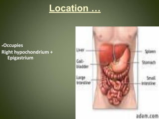
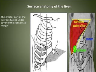
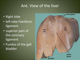
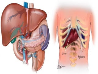
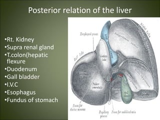
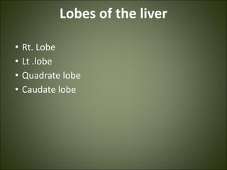
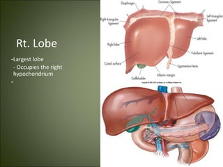
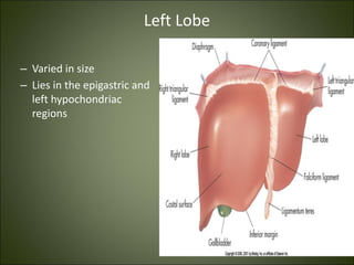
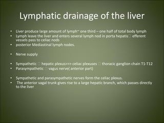
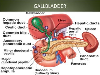
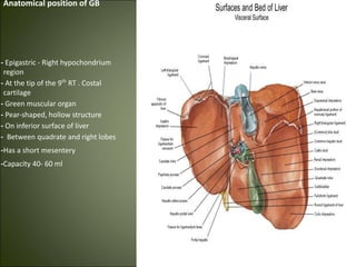
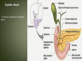
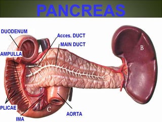
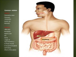
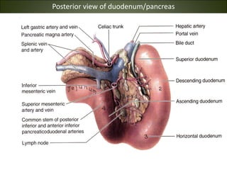
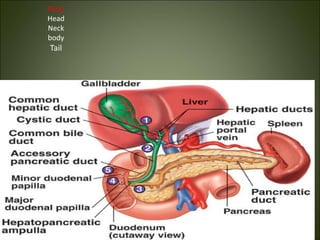
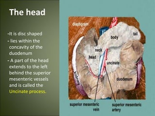
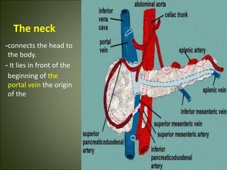
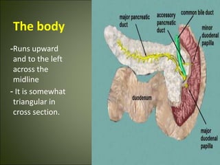
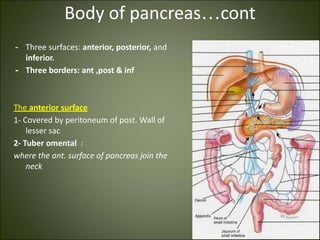
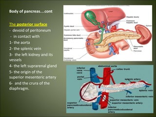
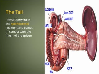
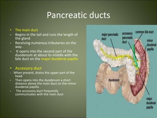
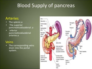
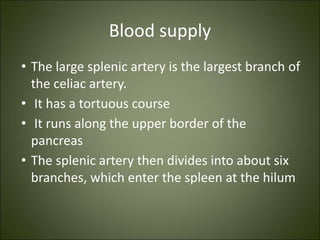
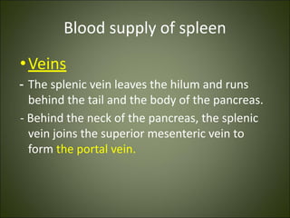
The liver, gallbladder, pancreas, and spleen are described. The liver is the largest gland and has many functions including bile production, carbohydrate and fat metabolism, and vitamin processing. The gallbladder stores and concentrates bile from the liver. The pancreas produces enzymes and hormones to aid digestion. The spleen filters blood and stores blood cells. All four organs have specific locations, blood supply from the hepatic and splenic arteries, and drainage into the portal vein and lymphatics.
To define the hepatobiliary system
To outline the embryological development and congenital anomalies of the hepatobiliary system.
To describe the gross anatomy and histology of the hepatobiliary system.
To outline the clinical anomalies associated with the hepatobiliary system
Composed of the liver and the bile ducts.
Mainly concerned with formation, transport, concentration and secretion of bile.
Bile is produced by the liver and transported by the bile ducts into the small intestines
MANAGEMENT OF ATRIOVENTRICULAR CONDUCTION BLOCK.pdfJim Jacob Roy
Cardiac conduction defects can occur due to various causes.
Atrioventricular conduction blocks ( AV blocks ) are classified into 3 types.
This document describes the acute management of AV block.
Pulmonary Thromboembolism - etilogy, types, medical- Surgical and nursing man...VarunMahajani
Disruption of blood supply to lung alveoli due to blockage of one or more pulmonary blood vessels is called as Pulmonary thromboembolism. In this presentation we will discuss its causes, types and its management in depth.
- Video recording of this lecture in English language: https://youtu.be/lK81BzxMqdo
- Video recording of this lecture in Arabic language: https://youtu.be/Ve4P0COk9OI
- Link to download the book free: https://nephrotube.blogspot.com/p/nephrotube-nephrology-books.html
- Link to NephroTube website: www.NephroTube.com
- Link to NephroTube social media accounts: https://nephrotube.blogspot.com/p/join-nephrotube-on-social-media.html
Report Back from SGO 2024: What’s the Latest in Cervical Cancer?bkling
Are you curious about what’s new in cervical cancer research or unsure what the findings mean? Join Dr. Emily Ko, a gynecologic oncologist at Penn Medicine, to learn about the latest updates from the Society of Gynecologic Oncology (SGO) 2024 Annual Meeting on Women’s Cancer. Dr. Ko will discuss what the research presented at the conference means for you and answer your questions about the new developments.
The prostate is an exocrine gland of the male mammalian reproductive system
It is a walnut-sized gland that forms part of the male reproductive system and is located in front of the rectum and just below the urinary bladder
Function is to store and secrete a clear, slightly alkaline fluid that constitutes 10-30% of the volume of the seminal fluid that along with the spermatozoa, constitutes semen
A healthy human prostate measures (4cm-vertical, by 3cm-horizontal, 2cm ant-post ).
It surrounds the urethra just below the urinary bladder. It has anterior, median, posterior and two lateral lobes
It’s work is regulated by androgens which are responsible for male sex characteristics
Generalised disease of the prostate due to hormonal derangement which leads to non malignant enlargement of the gland (increase in the number of epithelial cells and stromal tissue)to cause compression of the urethra leading to symptoms (LUTS
Prix Galien International 2024 Forum ProgramLevi Shapiro
June 20, 2024, Prix Galien International and Jerusalem Ethics Forum in ROME. Detailed agenda including panels:
- ADVANCES IN CARDIOLOGY: A NEW PARADIGM IS COMING
- WOMEN’S HEALTH: FERTILITY PRESERVATION
- WHAT’S NEW IN THE TREATMENT OF INFECTIOUS,
ONCOLOGICAL AND INFLAMMATORY SKIN DISEASES?
- ARTIFICIAL INTELLIGENCE AND ETHICS
- GENE THERAPY
- BEYOND BORDERS: GLOBAL INITIATIVES FOR DEMOCRATIZING LIFE SCIENCE TECHNOLOGIES AND PROMOTING ACCESS TO HEALTHCARE
- ETHICAL CHALLENGES IN LIFE SCIENCES
- Prix Galien International Awards Ceremony
Tom Selleck Health: A Comprehensive Look at the Iconic Actor’s Wellness Journeygreendigital
Tom Selleck, an enduring figure in Hollywood. has captivated audiences for decades with his rugged charm, iconic moustache. and memorable roles in television and film. From his breakout role as Thomas Magnum in Magnum P.I. to his current portrayal of Frank Reagan in Blue Bloods. Selleck's career has spanned over 50 years. But beyond his professional achievements. fans have often been curious about Tom Selleck Health. especially as he has aged in the public eye.
Follow us on: Pinterest
Introduction
Many have been interested in Tom Selleck health. not only because of his enduring presence on screen but also because of the challenges. and lifestyle choices he has faced and made over the years. This article delves into the various aspects of Tom Selleck health. exploring his fitness regimen, diet, mental health. and the challenges he has encountered as he ages. We'll look at how he maintains his well-being. the health issues he has faced, and his approach to ageing .
Early Life and Career
Childhood and Athletic Beginnings
Tom Selleck was born on January 29, 1945, in Detroit, Michigan, and grew up in Sherman Oaks, California. From an early age, he was involved in sports, particularly basketball. which played a significant role in his physical development. His athletic pursuits continued into college. where he attended the University of Southern California (USC) on a basketball scholarship. This early involvement in sports laid a strong foundation for his physical health and disciplined lifestyle.
Transition to Acting
Selleck's transition from an athlete to an actor came with its physical demands. His first significant role in "Magnum P.I." required him to perform various stunts and maintain a fit appearance. This role, which he played from 1980 to 1988. necessitated a rigorous fitness routine to meet the show's demands. setting the stage for his long-term commitment to health and wellness.
Fitness Regimen
Workout Routine
Tom Selleck health and fitness regimen has evolved. adapting to his changing roles and age. During his "Magnum, P.I." days. Selleck's workouts were intense and focused on building and maintaining muscle mass. His routine included weightlifting, cardiovascular exercises. and specific training for the stunts he performed on the show.
Selleck adjusted his fitness routine as he aged to suit his body's needs. Today, his workouts focus on maintaining flexibility, strength, and cardiovascular health. He incorporates low-impact exercises such as swimming, walking, and light weightlifting. This balanced approach helps him stay fit without putting undue strain on his joints and muscles.
Importance of Flexibility and Mobility
In recent years, Selleck has emphasized the importance of flexibility and mobility in his fitness regimen. Understanding the natural decline in muscle mass and joint flexibility with age. he includes stretching and yoga in his routine. These practices help prevent injuries, improve posture, and maintain mobilit
Couples presenting to the infertility clinic- Do they really have infertility...Sujoy Dasgupta
Dr Sujoy Dasgupta presented the study on "Couples presenting to the infertility clinic- Do they really have infertility? – The unexplored stories of non-consummation" in the 13th Congress of the Asia Pacific Initiative on Reproduction (ASPIRE 2024) at Manila on 24 May, 2024.
Flu Vaccine Alert in Bangalore Karnatakaaddon Scans
As flu season approaches, health officials in Bangalore, Karnataka, are urging residents to get their flu vaccinations. The seasonal flu, while common, can lead to severe health complications, particularly for vulnerable populations such as young children, the elderly, and those with underlying health conditions.
Dr. Vidisha Kumari, a leading epidemiologist in Bangalore, emphasizes the importance of getting vaccinated. "The flu vaccine is our best defense against the influenza virus. It not only protects individuals but also helps prevent the spread of the virus in our communities," he says.
This year, the flu season is expected to coincide with a potential increase in other respiratory illnesses. The Karnataka Health Department has launched an awareness campaign highlighting the significance of flu vaccinations. They have set up multiple vaccination centers across Bangalore, making it convenient for residents to receive their shots.
To encourage widespread vaccination, the government is also collaborating with local schools, workplaces, and community centers to facilitate vaccination drives. Special attention is being given to ensuring that the vaccine is accessible to all, including marginalized communities who may have limited access to healthcare.
Residents are reminded that the flu vaccine is safe and effective. Common side effects are mild and may include soreness at the injection site, mild fever, or muscle aches. These side effects are generally short-lived and far less severe than the flu itself.
Healthcare providers are also stressing the importance of continuing COVID-19 precautions. Wearing masks, practicing good hand hygiene, and maintaining social distancing are still crucial, especially in crowded places.
Protect yourself and your loved ones by getting vaccinated. Together, we can help keep Bangalore healthy and safe this flu season. For more information on vaccination centers and schedules, residents can visit the Karnataka Health Department’s official website or follow their social media pages.
Stay informed, stay safe, and get your flu shot today!
Anti ulcer drugs and their Advance pharmacology ||
Anti-ulcer drugs are medications used to prevent and treat ulcers in the stomach and upper part of the small intestine (duodenal ulcers). These ulcers are often caused by an imbalance between stomach acid and the mucosal lining, which protects the stomach lining.
||Scope: Overview of various classes of anti-ulcer drugs, their mechanisms of action, indications, side effects, and clinical considerations.
2. Liver
• The liver is the largest gland in the body and has a wide variety of
functions
• It is exocrine(bile) & endocrine organ(Albumen , prothrombin &
fibrinogen)
• Function of the liver
• Secretion of bile & bile salt
• Metabolism of carbohydrate, fat and protein
• Formation of heparin & anticoagulant substances
• Detoxication
• Storage of glycogen and vitamins
• Activation of vita .D
4. Surface anatomy of the liver
-The greater part of the
liver is situated under
cover of the right costal
margin
5. Ant. View of the liver
• Right lobe
• left lobe Falciform
ligament
• superior part of
the coronary
ligament
• Fundus of the gall
bladder
6. Postero- infero surface= visceral
surface
Relations
• I.V.C
• the esophagus
• the stomach
• the duodenum
• the right colic flexure
• the right kidney
• Rt. Suprarenal gland
• the gallbladder.
• Porta hepatic( bile duct,H.a.H.V)
• Fissure for lig. Venoosum & lesser omentum
• Tubular omentum
• Lig.teres
14. Left Lobe
– Varied in size
– Lies in the epigastric and
left hypochondriac
regions
15. Porta hepatis
-It is the hilum of the liver
-It is found on the
posteroinferior surface
- lies between the caudate
and quadrate lobes
Contents
- Gallbladder 🡪 ant.
- Hepatic Art + Heptic vein
nerve+ lymphatic node 🡪
middle.
- Portal vein 🡪 post.
17. Blood supply of the liver
• Proper hepatic artery 🡪
The right and left hepatic
arteries enter the porta
hepatis.
• The right hepatic artery
usually gives off the cystic
artery, which runs to the
neck of the gallbladder.
18. Vein drainage of the liver
• The portal vein divides
into right and left
terminal branches that
enter the porta hepatis
behind the arteries.
• The hepatic veins (three
or more) emerge from
the posterior surface of
the liver and drain into
the inferior vena cava.
19. Lymphatic drainage of the liver
• Liver produce large amount of lymph~ one third – one half of total body lymph
• Lymph leave the liver and enters several lymph nod in porta hepatis🡪 efferent
vessels pass to celiac nods
• posterior Mediastinal lymph nodes.
• Nerve supply
• Sympathetic 🡪 hepatic plexus>>> celiac plexuses 🡪 thoracic ganglion chain T1-T12
• Parasympathetic 🡪 vagus nerve( anterior part)
• Sympathetic and parasympathetic nerves form the celiac plexus.
• The anterior vagal trunk gives rise to a large hepatic branch, which passes directly
to the liver
21. Anatomical position of GB
- Epigastric - Right hypochondrium
region
- At the tip of the 9th
RT . Costal
cartilage
- Green muscular organ
- Pear-shaped, hollow structure
- On inferior surface of liver
- Between quadrate and right lobes
-Has a short mesentery
-Capacity 40- 60 ml
22. Structure of GB
Fundus
-Ant:ant.abdominal wall
- Post.inf: transverscolon
Body
sup: liver
post.inf: Tr.colon. End of 1st
part of doudenum ,
begins of 2nd
part of doudenum
Neck
- Form the cystic duct, 4cm
Hartmann’s Pouch
1. Lies between body and neck of gallbladder
2. A normal variation
3. If very large, may see cystic duct arising from pouch
24. Arterial Supply to the Gallbladder
• Cystic artery
• Right hepatic artery
• Proper hepatic artery
• Common hepatic artery
25. Blood supply of GB:
- Cystic artery🡪 branch of Rt.
Hepatic artery
- Cystic vein 🡪 end in portal
vein
- Small branches ( arteries and
veins run between liver and gall
bladder
Common Hepatic
Artery
Proper Hepatic
Artery
Gastroduodenal
Artery
26. Lymphatic drainage of GB
1. Terminate @ celiac nodes
2. Cystic node at neck of GB
3. Other lymph vessels also drain
into hepatic nodes
33. The head
-It is disc shaped
- lies within the
concavity of the
duodenum
- A part of the head
extends to the left
behind the superior
mesenteric vessels
and is called the
Uncinate process.
34. The neck
-connects the head to
the body.
- It lies in front of the
beginning of the
portal vein the origin
of the
35. The body
-Runs upward
and to the left
across the
midline
- It is somewhat
triangular in
cross section.
36. Body of pancreas…cont
- Three surfaces: anterior, posterior, and
inferior.
- Three borders: ant ,post & inf
The anterior surface
1- Covered by peritoneum of post. Wall of
lesser sac
2- Tuber omental :
where the ant. surface of pancreas join the
neck
37. Body of pancreas…cont
The posterior surface
- devoid of peritoneum
- in contact with
1- the aorta
2- the splenic vein
3- the left kidney and its
vessels
4- the left suprarenal gland
5- the origin of the
superior mesenteric artery
6- and the crura of the
diaphragm.
38. The Tail
- Passes forward in
the splenicorenal
ligament and comes
in contact with the
hilum of the spleen
39. Pancreatic ducts
• The main duct
- Begins in the tail and runs the length of
the gland
- Receiving numerous tributaries on the
way .
- It opens into the second part of the
duodenum at about its middle with the
bile duct on the major duodenal papilla
• Accessory duct
- When present, drains the upper part of the
head
- Then opens into the duodenum a short
distance above the main duct on the minor
duodenal papilla .
- The accessory duct frequently
communicates with the main duct
40. Blood Supply of pancreas
Arteries
• The splenic.a
• The superior
pancreaticoduodenal .a
• Inferior
pancreaticoduodenal
arteries.a
Veins
• The corresponding veins
drain into the portal
system.
42. Lymphatic drainage of pancreas
• Lymph nodes are
situated along
the arteries that
supply the gland.
• The efferent
vessels
ultimately drain
into the celiac
and superior
mesenteric
lymph nodes.
45. Spleen
Location and Description
• it is reddish & oval shaped
• the largest single mass of
lymphoid tissue in the body.
• and
• has a notched anterior border.
• location:
• Lt hypochondrium
- It lies just beneath the left half of the
diaphragm
- under the 9th, 10th, and 11th ribs.
- Its long axis parallel to the 10th rib
- Medial end is 4 cm away from mid line
post
- Lat.end is in left mid axillary line
46. Spleen
• Peritoneum
• The spleen is completely covered
with peritoneum 🡪 intraperitoneal
organ
• Two ligaments
1- the gastrosplenic omentum
(ligament)🡪 between the spleen &
the greater curvature of the stomach
(carrying the short gastric and left
gastroepiploic vessels)
2- splenicorenal ligament🡪 between
spleen & kidney (carrying the splenic
vessels and the tail of the pancreas).
47. Surfaces of spleen
2 surfaces
- Diaphragmatic surface
-Visceral surface.
-Anterior:
-Stomach, pancreas, left colic
flexures, left kidney.
-
Posterior :Diaphragm,
9,10,11 ribs
48. Spleen……cont
Gastric surface
- Extends forward, upward,
and medialward
- Broad and concave
- Related to stomach
Renal surface
- Directed medialward and
downward.
-It is somewhat flattened
- Related to Lt.kidney
50. Spleen..cont
• Borders of spleen
1- sup. Border
- It is free
- Sharp
- Thin
- Often notched(sup.notch)
, especially below
- It separates the
diaphragmatic surface
from the gastric surface
51. Spleen……cont
2- inferior border
- More rounded and blunter
- Separates the renal from the
diaphragmatic surface;
- It corresponds to the lower border of
the eleventh rib
- lies between the diaphragm and left
kidney. The
52. Spleen…cont
• 2 Ends
• Med.end🡪 sup &
back
• 4cm away from mid
line post
• Lat.end🡪 in left mid
axillary line
53. Blood supply
• The large splenic artery is the largest branch of
the celiac artery.
• It has a tortuous course
• It runs along the upper border of the
pancreas
• The splenic artery then divides into about six
branches, which enter the spleen at the hilum
54. Blood supply of spleen
•Veins
- The splenic vein leaves the hilum and runs
behind the tail and the body of the pancreas.
- Behind the neck of the pancreas, the splenic
vein joins the superior mesenteric vein to
form the portal vein.
55. Lymphatic Drainage of spleen
• The lymph vessels emerge from the hilum and
pass through a few lymph nodes along the
course of the splenic artery and then drain
into the celiac nodes.
56. Nerve Supply of spleen
• The nerves accompany the splenic artery and
are derived from the celiac plexus.