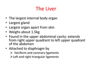
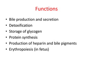
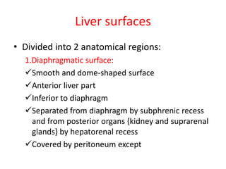
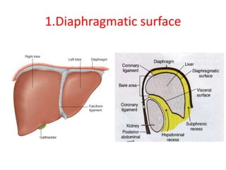
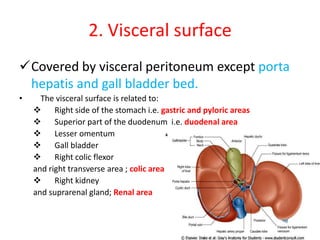
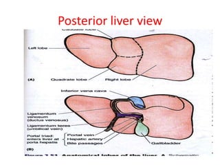
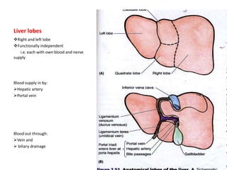
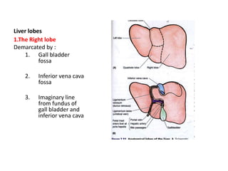
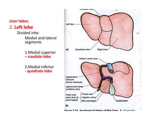
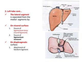
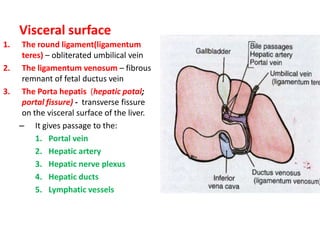
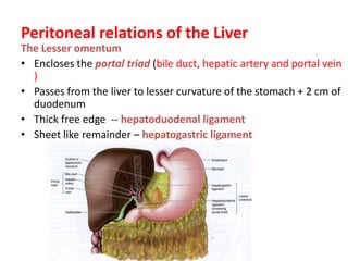
This is not a substitute for Books. Let it just help you understand some concepts in liver anatomy.
Continuation of this work will depend on your feedback. Stay Blessed.
anatomy of large intestine, its section, ceacum, ascending colon, transverse colon, descending colon, sigmoid colon, functions of large intestine , relations of each components of large intestine, carddinal siggns of large intestine, iliocecal junstion, difference between large and small intestine. abdominal angina, superior mesenteric and inferior mesenteric artery, lymphatic drainage, colonoscophy,
anatomy of stomach,functions of stomach, location, shape position and parts of stomach,orifices of stomach, curvature of stomach, relations of stomach, blood supply, innervation, lymphatic drainage, clinical relation , GERD, peptic ulcer,
It includes structure of stomach, stomach bed, function and internal structure.
Give your like & share with other nursing students.
The stomach is an important organ and the most dilated portion of the digestive system. The esophagus precedes it, and the small intestine follows. It is a large, muscular, and hollow organ allowing for a capacity to hold food. It is comprised of 4 main regions, the cardia, fundus, body, and pylorus.
Surgical Anatomy of the Liver : Ηepatectomies - Dimitris P. KorkolisDimitris P. Korkolis
- The liver is the largest gland in the body and has a wide variety of functions
- Weight: 1/50 of body weight in adult & 1/20 of body weight in infant
- It is exocrine(bile) & endocrine organ(Albumin , prothrombin & fibrinogen)
Function of the liver :
- Secretion of bile & bile salt
- Metabolism of carbohydrate, fat and protein
- Formation of heparin & anticoagulant substances
- Detoxication
- Storage of glycogen and vitamins
- Activation of vita .D
The gall bladder is located in the junction of the right ninth costal cartilage and lateral border of the rectus abdominis.
It is a pear shaped sac lying on the inferior surface of the liver in a fossa between the right and quadrate lobes with a capacity of about 30 to 50 mL.
Liver is the largest internal organ of the body weighing about 1500g in adults. It occupies the right hypochondrium and extends into the epigastrium and left hypochondrium .
Muscle is a specialized tissue which brings
xThe cells exhibit cross-striations under
functional unit of muscle fiber.
about movement by contraction.
xMuscle tissue is made up of cells called
myocytes. These usually appear as fibers
known as muscle fibers.
xEach muscle fiber is covered by a
membrane known as sarcolemma and a
cytoplasm known as sarcoplasm.
xCytoplasm of each muscle fiber contains
numerous longitudinal threadlike struc
tures called myofibrils, which are made
up of different types of muscle protein
(mainly actin and myosin).
xIt is also rich in mitochondria and
glycogen, which provide energy for it.
xNumerous mitochondria (sarcosomes)
and endoplasmic reticulum (sarcoplasmic
reticulum) are also seen.
Classification of Muscle
Tissue
Based on the appearance of contractile
cells, the muscle tissue is classified as the
following:
xSkeletal/striated/voluntary muscle/
striped muscle.
xCardiac/involuntary muscle.
xSmooth muscle/involuntary/visceral
muscle.
Skeletal Muscle
xSkeletal muscle is attached to bone and
is responsible for movement of axial and
light microscope; hence, it is called stri
ated muscle.
xSkeletal muscles have limited capacity of
regeneration.
Microscopic Structure (Longitudinal
Section) of Skeletal Muscle
xLongitudinal section of skeletal muscles
shows long unbranched cylindrical
muscle fibers running parallel to each
other (Figs.8.1 and 8.2).
xLength of muscle fiber is variable ranging
from few millimeter to many centimeter.
xEach muscle fiber shows multinucleated
flat oval nucleus located peripherally
underneath the sarcolemma.
xMultinucleated appearance of muscle
fiber is due to the fusion of multiple
myoblasts during the embryonic life.
xThe sarcoplasm contains numerous
myofibrils (Fig.8.3).
xUnder light microscope, myofibrils are
seen as dark and light bands.
xThe dark bands are A-bands (anisotropic
under polarized light) and light bands are
I-bands (isotropic under polarized light).
xStriated appearance is mainly due to the
regular arrangement of actin and myosin
myofilaments.
xThe middle of Aband has a light area
known as Hband.
xThe center of H-band has a dark line
known as M-line.
xThe center of I-band is bisected by Z-line.
xThe area between two Z-lines is called
sarcomere, which is the structural and
appendicular skeleton.
To define the hepatobiliary system
To outline the embryological development and congenital anomalies of the hepatobiliary system.
To describe the gross anatomy and histology of the hepatobiliary system.
To outline the clinical anomalies associated with the hepatobiliary system
Composed of the liver and the bile ducts.
Mainly concerned with formation, transport, concentration and secretion of bile.
Bile is produced by the liver and transported by the bile ducts into the small intestines
2. The Liver
• The largest internal body organ
• Largest gland
• Largest organ apart from skin
• Weighs about 1.5kg
• Found in the upper abdominal cavity: extends
from right upper quadrant to left upper quadrant
of the abdomen
• Attached to diaphragm by
falciform and coronary ligaments
Left and right triangular ligaments
3. Functions
• Bile production and secretion
• Detoxification
• Storage of glycogen
• Protein synthesis
• Production of heparin and bile pigments
• Erythropoiesis (in fetus)
4. Liver surfaces
• Divided into 2 anatomical regions:
1.Diaphragmatic surface:
Smooth and dome-shaped surface
Anterior liver part
Inferior to diaphragm
Separated from diaphragm by subphrenic recess
and from posterior organs {kidney and suprarenal
glands} by hepatorenal recess
Covered by peritoneum except
6. 2. Visceral surface
Covered by visceral peritoneum except porta
hepatis and gall bladder bed.
• The visceral surface is related to:
Right side of the stomach i.e. gastric and pyloric areas
Superior part of the duodenum i.e. duodenal area
Lesser omentum
Gall bladder
Right colic flexor
and right transverse area ; colic area
Right kidney
and suprarenal gland; Renal area
8. Liver lobes
Right and left lobe
Functionally independent
i.e. each with own blood and nerve
supply
Blood supply in by:
Hepatic artery
Portal vein
Blood out through:
Vein and
biliary drainage
9. Liver lobes
1.The Right lobe
Demarcated by :
1. Gall bladder
fossa
2. Inferior vena cava
fossa
3. Imaginary line
from fundus of
gall bladder and
inferior vena cava
10. Liver lobes
2. Left lobe
Divided into:
Medial and lateral
segments
1.Medial superior
– caudate lobe
2.Medial inferior
- quadrate lobe
11. 2. Left lobe cont…
The lateral segment
is separated from the
medial segments by:
On visceral surface:
1. fissure of
ligamentum teres
(round ligament)
2. fissure of
ligamentum
venosum
On diaphragmatic
surface:
1. Attachment of
falciform ligament
12. Visceral surface
1. The round ligament(ligamentum
teres) – obliterated umbilical vein
2. The ligamentum venosum – fibrous
remnant of fetal ductus vein
3. The Porta hepatis (hepatic potal;
portal fissure) - transverse fissure
on the visceral surface of the liver.
– It gives passage to the:
1. Portal vein
2. Hepatic artery
3. Hepatic nerve plexus
4. Hepatic ducts
5. Lymphatic vessels
13. Peritoneal relations of the Liver
The Lesser omentum
• Encloses the portal triad (bile duct, hepatic artery and portal vein
)
• Passes from the liver to lesser curvature of the stomach + 2 cm of
duodenum
• Thick free edge -- hepatoduodenal ligament
• Sheet like remainder – hepatogastric ligament
14. To be continued ….
• To be continued………………..