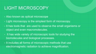
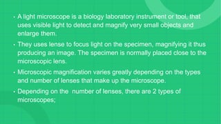
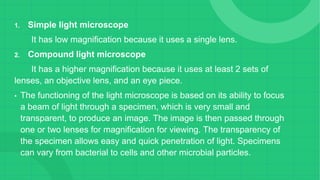
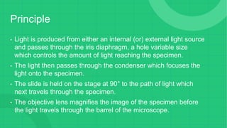
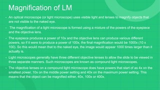
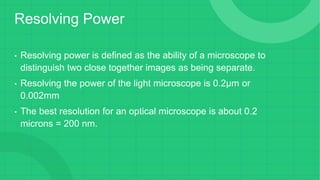
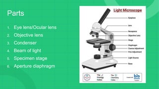
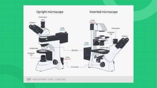
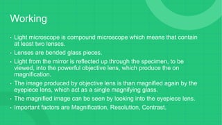
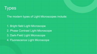
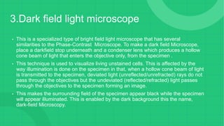
9953056974 Young Call Girls In Mahavir enclave Indian Quality Escort service
light microscopy seminar.pptx
1.
2. LIGHT MICROSCOPY
• Also known as optical microscope
• Light microscopy is the simplest form of microscopy.
• It has tools that are used to observe the small organisms or
object and even macromolecules.
• It has wide variety of microscopic tools for studying the
biomolecules and biological processes.
• It includes all forms of microscopic methods that use
electromagnetic radiation to achieve magnification.
3. • A light microscope is a biology laboratory instrument or tool, that
uses visible light to detect and magnify very small objects and
enlarge them.
• They uses lense to focus light on the specimen, magnifying it thus
producing an image. The specimen is normally placed close to the
microscopic lens.
• Microscopic magnification varies greatly depending on the types
and number of lenses that make up the microscope.
• Depending on the number of lenses, there are 2 types of
microscopes;
4. 1. Simple light microscope
It has low magnification because it uses a single lens.
2. Compound light microscope
It has a higher magnification because it uses at least 2 sets of
lenses, an objective lens, and an eye piece.
• The functioning of the light microscope is based on its ability to focus
a beam of light through a specimen, which is very small and
transparent, to produce an image. The image is then passed through
one or two lenses for magnification for viewing. The transparency of
the specimen allows easy and quick penetration of light. Specimens
can vary from bacterial to cells and other microbial particles.
5. Principle
• Light is produced from either an internal (or) external light source
and passes through the iris diaphragm, a hole variable size
which controls the amount of light reaching the specimen.
• The light then passes through the condenser which focuses the
light onto the specimen.
• The slide is held on the stage at 90° to the path of light which
next travels through the specimen.
• The objective lens magnifies the image of the specimen before
the light travels through the barrel of the microscope.
6. • Finally light is passes through
the eyepieces lens & into the
viewer’s eye which sends
impulses to the brain which in
turn interprets the image.
7. Magnification of LM
• An optical microscope (or light microscope) uses visible light and lenses to magnify objects that
are not visible to the naked eye.
• The magnification of a light microscope is formed using a mixture of the powers of the eyepiece
and the objective lens.
• The eyepiece produces a power of 10x and the objective lens can produce various different
powers, so if it were to produce a power of 100x, the final magnification would be 1000x (10 x
100). So this would mean that to the naked eye, the image would appear 1000 times larger than it
actually is.
• Light microscopes generally have three different objective lenses to allow the slide to be viewed in
three separate manners. Such microscopes are known as compound light microscopes.
• The objective lenses on a compound light microscope does have powers that start of as 4x on the
smallest power, 10x on the middle power setting and 40x on the maximum power setting. This
means that the object can be magnified either, 40x, 100x or 400x.
8. Resolving Power
• Resolving power is defined as the ability of a microscope to
distinguish two close together images as being separate.
• Resolving the power of the light microscope is 0.2μm or
0.002mm
• The best resolution for an optical microscope is about 0.2
microns = 200 nm.
10. • Objective lens – made up of six or more glasses, which make the
image clear from the object.
• Condenser – mounted below the stage which focuses a beam of light
onto the specimen. It can be fixed or movable, to adjust the quality of
light, but this entirely depends on the microscope.
• Stage – where the specimen is placed, allowing movement of the
specimen around for better viewing with the flexible knobs and it is
where the light is focused on.
• Light illuminator/mirror – found at the base or on the microbes of the
nosepiece.
• Aperture diaphragm – known as the contrast, which controls the
diameter of the beam of light that passes through the condenser.
11. • Light microscopes come in two designs: upright and inverted.
• Upright microscope: In an upright microscope, the objective
turret is usually fixed and the image is focused by moving the
sample stage up and down.
• Inverted microscope: In an inverted microscope, the sample
stage is fixed and objective turret is moved up and down to focus
the final image.
12.
13.
14. Working
• Light microscope is compound microscope which means that contain
at least two lenses.
• Lenses are bended glass pieces.
• Light from the mirror is reflected up through the specimen, to be
viewed, into the powerful objective lens, which produce the on
magnification.
• The image produced by objective lens is than magnified again by the
eyepiece lens, which act as a single magnifying glass.
• The magnified image can be seen by looking into the eyepiece lens.
• Important factors are Magnification, Resolution, Contrast.
15. Types
The modern types of Light Microscopes include:
1. Bright field Light Microscope
2. Phase Contrast Light Microscope
3. Dark-Field Light Microscope
4. Fluorescence Light Microscope
16. 1.Bright field light Microscope
• This is the most basic optical Microscope used in microbiology laboratories
which produces a dark image against a bright background.
• Made up of two lenses, it is widely used to view plant and animal cell
organelles including some parasites such as Paramecium after staining with
basic stains.
• Its functionality is based on being able to provide a high-resolution image,
which highly depends on the proper use of the microscope. This means that
an adequate amount of light will enable sufficient focusing of the image, to
produce a quality image.
• It is also known as a compound light microscope.
17. Parts of BLM
• It is composed of:
• Two lenses which include the objective lens and
the eyepiece or ocular lens.
• Objective lens is made up of six or more glasses,
which make the image clear from the object
• The condenser is mounted below the stage which
focuses a beam of light onto the specimen. It can
be fixed or movable, to adjust the quality of light,
but this entirely depends on the microscope.
• They are held together by a sturdy metallic
curved back used as an arm and a stand at the
bottom, known as the base, of the microscope.
The arm and the base hold all the parts of the
microscope.
18. •
• The stage where the specimen is placed, allowing movement of the specimen around for better
viewing with the flexible knobs and it is where the light is focused on.
• Two focusing knobs i.e the fine adjustment knob and the coarse adjustment knob, found on the
microscopes’ arm, which can move the stage or the nosepiece to focus on the image. The
sharpen the image clarity.
• It has a light illuminator or a mirror found at the base or on the microbes of the nosepiece.
• The nosepiece has about three to five objective lenses with different magnifying power. It can
move round to any position depending on the objective lens to focus on the image.
• An aperture diaphragm also is known as the contrast, which controls the diameter of the beam
of light that passes through the condenser, in that, when the condenser is almost closed, the
light comes through to the center of the condenser creating high contrast. But when the
condenser is widely open, the image is very bright with very low contrast.
19. Applications of BLM
• Vastly used in Microbiology, this microscope is used to view
fixed and live specimens, that have been stained with basic
stains.
• This gives contrast for easy visibility under the microscope.
• Therefore it can be used to identify basic bacteria cells and
parasitic protozoans such as Paramecium.
20. 2.Phase contrast light microscope
• This is a type of optical microscope whereby small light deviations known as phase
shifts occur during light penetration into the unstained specimen. These phase shifts
are converted into the image to mean, when light passes through the opaque
specimen, the phase shifts brighten the specimen forming an illuminated (bright)
image in the background.
• The phase-contrast microscope produces high contrast images when using a
transparent specimen more so those of microbial cultures, thin tissue fragments, cell
tissues, and subcellular particles.
• The principle behind the working of the phase-contrast microscope is the use of an
optical method to transform a specimen into an amplitude image, that’s viewed by the
eyepiece of the microscope.
21. • The PCM can be used to view unstained cells also known as the
phase objects, which means that the morphology of the cell is
maintained and the cells can be observed in their natural state, in high
contrast and efficient clarity. This is because if the specimens are
stained and fixed, they kill most cells, a characteristic that is uniquely
undone by the brightfield light microscope.
• The shifts that occur during light penetration, become converted to
changes in amplitude which causes the image contrast.
• Coupled with contrast-enhancing elements such as fluorescence, they
produce better visuals of the specimens’ image.
22. • The instrumentation of the Phase Contrast Microscope is based on its light pathways from
receiving the source of light to the visualization of the image.
• Therefore its sequentially made up of:
1. Light source (Mercury arc lamp)
2. Collective lens
3. Aperture
4. Condenser
5. Condenser annular
6. Specimen
7. Objective
8. Phase plate
9. Deflected light
10. Phase ring
23. Application PCM
• Determine morphologies of living cells such as plant and animal
cells
• Studying microbial motility and structures of locomotion
• To detect certain microbial elements such as the bacterial
endospores
24. 3.Dark field light microscope
• This is a specialized type of bright field light microscope that has several
similarities to the Phase-Contrast Microscope. To make a dark field Microscope,
place a darkfield stop underneath and a condenser lens which produces a hollow
cone beam of light that enters the objective only, from the specimen .
• This technique is used to visualize living unstained cells. This is affected by the
way illumination is done on the specimen in that, when a hollow cone beam of light
is transmitted to the specimen, deviated light (unreflected/unrefracted) rays do not
pass through the objectives but the undeviated (reflected/refracted) light passes
through the objectives to the specimen forming an image.
• This makes the surrounding field of the specimen appear black while the specimen
will appear illuminated. This is enabled by the dark background this the name,
dark-field Microscopy.
25. Applications of DFM
• It is used to visualize the internal organs of larger cells such as
the eukaryotic cells
• Identification of bacterial cells with distinctive shapes such as
Treponema pallidum, a causative agent of syphilis.
26. 4.The Fluorescent Microscope
• In the case of the fluorescent Microscope, the specimen emits light by
adding a dye molecule to the specimen.
• This dye molecule will normally become excited when it absorbs light
energy, hence it releases any trapped energy as light.
• The light energy that is released by the excited molecule has a long
wavelength compared to its radiating light.
• The dye molecule is normally a fluorochrome, that fluoresces when exposed
to the light of a certain specific wavelength. The image formed is a
fluorochrome-labeled image from the emitted light
27. • The principle behind this working mechanism is that the fluorescent
microscope will expose the specimen to ultra or violet or blue light,
which forms an image of the specimen that is emanated by the
fluorescent light.
• They have a mercury vapor arc lamp that produces an intense beam
of light that passes through an exciter filter.
• The exciter filter functions to transmit a specific wavelength to the
fluorochrome stained specimen, producing the fluorochrome-labeled
image, at the objective.
• After the objective, there is a barrier filter that functions primarily to
remove any ultraviolet radiation that may be harmful to the viewer’s
light, thus reducing the contrast of the image.
28. Applications of FM
• Used in the visualization of bacterial agents such as
Mycobacterium tuberculosis.
• Used to identify specific antibodies produced against bacterial
antigens/pathogens in immunofluorescence techniques by
labeling the antibodies with fluorochromes.
• Used in ecological studies to identify and observe
microorganisms labeled by the fluorochromes
• It can also be used to differentiate between dead and live
bacteria by the color they emit when treated with special stains
29. Applications of LM
1. A combination of staining and light microscopy can allow
scientists to identify different kinds of bacteria.
2. Light microscope play a large part in today’s biology.
3. Observe real time movement in cells and organisms.
4. Biologist use microbes to observe objects and details at a
cellular level to learn more about the building blocks of all
organisms.
30. Advantages LM
1. Relatively easy to use.
2. Small and Lightweight.
3. It offer high levels of observational quality.
4. It is unaffected by electromagnetic fields.
5. Do not require radiation to operate.
6. Require very little training.
7. Allow to observe living organisms.
8. Have minor maintenance cost compared to other models.
9. Fully adjustable to the comfort level of the user.
31. Disadvantages LM
1. Have a lower resolution.
2. Making it challenging to view living internal structures.
3. Cannot operate in darkness.
4. Cannot provide 3D renderings.
5. Very low magnification caps.