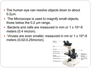
Dr. Sarah Abdulsalam
Microscopes
1st STAGE / LEC2/ Lab
Instrumentation theory
WHAT IS A MICROSCOPE ?
 Origin of A microscope word came: (from the Ancient
Greek: mikron- "small“ and scopeos-"to look")
 Microscope: is an instrument used to see objects that
are too small for the naked eye.
 A microscope is an instrument that produces an
accurately enlarged image of small objects.
 Microscopy: The science of investigating small objects
using such an instrument is called microscopy.
Historical background :
 1590 - Hans Janssen and his son Zacharias Janssen,
developed first microscope.
 1609 - Galileo Galilei - occhiolino or compound
microscope.
 1674 – Anton van Leeuwenhoek built a simple
microscope with only one lens to examine blood, yeast,
insects and many other tiny objects. Leeuwenhoek was
the first person to describe bacteria.
 1931 – Ernst Ruska co-invented the electron
Microscope for which he won the Nobel Prize in Physics
in 1986.
Anton van
leeuwenhoek
Robert hooke
 The human eye can resolve objects down to about
0.2μm.
 The Microscope is used to magnify small objects,
those below the 0.2 μm range.
 Bacteria and cells are measured in mm or 1 x 10^-6
meters (0.4 micron).
 Viruses are even smaller, measured in nm or 1 x 10^-9
meters (0.02-0.25micron).
CLASSIFICATION OF MICROSCOPES
 Depending on lens system:
I. Simple
II. Compound
 Depending on optical technique:
A. LIGHT MICROSCOPE : use sunlight or artificial light.
 A. Bright field microscope.
 B. Dark field microscope.
 C. Phase contrast microscope.
 D. Fluorescence microscope.
B. ELECTRON MICROSCOPE : use of electron.
 1. Transmission electron microscope.
 2. Scanning electron microscope.
Principles of Light Microscopy
 Microscopes provide three important qualities:
1. Magnification: Degree of enlargement.
 No of times the length, breadth or diameter, of an object is multiplied.
 It depends upon –
I. Optical tube length
II. Focal length of objective
III. Magnifying power of eye piece
 Our microscope has four objective lens settings (scanning lens, 4X; low power
lens,10X; high dry
power lens, 100X oil immersion lens
 TOTAL MAGNIFICATION = magnification of the eyepiece X magnification
of the objective.
 Power of objective X Power of ocular =Total magnification
 10× low power objective × 10× = 100×
 40× high dry objective × 10× = 400×
 100× oil immersion objective × 10× = 1,000×
2. Resolution :Ability to reveal closely adjacent structural details as separate and
distinct.
3. Contrast: Differences in intensity between two objects, or between an object
and background.
Oil immersion:
 The white light used in a compound light microscope
has relatively long wave length and cannot resolve
structures smaller than about 0.2 μm.
 Oil immersion is placed between the glass and
objective lens.
 The oil immersion has the same refractive index as
glass of the microscope.
 The oil enhances the resolution by preventing light rays
from dispersing and changing wave length after passing
through the specimens.
SIMPLE MICROSCOPE
 Because of the limited ability of the eye's lens to change
its shape, objects brought very close to the eye cannot
have their images brought to focus on the retina.
 The "simple microscope" or magnifying glass reached
its highest state of perfection, in the 1600's, in the work
of Anton von Leeuwenhoek who was able to see single-
celled animals ("animalcules") and even some larger
bacteria.
LIGHT MICROSCOPY
THE COMPOUND MICROSCOPE
 In light microscopy, light typically passes through a
specimen and then through a series of magnifying
lenses.
 Oil immersion lens increases resolution.
 Have one or two ocular lenses.
 Light is transmitted and focussed by mirror and
condenser.
 The refracted light is collected by an objective where
primary image of the object is formed, it is real, inverted
enlarged image of the object.
 The eyepiece further magnifies this primary image into
virtual, erect enlarged image, this is the final image that
Brightfield Microscope (Compound Light
Microscope)
 Brightfield Microscope is also known as
the Compound Light Microscope. It is an optical
microscope that uses light rays to produce a dark image
against a bright background. It is the standard
microscope that is used in Biology, Cellular Biology, and
Microbiological Laboratory studies.
 This microscope is used to view fixed and live
specimens, that have been stained with basic stains
which gives a contrast between the image and the
image background. It is specially designed with
magnifying glasses known as lenses that modify the
specimen to produce an image seen through the
 The functioning of the microscope is based on its ability
to produce a high-resolution image from an adequately
provided light source, focused on the image, producing
a high-quality image.
 The specimen which is placed on a microscopic slide is
viewed under oil immersion or/and covered with a
coverslip.
Principle :
 The rays emitted from the light source pass through
the iris diaphragm and fall on the specimen.
 The rays passing through the specimen is gathered
by the objective and a magnified image is formed.
 This image is further magnified by the ocular lens to
produce the final magnified virtual image.
:
Part of a Microscope
A. SUPPORT SYSTEM:
 Base: It holds various part of microscope, such as the light
source, the fine and coarse adjustment knobs.
 Arm: the C-shaped upright structure.
B. FOCUSSING SYSTEM:
I. Course adjustment screw: Used to make relatively wide
focusing adjustments to the microscope.
II. Fine adjustment screw: used to focus the lenses by
moving the body tube, but by a much smaller magnitude.
C. THE STAGE:
I. Fixed Stage: the plate on which the specimens are
placed.
II. Mechanical stage: The arm bears a stage with stage clips
to hold the slides and the stage control knobs to move the
D. OPTICAL SYSTEM:
I. Body Tube: the hollow tube on which the objectives and
eyepiece lenses are mounted.
II. Nose piece : The upper part of a compound microscope that
holds the objective lens. Also called a revolving nosepiece.
III. Objective lenses: the lens closest to the specimen; The arm
also contains a revolving nose piece that bears three to five
objectives with lenses of differing magnifying power
(4X,10X,40X, and 100X).
IV. Eye piece or Ocular Lens: The arm contains an eye piece that
bears an ocular lens of 10X magnification power.
E. ILLUMINATION SYSTEM
I. Source of light: Light is an essential part of the system.
Sunlight, low voltage electric lamps,etc.
II. The mirror: is used to focus light up through the hole in
the microscope's stage, or slide platform.
III. The condenser: It is mounted beneath the stage which focuses
a cone of light on the slide.
IV. Iris diaphragm: it control the light pass through the condenser.
اجهزة مختبرية -م2.pptx

اجهزة مختبرية -م2.pptx

  • 1.
    Dr. Sarah Abdulsalam Microscopes 1stSTAGE / LEC2/ Lab Instrumentation theory
  • 2.
    WHAT IS AMICROSCOPE ?  Origin of A microscope word came: (from the Ancient Greek: mikron- "small“ and scopeos-"to look")  Microscope: is an instrument used to see objects that are too small for the naked eye.  A microscope is an instrument that produces an accurately enlarged image of small objects.  Microscopy: The science of investigating small objects using such an instrument is called microscopy.
  • 3.
    Historical background : 1590 - Hans Janssen and his son Zacharias Janssen, developed first microscope.  1609 - Galileo Galilei - occhiolino or compound microscope.  1674 – Anton van Leeuwenhoek built a simple microscope with only one lens to examine blood, yeast, insects and many other tiny objects. Leeuwenhoek was the first person to describe bacteria.  1931 – Ernst Ruska co-invented the electron Microscope for which he won the Nobel Prize in Physics in 1986.
  • 4.
  • 5.
     The humaneye can resolve objects down to about 0.2μm.  The Microscope is used to magnify small objects, those below the 0.2 μm range.  Bacteria and cells are measured in mm or 1 x 10^-6 meters (0.4 micron).  Viruses are even smaller, measured in nm or 1 x 10^-9 meters (0.02-0.25micron).
  • 6.
    CLASSIFICATION OF MICROSCOPES Depending on lens system: I. Simple II. Compound  Depending on optical technique: A. LIGHT MICROSCOPE : use sunlight or artificial light.  A. Bright field microscope.  B. Dark field microscope.  C. Phase contrast microscope.  D. Fluorescence microscope. B. ELECTRON MICROSCOPE : use of electron.  1. Transmission electron microscope.  2. Scanning electron microscope.
  • 7.
    Principles of LightMicroscopy  Microscopes provide three important qualities: 1. Magnification: Degree of enlargement.  No of times the length, breadth or diameter, of an object is multiplied.  It depends upon – I. Optical tube length II. Focal length of objective III. Magnifying power of eye piece  Our microscope has four objective lens settings (scanning lens, 4X; low power lens,10X; high dry power lens, 100X oil immersion lens  TOTAL MAGNIFICATION = magnification of the eyepiece X magnification of the objective.  Power of objective X Power of ocular =Total magnification  10× low power objective × 10× = 100×  40× high dry objective × 10× = 400×  100× oil immersion objective × 10× = 1,000× 2. Resolution :Ability to reveal closely adjacent structural details as separate and distinct. 3. Contrast: Differences in intensity between two objects, or between an object and background.
  • 8.
    Oil immersion:  Thewhite light used in a compound light microscope has relatively long wave length and cannot resolve structures smaller than about 0.2 μm.  Oil immersion is placed between the glass and objective lens.  The oil immersion has the same refractive index as glass of the microscope.  The oil enhances the resolution by preventing light rays from dispersing and changing wave length after passing through the specimens.
  • 10.
    SIMPLE MICROSCOPE  Becauseof the limited ability of the eye's lens to change its shape, objects brought very close to the eye cannot have their images brought to focus on the retina.  The "simple microscope" or magnifying glass reached its highest state of perfection, in the 1600's, in the work of Anton von Leeuwenhoek who was able to see single- celled animals ("animalcules") and even some larger bacteria.
  • 11.
    LIGHT MICROSCOPY THE COMPOUNDMICROSCOPE  In light microscopy, light typically passes through a specimen and then through a series of magnifying lenses.  Oil immersion lens increases resolution.  Have one or two ocular lenses.  Light is transmitted and focussed by mirror and condenser.  The refracted light is collected by an objective where primary image of the object is formed, it is real, inverted enlarged image of the object.  The eyepiece further magnifies this primary image into virtual, erect enlarged image, this is the final image that
  • 12.
    Brightfield Microscope (CompoundLight Microscope)  Brightfield Microscope is also known as the Compound Light Microscope. It is an optical microscope that uses light rays to produce a dark image against a bright background. It is the standard microscope that is used in Biology, Cellular Biology, and Microbiological Laboratory studies.  This microscope is used to view fixed and live specimens, that have been stained with basic stains which gives a contrast between the image and the image background. It is specially designed with magnifying glasses known as lenses that modify the specimen to produce an image seen through the
  • 13.
     The functioningof the microscope is based on its ability to produce a high-resolution image from an adequately provided light source, focused on the image, producing a high-quality image.  The specimen which is placed on a microscopic slide is viewed under oil immersion or/and covered with a coverslip.
  • 14.
    Principle :  Therays emitted from the light source pass through the iris diaphragm and fall on the specimen.  The rays passing through the specimen is gathered by the objective and a magnified image is formed.  This image is further magnified by the ocular lens to produce the final magnified virtual image.
  • 16.
    : Part of aMicroscope A. SUPPORT SYSTEM:  Base: It holds various part of microscope, such as the light source, the fine and coarse adjustment knobs.  Arm: the C-shaped upright structure. B. FOCUSSING SYSTEM: I. Course adjustment screw: Used to make relatively wide focusing adjustments to the microscope. II. Fine adjustment screw: used to focus the lenses by moving the body tube, but by a much smaller magnitude. C. THE STAGE: I. Fixed Stage: the plate on which the specimens are placed. II. Mechanical stage: The arm bears a stage with stage clips to hold the slides and the stage control knobs to move the
  • 18.
    D. OPTICAL SYSTEM: I.Body Tube: the hollow tube on which the objectives and eyepiece lenses are mounted. II. Nose piece : The upper part of a compound microscope that holds the objective lens. Also called a revolving nosepiece. III. Objective lenses: the lens closest to the specimen; The arm also contains a revolving nose piece that bears three to five objectives with lenses of differing magnifying power (4X,10X,40X, and 100X). IV. Eye piece or Ocular Lens: The arm contains an eye piece that bears an ocular lens of 10X magnification power. E. ILLUMINATION SYSTEM I. Source of light: Light is an essential part of the system. Sunlight, low voltage electric lamps,etc. II. The mirror: is used to focus light up through the hole in the microscope's stage, or slide platform. III. The condenser: It is mounted beneath the stage which focuses a cone of light on the slide. IV. Iris diaphragm: it control the light pass through the condenser.