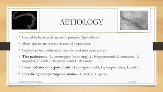
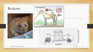
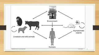
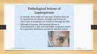
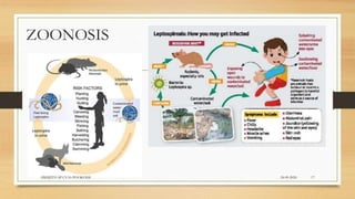
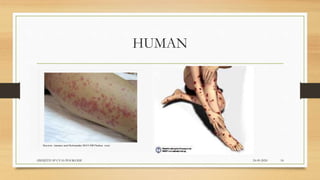
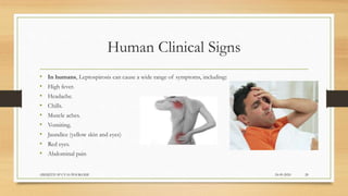
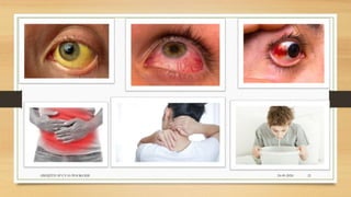
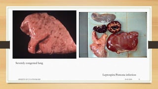
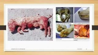
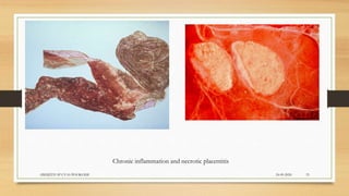
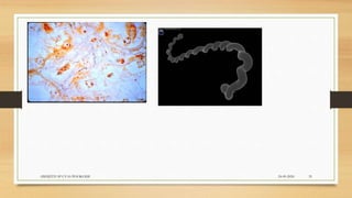
2. INTRODUCTION
ā¢ Leptospirosis is a blood infection caused by the bacteria Leptospira.
ā¢ It affects both humans and animals.
ā¢ It can pass from animals to humans and vice versa.
ā¢ In humans, it can cause a wide range of symptoms, some of which may be
mistaken for other diseases. Some infected persons, however, may have no
symptoms at all.
ā¢ 7-10 millions are affected annually.
24-09-2020ABHIJITH SP CVAS POOKODE 2
3. HISTORY
ā¢ Adolf Weil described the first case of leptospirosis in man in 1886.
ā¢ Stimson demonstrated by silver staining the presence of clumps of
spirochetes in the kidney tubules of a patient who reportedly died of yellow
fever in 1907.
ā¢ The causative agent of Weilās disease was isolated by Inada and Ido in 1915.
ā¢ Noguchi proposed the name āLeptospiraā (thin spirals) in 1918.
24-09-2020ABHIJITH SP CVAS POOKODE 3
4. AETIOLOGY
ā¢ Caused by bacteria of genus-Leptospira (Spirochetes).
ā¢ Many species are present in case of Leptospira.
ā¢ Leptospira has traditionally been divided into three groups
ā¢ The pathogenic - L. interrogans (most imp.), L. borgpetersenii, L. santarosai, L.
noguchii, L. weilli, L. kirschneri and L. alexanderi
ā¢ Intermediates or opportunistic - Leptospira inadai, Leptospira fainei, L. wolffii
ā¢ Free-living non-pathogenic strains - L. biflexa, L. parva
24-09-2020ABHIJITH SP CVAS POOKODE 4
5. AETIOLOGY
ā¢ The genus Leptospira is divided into 20 species based on DNA hybridization studies.
ā¢ All recognized species of Leptospira interrogans are categorized into 25 serogroups
and 250 serovars based on surface-exposed lipopolysaccharide (LPS).
ā¢ The common Leptospira interrogans serovars are:
ā¢ 1.L.interrogans serovar pomona,
ā¢ 2.L. interrogans serovar hardojobovis,
ā¢ 3.L.interrogans serovar icterohaemorrhagiae,
ā¢ 4.L.interrogans serovar canicola,
ā¢ 5.L.interrogans serovar grippotyphosa,
ā¢ 6.L.interrogans serovar bratislava,
ā¢ 7.L. interrogans serovar tarassovi
24-09-2020ABHIJITH SP CVAS POOKODE 5
8. EPIDEMIOLOGY
ā¢ Leptospirosis is a worldwide zoonotic infection with a much greater
incidence in tropical regions and has now been identified as one of the
emerging infectious diseases.
ā¢ The epidemiology of leptospirosis has been modified by changes in animal
husbandry, climate, and human behavior.
ā¢ A modeling exercise by the World Health Organization's (WHO's)
Leptospirosis Burden Epidemiology Group estimated that there were
873,000 cases worldwide annually with 48,600 deaths.
24-09-2020ABHIJITH SP CVAS POOKODE 8
9. MORPHOLOGY
ā¢ Leptospires are actively motile, helically shaped, slender spirochetes possessing large number
of light and fine spirals.
ā¢ Although cytochemically gram-negative, they do not stain well with conventional
bacteriological dyes and are usually visualized using dark-field microscopy.
ā¢ Silver impregnation (Levaditiās stain) staining techniques are used to demonstrate
Leptospira in tissues and Fontana staining for smear on slides.
ā¢ Characteristically, Leptospira have hooked ends and two periplasmic flagella (PF), also
known as axial filament and endoflagella which arise from each end of bacteria.
ā¢ Rotation of the PF results in the distinct spinning mobility of Leptospira.
24-09-2020ABHIJITH SP CVAS POOKODE 9
10. CULTURAL CHARACTERS
ā¢ They grow very well in media enriched with rabbit plasma (rabbit plasma contain high
concentration of bound vitamin B12).
ā¢ Several liquid and semisolid media are available.
ā¢ Liquid media- Korthofās medium, Stuartās medium, Vertwoortās medium
ā¢ Semi solid medium -Fletcher medium, EMJH, protein free medium, Ellis medium.
ā¢ In semisolid media, growth occurs characteristically 0.5 cm below the surface-.Dingerās
ring/disk.
ā¢ Addition of 5-flurouracil (100 mg/ml) in medium is inhibitory for most of the microorganism
but not for Leptospires
ā¢ Leptospira are highly susceptible to heat, being killed in 10 mts at 50ĀŗC and in 10 sec at 60ĀŗC. .
24-09-2020ABHIJITH SP CVAS POOKODE 10
16. INCUBATION PERIOD
ā¢ The incubation period is usually 5-14 days, with a range of 2-30 days.
ā¢ Animals also the same .
ā¢ Depends on the strain, serovar and the bacterial load and health status.
24-09-2020ABHIJITH SP CVAS POOKODE 16
20. Human Clinical Signs
ā¢ In humans, Leptospirosis can cause a wide range of symptoms, including:
ā¢ High fever.
ā¢ Headache.
ā¢ Chills.
ā¢ Muscle aches.
ā¢ Vomiting.
ā¢ Jaundice (yellow skin and eyes)
ā¢ Red eyes.
ā¢ Abdominal pain
24-09-2020ABHIJITH SP CVAS POOKODE 20
24. Cattle, Sheep- Leptospirosis
ā¢ Leptospira interrogans hardjobovis and L. borgpetersenii serovar hardjo is the host-
adapted lepto serovar in cattle and sheep and can infect animals at any age, including young
calves.
ā¢ Infection with this serovar will often produce a carrier state in the kidneys associated with long-
term urinary shedding. In addition, infections with hardjobovis can persist in the reproductive tract.
ā¢ red urine (red water), jaundice
ā¢ Lactating cows produce less milk, and, for a week or more, the milk they produce is thick and
yellow.
ā¢ However, unlike many other udder infections, leptospirosis does not usually cause any firmness
of the udder and it causes Flaccid mastitis.
24-09-2020ABHIJITH SP CVAS POOKODE 24
25. Equine Leptospirosis
ā¢ The Infection with serovar bratislava, which has been associated with
abortions and stillbirths in horses.
ā¢ Clinical disease most often results from incidental infection with serovar
pomona.
ā¢ Signs include abortion in mares and renal disease in young horses.
An immune-mediated anterior uveitis (periodic ophthalmia, 'moon
blindness') may be a manifestation of chronic leptospirosis in horses.
24-09-2020ABHIJITH SP CVAS POOKODE 25
26. Canine Leptospirosis
ā¢ Dogs: The acute form of leptospirosis is known as Stuttgart disease.
ā¢ The serovars associated with leptospirosis in dogs are canicola and
icterohaemorrhagiae .
ā¢ Serovar canicola, which is host-adapted for dogs, causes severe renal disease in
pups. In animals which survive the acute phase, a chronic uremic syndrome may
subsequently develop.
ā¢ Nephritis with petechial hemorrhage and degeneration of tubules is the main lesion
in acute form. While in chronic form areas of fibrosis, thickening of Bowmanās
capsule are the main lesions.
24-09-2020ABHIJITH SP CVAS POOKODE 26
27. Porcine Leptospirosis
ā¢ Acute leptospirosis in pigs is usually caused by rodent adapted serovars such as
icterohaemorrhagiae and copenhagenii .
ā¢ These serovars cause serious, sometimes fatal, disease in young pigs. It causes
hemoglobinuria, jaundice and fever.
ā¢ In many parts of the world, the principal host-adapted serovar is pomona.
ā¢ Pigs also serve as maintenance hosts for serovars tarassovi and bratislava, which may also
cause reproductive failure. Abortion occurs in the last 3rd of pregnancy and stillbirth.
ā¢ Lesions include icterus, petechial hemorrhage of liver, kidney and intestine, grayish white
areas surrounded by zone of congestion in kidneys.
24-09-2020ABHIJITH SP CVAS POOKODE 27
36. DIAGNOSIS
ā¢ 1.Tentative Diagnosis: Clinical signs with the history of exposure to contaminated urine.
ā¢ 2. Direct microscopy :Leptospires can be demonstrated in blood and CSF (during first 3-7 days),
urine (after 10 days of infection), by darkfield microscopy (DFM), Fontana staining and by FAT.
ā¢ 3.In tissue by Levaditiās staining and FAT.
ā¢ 4. Isolation in culture media
ā¢ 5. Animal inoculation āGuinea pigs
ā¢ 6.Microscopic Agglutination test (MAT), using live organisms- is the gold standard test for
diagnosis and is serovar specific) āGST.
ā¢ 7.ELISA IgM and slide agglutination tests (SAT).
24-09-2020ABHIJITH SP CVAS POOKODE 36
37. TREATMENT
ā¢ Penicillin G āDrug of Choice.
ā¢ Ampicillin, Doxycycline are also used.
24-09-2020ABHIJITH SP CVAS POOKODE 37
38. PREVENTION AND CONTROL
ā¢ Proper hygiene measure should be practiced.
ā¢ Urine should be cleaned from sheds.
ā¢ Animals should be taken care of.
ā¢ Proper vaccination practice.
ā¢ Control the rodent population
ā¢ Use of rodenticides.
24-09-2020ABHIJITH SP CVAS POOKODE 38