Download to read offline


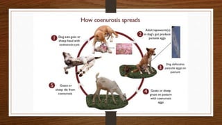

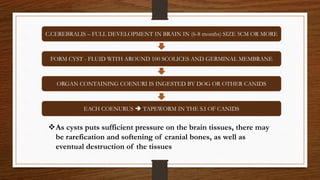


Coenurosis, caused by the tapeworm Taenia multiceps, primarily affects sheep, goats, and occasionally humans, leading to severe neurological symptoms. The disease's transmission cycle involves dogs and domestic herbivores, where eggs passed in dog feces infect the intermediate hosts, developing into cysts in the brain or spinal cord. Diagnosis is through examination of parasites and imaging, with treatments often including surgical removal and anthelmintics, while control measures focus on managing tapeworms in dogs.


