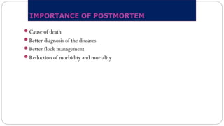
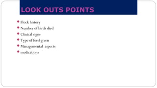
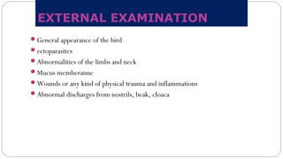
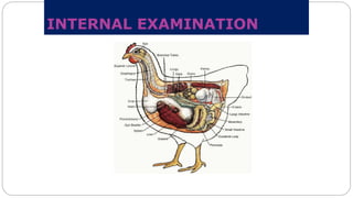
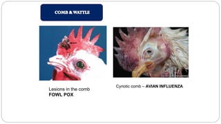
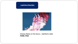
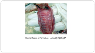
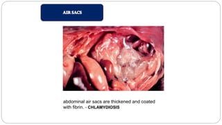
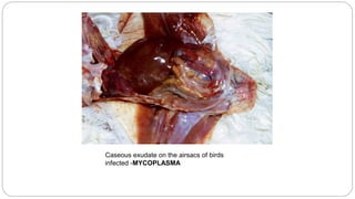
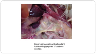
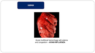
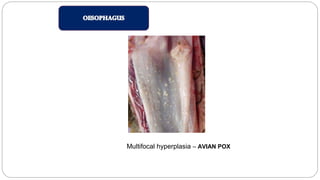
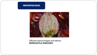
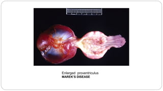
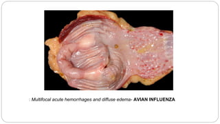
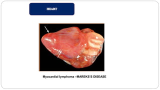
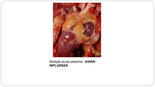
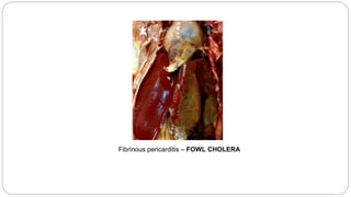
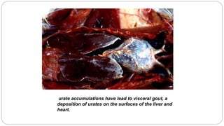
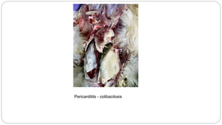
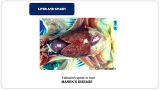
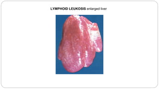
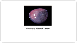
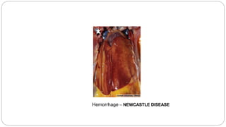
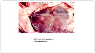
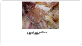
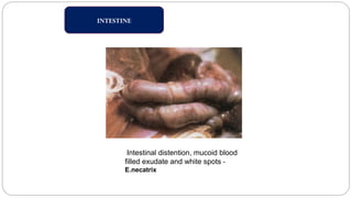
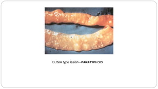
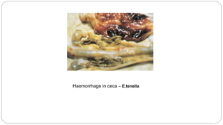
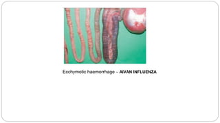
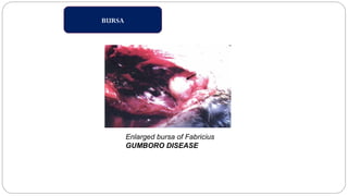
By
Ranjini Manuel
AVIAN POSTMORTEM
IMPORTANCE OF POSTMORTEM
Cause of death
Better diagnosis of the diseases
Better flock management
Reduction of morbidity and mortality
LOOK OUTS POINTS
Flock history
Number of birds died
Clinical signs
Type of feed given
Managemental aspects
medications
EXAMINATION PROCEDURE
STEPS
 Obtain flock history
 Examine the bird externally
 Open the body; observe organs in situ
 Remove the organs for detailed examination and sampling
 Examine and sample the organs
 Consider observations for diagnosis and write the report
EXTERNAL EXAMINATION
General appearance of the bird
ectoparasites
Abnormalities of the limbs and neck
Mucus memberanne
Wounds or any kind of physical trauma and inflammations
Abnormal discharges from nostrils, beak, cloaca
INTERNAL EXAMINATION
Lesions in the comb
FOWL POX
Cynotic comb – AVIAN INFLUENZA
Cheesy flakes on the larynx – diphtheric state
FOWL POX
Haemorrhages of the trachea – AVIAN INFLUENZA
abdominal air sacs are thickened and coated
with fibrin. - CHLAMYDIOSIS
Caseous exudate on the airsacs of birds
infected -MYCOPLASMA
Severe airsacculitis with abundant
foam and aggregates of caseous
exudate.
Acute multifocal hemorrhage with edema
and congestion – AVIAN INFLUENZA
Pulmonary congestion and edema,-
NEWCASTLE DISEASE
accumulation of fibrinous exudate – FOWL CHOLERA
lymphomatous lesions- MAREK’S DISEASE
fibrinous pneumonia -MYCOPLASMA
Multifocal hyperplasia – AVIAN POX
Diffused haemorrhages and edema
NEWCASTLE DISEASES
Enlarged proventriculus
MAREK’S DISEASE
: Multifocal acute hemorrhages and diffuse edema- AVIAN INFLUENZA
Myocardial lymphoma –MAREKS’S DISEASE
Multiple acute petechie –AVIAN
INFLUENZA
Fibrinous pericarditis – FOWL CHOLERA
urate accumulations have lead to visceral gout, a
deposition of urates on the surfaces of the liver and
heart.
Pericarditits - colibacilosis
Yellowish spots in liver
MAREK’S DISEASE
LYMPHOID LEUKOSIS enlarged liver
Splenomegaly - COLISEPTICAEMIA
Hemorrhage – NEWCASTLE DISEASE
fibrinous perihepatitis.-
CHLAMYDIOSIS
enlarged, pale, and friable,-
MYCOTOXICOSIS
INTESTINE
Intestinal distention, mucoid blood
filled exudate and white spots -
E.necatrix
Button type lesion - PARATYPHOID
Haemorrhage in ceca – E.tenella
Ecchymotic haemorrhage – AIVAN INFLUENZA
BURSA
Enlarged bursa of Fabricius
GUMBORO DISEASE
MUSCLES
Thigh muscle showing haemorrhage
GUMBORO DISEASE
REPRODUCTIVE TRACT
INFECTIOUS BRONCHITITS – shell less egg
Avian postmortem

Avian postmortem