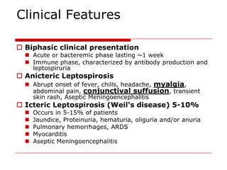
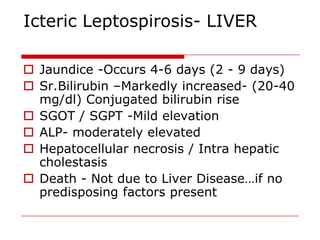
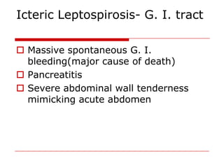
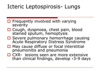
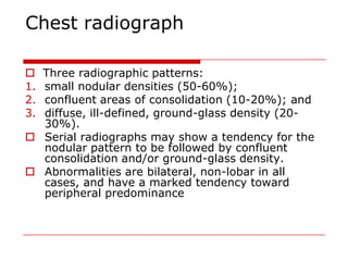
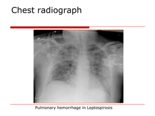
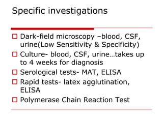
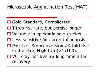
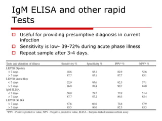
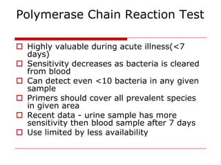
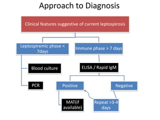
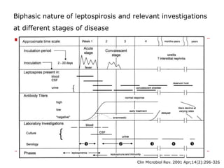
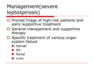
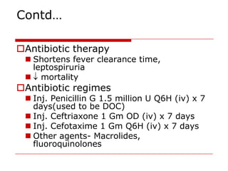
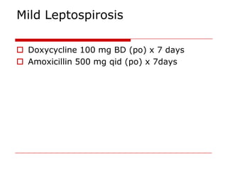
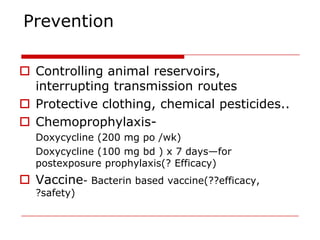
Leptospirosis


                DR. ANKIT RAIYANI

                           LTMGH
Introduction

 Most common, underreported and under
  diagnosed zoonosis.
 India - Cases reported from Maharashtra,
  Gujarat , Kerala, Tamil Nadu, Karnataka,
  Andaman.
 Source –Animals ( rodents and domestic
  animals) or an environment contaminated by
  their urine
 Causative org. can survive for months in water
  & damp soil
Organism

   Order-Spirochaetales
   Family-Leptospiraceae
   Genus-Leptospira
   Species-17(11 pathogenic-L. interrogans sensu lato)
    & (6 non-pathogenic-L. biflexa sensu lato)
   Pathogenic leptospira divided into ›26 serogroups & ›250
    serovars.
   Leptospires are coiled thin highly motile organisms, 6-
    20µm long and ~0.1µm wide, obligate aerobes
   Stain poorly… but seen by dark-field microscopy & by
    silver impregnation tech.
   Culture media- EMJH, Fletcher, Korthof
Leptospira interrogans


L. Interrogans scanning electron microscope


                                                    L. Interrogans Silver impregnation


                                        L. Interrogans Dark-field microscopy
Epidemiology

 Etiology: L interrogans
 Most widespread zoonosis in the world
 Peak incidence during rainy season
 Occupational & recreational exposures
 Source of infection in humans: direct or
  indirect contact with the urine of an infected
  animal
 Portal of entry: abrasions or cuts on skin,
  conjunctiva & other mucous membranes.
 Inc. period: 2-30 days(commonly 5-14 days)
Epidemiology

  Rainfall
  Contaminated environment
  Poor Sanitation
  Inadequate drainage facilities
  Presence of Rodents, cattle& stray dogs
  Walking bare foot
  Specific source of infection cannot be
  pinpointed with certainty.
 Males>Females… more chance of exposure
Transmission


           Rodents
           (Urine)


                                    Humans

Domestic             Contaminated
 animals             environment
PATHOGENESIS

 Pathogenesis not completely understood
1. Leptospiremia...multiplication in blood, tissues.
2. Vasculitis– responsible for most of the
   manifestations… LOS TNF-α
3. Kidneys- interstitial nephritis, tubular injury
4. Liver- centrilobular necrosis(usually not
   extensive)
5. Lungs- involvement due to hemorrhage, not to
   inflammation
6. Microcirculation damage, increased vascular
   permeability….hypovolemia
7. Skeletal muscles-necrosis
Clinical Features

 Biphasic clinical presentation
   Acute or bacteremic phase lasting ~1 week
   Immune phase, characterized by antibody production and
    leptospiruria
 Anicteric Leptospirosis
   Abrupt onset of fever, chills, headache, myalgia,
    abdominal pain, conjunctival suffusion, transient
    skin rash, Aseptic Meningoencephalitis
 Icteric Leptospirosis (Weil’s disease) 5-10%
     Occurs in 5-15% of patients
     Jaundice, Proteinuria, hematuria, oliguria and/or anuria
     Pulmonary hemorrhages, ARDS
     Myocarditis
     Aseptic Meningoencephalitis
Icteric Leptospirosis- LIVER

 Jaundice -Occurs 4-6 days (2 - 9 days)
 Sr.Bilirubin –Markedly increased- (20-40
  mg/dl) Conjugated bilirubin rise
 SGOT / SGPT -Mild elevation
 ALP- moderately elevated
 Hepatocellular necrosis / Intra hepatic
  cholestasis
 Death - Not due to Liver Disease…if no
  predisposing factors present
Icteric Leptospirosis- G. I. tract

 Massive spontaneous G. I.
  bleeding(major cause of death)
 Pancreatitis
 Severe abdominal wall tenderness
  mimicking acute abdomen
Icteric Leptospirosis- Lungs

 Frequently involved with varying
  severity
 Cough, dyspnoea, chest pain, blood
  stained sputum, hemoptysis
 Severe pulmonary hemorrhage causing
  Acute Respiratory Distress Syndrome
 May cause diffuse or focal interstitial
  pneumonitis and pneumonia
 CXR- lesions seen are more extensive
  than clinical findings, develop ›3-9 days
Microscopy
Hematological disturbances

 Severe thrombocytopenia…bleeding
 Anemia- d/t bloodloss, &/or hemolysis
 Petechiae, purpura, ecchymoses
  common
 PT & PTTK time may be normal to
  moderately elevated…can be corrected
  with Vit K supplementation.
 D.I.C.-rare as compared to Gm(-) sepsis
 Jarisch-Herxheimer reaction- rare
Icteric Leptospirosis- Kidneys

 Invariably involved --Mild to Severe
 Urinalysis : Hematuria / Pyuria /Proteinuria
 Renal Failure: initially non-oliguric with K+
  wasting….may become oliguric


          Prerenal                    ATN
           Hypotension
                                    Endotoxins
       Fluid loss/ Fluid shift
                                 Toxic Metabolites
            Myocarditis
Icteric Leptospirosis- Heart


Hemorrhagic Myocarditis- Arrhythmias
,Cardiac failure, Hypotension / Death


Arrhythmias- Atrial fibrillation / Conduction
defects (seen in 20-40%)


ECG changes- Non Specific ST-T changes, Low
voltage complexes
Icteric Leptospirosis- CNS

 Aseptic MeningoEncephalitis-
    Occurs in the Immune phase
    CSF – raised proteins , -
    lymphocytes
 Rare-
    Convulsions, Encephalitis
    Myelitis & Polyneuropathy.
Other manifestations

 Skeletal Ms involvement- myalgia,
  raised CPK levels
 Conjunctival suffusion
  (pathognomic)
 Anterior uveal tract inflammation
  Iritis / Iridocyclitis / chorioretinitis
 Occurs 2 weeks – 1 yr (6 months)
Modified Faine’s criteria
Diff. Diagnosis

 Fever-Viral fever/Malaria/Typhoid /
  UTI
 Jaundice-Malaria, Viral hepatitis,
  Sepsis
 Renal failure-Malaria, Hanta Virus,
  Sepsis
 Meningitis-Bacterial / Viral
 Hemorrhagic Fever-Dengue, Hanta
  virus, Typhus
Investigations- Routine

 CBC- low Hb, thrombocytopenia, leucocytosis
 LFTs-      conjugated bilirubinemia, raised ALP,
  mild elevation of AST/ALT
 RFTs-      raised BUN/Creat…Ratio
 SE- hypokalemia, hypocalcemia,
  hypomagnesaemia
 CPK-       elevated
 PT/PTTK-mild elevation
 Pl. fibrinogen- normal
 Urine(R/M)- active sediments, proteinuria,
  hematuria, Hb/Mb-uria.
 ECG- conduction defects, non-specific ST changes
Chest radiograph

 Three radiographic patterns:
1.small nodular densities (50-60%);
2.confluent areas of consolidation (10-20%); and
3.diffuse, ill-defined, ground-glass density (20-
  30%).
 Serial radiographs may show a tendency for the
  nodular pattern to be followed by confluent
  consolidation and/or ground-glass density.
 Abnormalities are bilateral, non-lobar in all
  cases, and have a marked tendency toward
  peripheral predominance
Chest radiograph


     Pulmonary hemorrhage in Leptospirosis
Specific investigations

 Dark-field microscopy –blood, CSF,
  urine(Low Sensitivity & Specificity)
 Culture- blood, CSF, urine…takes up
  to 4 weeks for diagnosis
 Serological tests- MAT, ELISA
 Rapid tests- latex agglutination,
  ELISA
 Polymerase Chain Reaction Test
Microscopic Agglutination Test(MAT)


 Gold Standard, Complicated
 Titres rise late, but persist longer
 Valuable in epidemiologic studies
 Less sensitive for current diagnosis
 Positive- Seroconversion / 4 fold rise
  in the titre, High titre(>1:100).
 Will stay positive for long time after
  recovery
IgM ELISA and other rapid
Tests
 Useful for providing presumptive diagnosis in current
  infection
 Sensitivity is low- 39-72% during acute phase illness
 Repeat sample after 3-4 days.
Polymerase Chain Reaction Test

 Highly valuable during acute illness(<7
  days)
 Sensitivity decreases as bacteria is cleared
  from blood
 Can detect even <10 bacteria in any given
  sample
 Primers should cover all prevalent species
  in given area
 Recent data - urine sample has more
  sensitivity then blood sample after 7 days
 Use limited by less availability
Approach to Diagnosis
  Clinical features suggestive of current leptospirosis


Leptospiremic phase <       Immune phase > 7 days
        7days


     Blood culture              ELISA / Rapid IgM


     PCR             Positive                    Negative


                       MAT(if         Repeat >3-4
                      available)         days
Biphasic nature of leptospirosis and relevant investigations
at different stages of disease


                                      Clin Microbiol Rev. 2001 Apr;14(2):296-326
Management(severe
leptospirosis)
 Prompt triage of high-risk patients and
  early supportive treatment
 General management and supportive
  therapy
 Specific treatment of various organ
  system failure
     Hemat
     RS
     Renal
     Liver
Contd…

Antibiotic therapy
  Shortens fever clearance time,
   leptospiruria
  mortality
Antibiotic regimes
  Inj. Penicillin G 1.5 million U Q6H (iv) x 7
   days(used to be DOC)
  Inj. Ceftriaxone 1 Gm OD (iv) x 7 days
  Inj. Cefotaxime 1 Gm Q6H (iv) x 7 days
  Other agents- Macrolides,
   fluoroquinolones
Mild Leptospirosis

 Doxycycline 100 mg BD (po) x 7 days
 Amoxicillin 500 mg qid (po) x 7days
Prognosis

 Mortality is high in patients with
  severe disease
 Weil’s disease- 5 to 40%
 Severe pulmonary hemorrhage->
  50%
 Poor prognostic indicators- age>40
  yrs, oliguria, respiratory distress,
  pulmonary hemorrhage, arrhythmias,
  altered sensorium, alcoholic patients
Prevention

 Controlling animal reservoirs,
  interrupting transmission routes
 Protective clothing, chemical pesticides..
 Chemoprophylaxis-
   Doxycycline (200 mg po /wk)
   Doxycycline (100 mg bd ) x 7 days—for
   postexposure prophylaxis(? Efficacy)
 Vaccine- Bacterin based vaccine(??efficacy,
   ?safety)
Thank you!!

Leptospirosis

  • 1.
    Leptospirosis DR. ANKIT RAIYANI LTMGH
  • 2.
    Introduction  Most common,underreported and under diagnosed zoonosis.  India - Cases reported from Maharashtra, Gujarat , Kerala, Tamil Nadu, Karnataka, Andaman.  Source –Animals ( rodents and domestic animals) or an environment contaminated by their urine  Causative org. can survive for months in water & damp soil
  • 3.
    Organism  Order-Spirochaetales  Family-Leptospiraceae  Genus-Leptospira  Species-17(11 pathogenic-L. interrogans sensu lato) & (6 non-pathogenic-L. biflexa sensu lato)  Pathogenic leptospira divided into ›26 serogroups & ›250 serovars.  Leptospires are coiled thin highly motile organisms, 6- 20µm long and ~0.1µm wide, obligate aerobes  Stain poorly… but seen by dark-field microscopy & by silver impregnation tech.  Culture media- EMJH, Fletcher, Korthof
  • 4.
    Leptospira interrogans L. Interrogansscanning electron microscope L. Interrogans Silver impregnation L. Interrogans Dark-field microscopy
  • 5.
    Epidemiology  Etiology: Linterrogans  Most widespread zoonosis in the world  Peak incidence during rainy season  Occupational & recreational exposures  Source of infection in humans: direct or indirect contact with the urine of an infected animal  Portal of entry: abrasions or cuts on skin, conjunctiva & other mucous membranes.  Inc. period: 2-30 days(commonly 5-14 days)
  • 6.
    Epidemiology  Rainfall  Contaminated environment  Poor Sanitation  Inadequate drainage facilities  Presence of Rodents, cattle& stray dogs  Walking bare foot  Specific source of infection cannot be pinpointed with certainty.  Males>Females… more chance of exposure
  • 7.
    Transmission Rodents (Urine) Humans Domestic Contaminated animals environment
  • 8.
    PATHOGENESIS Pathogenesis notcompletely understood 1. Leptospiremia...multiplication in blood, tissues. 2. Vasculitis– responsible for most of the manifestations… LOS TNF-α 3. Kidneys- interstitial nephritis, tubular injury 4. Liver- centrilobular necrosis(usually not extensive) 5. Lungs- involvement due to hemorrhage, not to inflammation 6. Microcirculation damage, increased vascular permeability….hypovolemia 7. Skeletal muscles-necrosis
  • 9.
    Clinical Features  Biphasicclinical presentation  Acute or bacteremic phase lasting ~1 week  Immune phase, characterized by antibody production and leptospiruria  Anicteric Leptospirosis  Abrupt onset of fever, chills, headache, myalgia, abdominal pain, conjunctival suffusion, transient skin rash, Aseptic Meningoencephalitis  Icteric Leptospirosis (Weil’s disease) 5-10%  Occurs in 5-15% of patients  Jaundice, Proteinuria, hematuria, oliguria and/or anuria  Pulmonary hemorrhages, ARDS  Myocarditis  Aseptic Meningoencephalitis
  • 10.
    Icteric Leptospirosis- LIVER Jaundice -Occurs 4-6 days (2 - 9 days)  Sr.Bilirubin –Markedly increased- (20-40 mg/dl) Conjugated bilirubin rise  SGOT / SGPT -Mild elevation  ALP- moderately elevated  Hepatocellular necrosis / Intra hepatic cholestasis  Death - Not due to Liver Disease…if no predisposing factors present
  • 11.
    Icteric Leptospirosis- G.I. tract  Massive spontaneous G. I. bleeding(major cause of death)  Pancreatitis  Severe abdominal wall tenderness mimicking acute abdomen
  • 12.
    Icteric Leptospirosis- Lungs Frequently involved with varying severity  Cough, dyspnoea, chest pain, blood stained sputum, hemoptysis  Severe pulmonary hemorrhage causing Acute Respiratory Distress Syndrome  May cause diffuse or focal interstitial pneumonitis and pneumonia  CXR- lesions seen are more extensive than clinical findings, develop ›3-9 days
  • 13.
  • 14.
    Hematological disturbances  Severethrombocytopenia…bleeding  Anemia- d/t bloodloss, &/or hemolysis  Petechiae, purpura, ecchymoses common  PT & PTTK time may be normal to moderately elevated…can be corrected with Vit K supplementation.  D.I.C.-rare as compared to Gm(-) sepsis  Jarisch-Herxheimer reaction- rare
  • 15.
    Icteric Leptospirosis- Kidneys Invariably involved --Mild to Severe  Urinalysis : Hematuria / Pyuria /Proteinuria  Renal Failure: initially non-oliguric with K+ wasting….may become oliguric Prerenal ATN Hypotension Endotoxins Fluid loss/ Fluid shift Toxic Metabolites Myocarditis
  • 16.
    Icteric Leptospirosis- Heart HemorrhagicMyocarditis- Arrhythmias ,Cardiac failure, Hypotension / Death Arrhythmias- Atrial fibrillation / Conduction defects (seen in 20-40%) ECG changes- Non Specific ST-T changes, Low voltage complexes
  • 17.
    Icteric Leptospirosis- CNS Aseptic MeningoEncephalitis- Occurs in the Immune phase CSF – raised proteins , - lymphocytes  Rare- Convulsions, Encephalitis Myelitis & Polyneuropathy.
  • 18.
    Other manifestations  SkeletalMs involvement- myalgia, raised CPK levels  Conjunctival suffusion (pathognomic)  Anterior uveal tract inflammation Iritis / Iridocyclitis / chorioretinitis  Occurs 2 weeks – 1 yr (6 months)
  • 19.
  • 20.
    Diff. Diagnosis  Fever-Viralfever/Malaria/Typhoid / UTI  Jaundice-Malaria, Viral hepatitis, Sepsis  Renal failure-Malaria, Hanta Virus, Sepsis  Meningitis-Bacterial / Viral  Hemorrhagic Fever-Dengue, Hanta virus, Typhus
  • 21.
    Investigations- Routine  CBC-low Hb, thrombocytopenia, leucocytosis  LFTs- conjugated bilirubinemia, raised ALP, mild elevation of AST/ALT  RFTs- raised BUN/Creat…Ratio  SE- hypokalemia, hypocalcemia, hypomagnesaemia  CPK- elevated  PT/PTTK-mild elevation  Pl. fibrinogen- normal  Urine(R/M)- active sediments, proteinuria, hematuria, Hb/Mb-uria.  ECG- conduction defects, non-specific ST changes
  • 22.
    Chest radiograph  Threeradiographic patterns: 1.small nodular densities (50-60%); 2.confluent areas of consolidation (10-20%); and 3.diffuse, ill-defined, ground-glass density (20- 30%).  Serial radiographs may show a tendency for the nodular pattern to be followed by confluent consolidation and/or ground-glass density.  Abnormalities are bilateral, non-lobar in all cases, and have a marked tendency toward peripheral predominance
  • 23.
    Chest radiograph Pulmonary hemorrhage in Leptospirosis
  • 24.
    Specific investigations  Dark-fieldmicroscopy –blood, CSF, urine(Low Sensitivity & Specificity)  Culture- blood, CSF, urine…takes up to 4 weeks for diagnosis  Serological tests- MAT, ELISA  Rapid tests- latex agglutination, ELISA  Polymerase Chain Reaction Test
  • 25.
    Microscopic Agglutination Test(MAT) Gold Standard, Complicated  Titres rise late, but persist longer  Valuable in epidemiologic studies  Less sensitive for current diagnosis  Positive- Seroconversion / 4 fold rise in the titre, High titre(>1:100).  Will stay positive for long time after recovery
  • 26.
    IgM ELISA andother rapid Tests  Useful for providing presumptive diagnosis in current infection  Sensitivity is low- 39-72% during acute phase illness  Repeat sample after 3-4 days.
  • 27.
    Polymerase Chain ReactionTest  Highly valuable during acute illness(<7 days)  Sensitivity decreases as bacteria is cleared from blood  Can detect even <10 bacteria in any given sample  Primers should cover all prevalent species in given area  Recent data - urine sample has more sensitivity then blood sample after 7 days  Use limited by less availability
  • 28.
    Approach to Diagnosis Clinical features suggestive of current leptospirosis Leptospiremic phase < Immune phase > 7 days 7days Blood culture ELISA / Rapid IgM PCR Positive Negative MAT(if Repeat >3-4 available) days
  • 29.
    Biphasic nature ofleptospirosis and relevant investigations at different stages of disease Clin Microbiol Rev. 2001 Apr;14(2):296-326
  • 30.
    Management(severe leptospirosis)  Prompt triageof high-risk patients and early supportive treatment  General management and supportive therapy  Specific treatment of various organ system failure  Hemat  RS  Renal  Liver
  • 31.
    Contd… Antibiotic therapy Shortens fever clearance time, leptospiruria  mortality Antibiotic regimes  Inj. Penicillin G 1.5 million U Q6H (iv) x 7 days(used to be DOC)  Inj. Ceftriaxone 1 Gm OD (iv) x 7 days  Inj. Cefotaxime 1 Gm Q6H (iv) x 7 days  Other agents- Macrolides, fluoroquinolones
  • 32.
    Mild Leptospirosis  Doxycycline100 mg BD (po) x 7 days  Amoxicillin 500 mg qid (po) x 7days
  • 33.
    Prognosis  Mortality ishigh in patients with severe disease  Weil’s disease- 5 to 40%  Severe pulmonary hemorrhage-> 50%  Poor prognostic indicators- age>40 yrs, oliguria, respiratory distress, pulmonary hemorrhage, arrhythmias, altered sensorium, alcoholic patients
  • 34.
    Prevention  Controlling animalreservoirs, interrupting transmission routes  Protective clothing, chemical pesticides..  Chemoprophylaxis- Doxycycline (200 mg po /wk) Doxycycline (100 mg bd ) x 7 days—for postexposure prophylaxis(? Efficacy)  Vaccine- Bacterin based vaccine(??efficacy, ?safety)
  • 35.