Leptospirosis is a zoonotic disease caused by Leptospira bacteria transmitted through contact with infected animal urine. It is endemic in tropical and subtropical regions including parts of India. The disease affects both animals and humans. In animals, it can cause reproductive issues like abortions. In humans, symptoms range from flu-like illness to severe symptoms involving multiple organ failure. Diagnosis involves microscopic examination of samples, culture, serological tests and PCR. Control relies on rodent control, sanitation measures, vaccination of animals, and personal protective measures for humans.
etiology, local names, definition, transmission, source of infection, epidemiology, pathogenesis, clinical signs, diagnosis, differential diagnosis, treatment prevention and control
Leptospirosis is a worldwide public health problem. In humid tropical and subtropical areas, where most developing
countries are found, it is a greater problem than in those with a temperate climate. The magnitude of the problem in
tropical and subtropical regions can be largely attributed to climatic and environmental conditions but also to the
great likelihood of contact with a Leptospira-contaminated environment caused by, for example, local agricultural
practices and poor housing and waste disposal, all of which give rise to many sources of infection. In countries with
temperate climates, in addition to locally acquired leptospirosis, the disease may also be acquired by travellers
abroad, and particularly by those visiting the tropics.
Leptospirosis is a potentially serious but treatable disease. Its symptoms may mimic those of a number of other
unrelated infections such as influenza, meningitis, hepatitis, dengue or viral haemorrhagic fevers. Some of these
infections, in particular dengue, may give rise to large epidemics, and cases of leptospirosis that occur during such
epidemics may be overlooked. For this reason, it is important to distinguish leptospirosis from dengue and viral
haemorrhagic fevers, etc. in patients acquiring infections in countries where these diseases are endemic. At present,
this is still difficult, but new developments may reduce the technical problems in the near future. It is necessary,
therefore, to increase awareness and knowledge of leptospirosis as a public health threat.
Tuberculosis hardly excuse anyone irrespective of its shape, size, colour, cast, creed, breed, species or genus having a little warmth in blood. Therefore, elephants e not exception, rather very prone for this disease which have taken many times more lives than any of the war.
Brucellosis: Epidemiology and Control in indiaBhoj Raj Singh
Brucellosis is an important endemic infectious disease in animals in India. In India brucellosis was first recognized in 1942 by Polding. It causes economic loss to the tune of nearly Rs. 350 million/year. Bovine brucellosis is caused by the bacterium Brucella abortus. In countries where cattles are kept in close association with sheep and goat it can also be caused by B. melitensis. Occasionally B. suis may also cause disease in mammary gland of cattle but it has not been reported to cause abortion and usually does not spread to other animals. Principal manifestations of animal brucellosis are reproductive failure, i.e., abortion, still births and birth of unthrifty offspring in females, and orchitis and epididymitis in males. Genus Brucella has six recognized species on the basis of host specificity viz. B. abortus, B. melitensis, B. ovis, B. suis, B. canis and B. neotome, infecting cattle, goats and sheep, sheep, pig, dog and rats, respectively. All Brucella species may also infect wildlife species. Classical Brucella species have been isolated from a great variety of wildlife species such as bison, elk, feral swine, wild boar, fox, hare, African buffalo, reindeer, and caribou. Infection in wildlife can hinder eradication efforts in cattle.
The classical species viz., B. abortus, B. melitensis, and B. suis have been identified as category B bioterrorism agents (Rotz et al. 2002, CDC 2005) because they are zoonotic and capable of causing considerable morbidity with low mortality if used in a mass event.
It is highly contagious disease primarily of cattle, camels, sheep, goats and swine and secondarily in other animals and man
Characterized clinically by inflammation of the genital organs and fetal membrane, abortion with retained placenta and a subsequent high rate of infertility.
Bovine tuberculosis epidemiology & control in indiaBhoj Raj Singh
Tuberculosis in India is in hyperendemic state both in human and animals. No DOTS can help in control of human tuberculosis unless tuberculosis is controlled in animals. Control of tuberculosis in animals is a far reacheachable dream in India and thus the Tuberculosis will persist in India till the dooms day.
Prevalence of canine leptospirosis has increased in recent years.
As many as 8.2% of dogs are shedding leptospires, some asymptomatically.49
Weather changes, population growth, and habitat encroachment have all increased human and canine exposure to pathogens and their carriers.
Transmission of leptospirosis can occur through direct contact or indirectly through environmental exposure.
Leptospires enter the body through mucous membranes in the mouth, eyes, or nose, or through abraded or water-softened skin.
Leptospires multiply in a host animal's bloodstream.
Leptospires move from the bloodstream to the kidneys and other tissues to continue reproducing.
Leptospires pass from the kidneys into the urine; then are shed back into the environment.
Other dogs, wild animals, or people can become infected through direct or indirect contact.
clinical signs
Fever
Lethargy
Weight loss
Anorexia
Depression
Acute renal failure
Jaundice
Abdominal discomfort
Vomiting and diarrhea
Blood in urine is uncommon, but may occur
Respiratory distress
Dogs at risk
Dogs at risk for developing leptospirosis include those with
Access to ponds, lakes, streams, or standing water
Exposure to urine from other infected animals, including:
Other dogs in shelters or other pet care facilities
Wildlife (e.g. rodents, racoons, opossum, deer), either through direct contact with urine or through contaminated water
Morbidity threats
As leptospirosis progresses, it can result in
Leptospiremia
Leptospires can multiply in the bloodstream and spread to many tissues and organs
Vascular damage/thrombocytopenia
Can lead to kidney failure and interfere with liver function
Contributes to coagulatory abnormalities and hemorrhages
Severe kidney and liver damage
Acute renal failure occurs in dogs with severe clinical signs
Acute hepatic dysfunction or chronic hepatitis have been caused by specific serovars
Kyasanur forest disease, KFD is a febrile disease associated with haemorrhage caused by kyasanur forest disease virus, a member of virus family of arbovirus & flavivirus and transmitted to man by bite of infected ticks.
Fowl typhoid is a septicemic acute or chronic disease of domesticated birds.
The disease is worldwide distributed and natural outbreaks occur in chickens, turkeys, guinea fowl, peafowl, duckling and game birds such as quail, grouse and pheasant.
This can cause mortality in birds of any age.
Broiler parents and brown-shell egg layers are especially susceptible.
A short presentation about leptospirosis. It can cover almost all the basic aspects of leptospirosis such as distribution, incidence, microbiology, clinical features, diagnosis and treatment.
Leptospirosis an emerging public health problem. I have give an overview and skipped Pathogenesis & Surviellance. Tried to keep it short & informative.
Important Zoonotic disease and its prevention and control By: Dr.Manoj karkimanojj123
Zoonosis are those disease and infection which are naturally transmitted between animals and human. (WHO & FAO, 1959).
Zoonosis word derived from Greek word “ZOO” means Animals and “NOSES” means Disease.
One Health is not a new concept, but it has become more important in recent years because many factors have changed the interaction among human, animals and the environment. These changes have caused the emergence and re-emergence of many disease.
Similar to Leptospirosis: Its Epidemiology, Diagnosis and Control (20)
Global travel and spread of COVID 19: Current epidemiological statusChandrani Goswami
International departures and arrivals across the world have increased significantly in just 10 years, contributing to the rapid spread of coronavirus globally
OVERVIEW OF coronavirus
Epidemiology CoVs:
COVID-19 Transmission
COVID-19 Distribution
SARS-CoV-2 Strain
Heart failure (HF), often used to mean chronic heart failure (CHF), occurs when the heart is unable to pump sufficiently to maintain blood flow to meet the needs of the body.
It is defined simply as a technique to efficiently and stably introduce foreign genes into the genome of target cells.
The insertion of unrelated, therapeutic genetic information in the form of DNA into target cells
Somatic cells are mainly epithelial cells that has been shed from the epithelial lining of the gland and White blood cells (leukocyte) has entered the mammary gland in response to injury or infection.
Refers to inflammation of the mammary gland, which is characterized by physical, chemical as well as bacteriological changes in the milk and pathological changes in the udder tissues.
Major agents and their characteristics which has beenChandrani Goswami
Biological weapon, also called germ weapon
“Any of a number of disease-producing agents such as bacteria, viruses, rickettsia, fungi, toxins, or other biological agents that may be utilized as weapons against humans, animals, or plants”.
Richard's aventures in two entangled wonderlandsRichard Gill
Since the loophole-free Bell experiments of 2020 and the Nobel prizes in physics of 2022, critics of Bell's work have retreated to the fortress of super-determinism. Now, super-determinism is a derogatory word - it just means "determinism". Palmer, Hance and Hossenfelder argue that quantum mechanics and determinism are not incompatible, using a sophisticated mathematical construction based on a subtle thinning of allowed states and measurements in quantum mechanics, such that what is left appears to make Bell's argument fail, without altering the empirical predictions of quantum mechanics. I think however that it is a smoke screen, and the slogan "lost in math" comes to my mind. I will discuss some other recent disproofs of Bell's theorem using the language of causality based on causal graphs. Causal thinking is also central to law and justice. I will mention surprising connections to my work on serial killer nurse cases, in particular the Dutch case of Lucia de Berk and the current UK case of Lucy Letby.
The increased availability of biomedical data, particularly in the public domain, offers the opportunity to better understand human health and to develop effective therapeutics for a wide range of unmet medical needs. However, data scientists remain stymied by the fact that data remain hard to find and to productively reuse because data and their metadata i) are wholly inaccessible, ii) are in non-standard or incompatible representations, iii) do not conform to community standards, and iv) have unclear or highly restricted terms and conditions that preclude legitimate reuse. These limitations require a rethink on data can be made machine and AI-ready - the key motivation behind the FAIR Guiding Principles. Concurrently, while recent efforts have explored the use of deep learning to fuse disparate data into predictive models for a wide range of biomedical applications, these models often fail even when the correct answer is already known, and fail to explain individual predictions in terms that data scientists can appreciate. These limitations suggest that new methods to produce practical artificial intelligence are still needed.
In this talk, I will discuss our work in (1) building an integrative knowledge infrastructure to prepare FAIR and "AI-ready" data and services along with (2) neurosymbolic AI methods to improve the quality of predictions and to generate plausible explanations. Attention is given to standards, platforms, and methods to wrangle knowledge into simple, but effective semantic and latent representations, and to make these available into standards-compliant and discoverable interfaces that can be used in model building, validation, and explanation. Our work, and those of others in the field, creates a baseline for building trustworthy and easy to deploy AI models in biomedicine.
Bio
Dr. Michel Dumontier is the Distinguished Professor of Data Science at Maastricht University, founder and executive director of the Institute of Data Science, and co-founder of the FAIR (Findable, Accessible, Interoperable and Reusable) data principles. His research explores socio-technological approaches for responsible discovery science, which includes collaborative multi-modal knowledge graphs, privacy-preserving distributed data mining, and AI methods for drug discovery and personalized medicine. His work is supported through the Dutch National Research Agenda, the Netherlands Organisation for Scientific Research, Horizon Europe, the European Open Science Cloud, the US National Institutes of Health, and a Marie-Curie Innovative Training Network. He is the editor-in-chief for the journal Data Science and is internationally recognized for his contributions in bioinformatics, biomedical informatics, and semantic technologies including ontologies and linked data.
Observation of Io’s Resurfacing via Plume Deposition Using Ground-based Adapt...Sérgio Sacani
Since volcanic activity was first discovered on Io from Voyager images in 1979, changes
on Io’s surface have been monitored from both spacecraft and ground-based telescopes.
Here, we present the highest spatial resolution images of Io ever obtained from a groundbased telescope. These images, acquired by the SHARK-VIS instrument on the Large
Binocular Telescope, show evidence of a major resurfacing event on Io’s trailing hemisphere. When compared to the most recent spacecraft images, the SHARK-VIS images
show that a plume deposit from a powerful eruption at Pillan Patera has covered part
of the long-lived Pele plume deposit. Although this type of resurfacing event may be common on Io, few have been detected due to the rarity of spacecraft visits and the previously low spatial resolution available from Earth-based telescopes. The SHARK-VIS instrument ushers in a new era of high resolution imaging of Io’s surface using adaptive
optics at visible wavelengths.
THE IMPORTANCE OF MARTIAN ATMOSPHERE SAMPLE RETURN.Sérgio Sacani
The return of a sample of near-surface atmosphere from Mars would facilitate answers to several first-order science questions surrounding the formation and evolution of the planet. One of the important aspects of terrestrial planet formation in general is the role that primary atmospheres played in influencing the chemistry and structure of the planets and their antecedents. Studies of the martian atmosphere can be used to investigate the role of a primary atmosphere in its history. Atmosphere samples would also inform our understanding of the near-surface chemistry of the planet, and ultimately the prospects for life. High-precision isotopic analyses of constituent gases are needed to address these questions, requiring that the analyses are made on returned samples rather than in situ.
Slide 1: Title Slide
Extrachromosomal Inheritance
Slide 2: Introduction to Extrachromosomal Inheritance
Definition: Extrachromosomal inheritance refers to the transmission of genetic material that is not found within the nucleus.
Key Components: Involves genes located in mitochondria, chloroplasts, and plasmids.
Slide 3: Mitochondrial Inheritance
Mitochondria: Organelles responsible for energy production.
Mitochondrial DNA (mtDNA): Circular DNA molecule found in mitochondria.
Inheritance Pattern: Maternally inherited, meaning it is passed from mothers to all their offspring.
Diseases: Examples include Leber’s hereditary optic neuropathy (LHON) and mitochondrial myopathy.
Slide 4: Chloroplast Inheritance
Chloroplasts: Organelles responsible for photosynthesis in plants.
Chloroplast DNA (cpDNA): Circular DNA molecule found in chloroplasts.
Inheritance Pattern: Often maternally inherited in most plants, but can vary in some species.
Examples: Variegation in plants, where leaf color patterns are determined by chloroplast DNA.
Slide 5: Plasmid Inheritance
Plasmids: Small, circular DNA molecules found in bacteria and some eukaryotes.
Features: Can carry antibiotic resistance genes and can be transferred between cells through processes like conjugation.
Significance: Important in biotechnology for gene cloning and genetic engineering.
Slide 6: Mechanisms of Extrachromosomal Inheritance
Non-Mendelian Patterns: Do not follow Mendel’s laws of inheritance.
Cytoplasmic Segregation: During cell division, organelles like mitochondria and chloroplasts are randomly distributed to daughter cells.
Heteroplasmy: Presence of more than one type of organellar genome within a cell, leading to variation in expression.
Slide 7: Examples of Extrachromosomal Inheritance
Four O’clock Plant (Mirabilis jalapa): Shows variegated leaves due to different cpDNA in leaf cells.
Petite Mutants in Yeast: Result from mutations in mitochondrial DNA affecting respiration.
Slide 8: Importance of Extrachromosomal Inheritance
Evolution: Provides insight into the evolution of eukaryotic cells.
Medicine: Understanding mitochondrial inheritance helps in diagnosing and treating mitochondrial diseases.
Agriculture: Chloroplast inheritance can be used in plant breeding and genetic modification.
Slide 9: Recent Research and Advances
Gene Editing: Techniques like CRISPR-Cas9 are being used to edit mitochondrial and chloroplast DNA.
Therapies: Development of mitochondrial replacement therapy (MRT) for preventing mitochondrial diseases.
Slide 10: Conclusion
Summary: Extrachromosomal inheritance involves the transmission of genetic material outside the nucleus and plays a crucial role in genetics, medicine, and biotechnology.
Future Directions: Continued research and technological advancements hold promise for new treatments and applications.
Slide 11: Questions and Discussion
Invite Audience: Open the floor for any questions or further discussion on the topic.
Seminar of U.V. Spectroscopy by SAMIR PANDASAMIR PANDA
Spectroscopy is a branch of science dealing the study of interaction of electromagnetic radiation with matter.
Ultraviolet-visible spectroscopy refers to absorption spectroscopy or reflect spectroscopy in the UV-VIS spectral region.
Ultraviolet-visible spectroscopy is an analytical method that can measure the amount of light received by the analyte.
A brief information about the SCOP protein database used in bioinformatics.
The Structural Classification of Proteins (SCOP) database is a comprehensive and authoritative resource for the structural and evolutionary relationships of proteins. It provides a detailed and curated classification of protein structures, grouping them into families, superfamilies, and folds based on their structural and sequence similarities.
Professional air quality monitoring systems provide immediate, on-site data for analysis, compliance, and decision-making.
Monitor common gases, weather parameters, particulates.
2. INTRODUCTION
Leptospirosis is a zoonosis caused by pathogenic spirochetes of the genus
Leptospira.
Disease was first described by Adolf Weil in 1886
In 1908, a Japanese research group led by Ryokichi Inada and Yutaka to first
identified the bacterium as the causative agent of leptospirosis and noted its
presence in rats in 1916
Generally it is transmitted by the infected urine of rodents.
Leptospirosis is in the group of 17 neglected tropical diseases, categorized by
WHO.
Leptospirosis is an underreported disease, and there are no reliable global
incidence figures (WHO, 2015)
Synonyms: Weil's Syndrome, Weil-Vasiliev disease, Swineherd's disease, Rice-
field fever, Waterborne fever, Nanukayami fever, Cane-cutter fever, Swamp fever,
Mud fever, Stuttgart disease, and Canicola fever.
3. Etiology
Aerobic, gram-negative, slow growing
Spirochetes that are fastidious
Characteristic corkscrew-like motility
Leptospira were divided into two groups;
The pathogenic Leptospira were all
classified as members of L. interrogans
The Saprophytic Leptospira were classified
as L biflexa
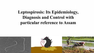
Long, Thin, Highly Coiled
Under Dark Field Microscopy
Source: Merck Veterinary Manual
Source: Wikipedia
5. CURRENT STATUS OF THE DISEASE
Worldwide major outbreaks were reported from the Latin America and the
Caribbean region (36%), followed by Southern Asia (13%), and North America
(11%) (Claudia et. al., 2020)
In India, the disease is prevalent in almost all the species of domestic animals and
in several rodent species It has been reported more frequently from southern states
although few reports are also available from western states (Gujarat and Rajasthan)
(WHO, 2015)
Human Leptospirosis is reported to be of endemic importance from many States of
India which includes Andaman and Nicobar Islands (since 1988), Gujarat (since
1994), Maharashtra ( since 1998), Kerala and Tamil nadu. (WHO, 2015)
Few reports from Assam. Kader et.al., 2018 carried out a study in cattle from few
districts of lower Brahmaputra valley, Assam and recorded a seropositivity of 11.58
%.
6. Three epidemiological patterns of leptospirosis were defined by
Faine (1994)
• The first occurs in temperate climates where few serovars are involved
and human infection almost invariably occurs by direct contact with
infected animals though farming of cattle and pigs
• The second occurs in tropical wet areas, within which there are many
more serovars infecting humans and animals and larger numbers of
reservoir species, including rodents, farm animals, and dogs
• The third pattern comprises rodent-borne infection in the urban
environment (slums)
EPIDEMIOLOGY
7. EPIDEMIOLOGY
HOST RANGE
1.Reservoir host: wide range of natural rodent and non-
rodent reservoir hosts which include foxes, rabbits, rat,
mice, moles etc.
2.Carrier hosts: Domestic animals includes cattle,
sheep, goat, buffalo, horse and dogs.
3.Accidental/ Incidental host: Human act as accidental
host of disease.
MODE OF TRANSMISSION
1.Direct contact with an infected animal.
No human to human transmission is reported
2.Indirect contact via soil, water and food contaminated with urine from an infected animal
3.Transplacental transmission may occur if infection occurs during pregnancy
4.Droplet infection –breathing air polluted with droplets of urine, eg. During milking of
cattle and buffalo.
8. FACTORS FAVOURS EMERGENCE OF LEPTOSPIROSIS
Reservoir and carrier host
Soil salinization -The salinity of the soil provides favourable environment for survival of
leptospires for months together
Climate change - These factors include rainfall, humidity, ambient temperature, water
retaining capacity of the soil, pH and salinity of the soil and surface waters and forest cover
Seasonal variation - It is usually a seasonal disease that starts at the onset of the rainy
season and declines as the rains recede. Sporadic cases may occur throughout the year
Rodent density
Population size of the farm and other domestic animals
Sanitation of animal habitats
Availability of veterinary/ medical services for prompt detection and treatment of animal
leptospirosis and control programs
Personal hygiene
9. HIGH RISK GROUPS
●Occupational exposure – Farmers, ranchers,
abattoir workers, trappers, veterinarians,
loggers, sewer workers, rice farmers, pet
traders, military personnel, laboratory workers
●Recreational activities – Freshwater
swimming, canoeing, kayaking, trail biking
●Household exposure – Pet dogs,
domesticated livestock, rainwater catchment
systems, infestation by infected rodents
●Other – Walking barefoot through surface
water, skin lesions, contact with wild rodents,
accidental laboratory exposure
10. Occupational vulnerability: about 75% of
leptospirosis cases are farmers
Recreational vulnerability affecting a
wider range of rural populations
Leptospira interrogans,
transported by water in
the environment
Vectors of leptospirosis:
Rice field rodents
(Bandicota sp. And Rattus
sp.)
Leptospirosis: a Zoonoses amplified by seasonal flooding
11. TRANSMISSION CYCLE AND PATHOGENESIS OF LEPTOSPIROSIS
Faisal et. al., 2012
Leptospires invade the body
Via penetrating exposed mucous
membranes or damaged skin
After a variable incubation period
(4–20 days)
leptospires circulate in the blood
replicate in many tissues
including the liver, kidneys,
lungs, genital tract, and CNS
leading to various manifestation
12. DISEASES IN ANIMALS
In Cattle: Incubation period (I.P) : 2–12 weeks
(A)Acute form: Sudden onset of elevated temperature (40-41.1°C), anorexia,
anaemia/jaundice, hemoglobinuria, abortion and depression may be seen
(B) Subacute form: Onset is slow, 'milk drop syndrome' due to mastitis resulting
in reduced yield, thick flaking like of colostrum) and yellow to blood coloration
of milk is common. The symptoms lasts for about weeks. Jaundice may be seen
in some cases
(C)Chronic form: Abortions, still births, foetal deaths, weak calves and retained
placenta cases are common in herds.
13. DISEASES IN ANIMALS
Dogs: I.P: 5–15 days
Elevated body temperature, depression, deep
sunken eyes, anorexia, muscle tenderness,
vomiting, intussuseption, foul breath, ulcerated
gums are usually associated with anaemia and
extensive jaundice that may lead to death.
The acute form of disease, known as “Stuttgart
disease” characterized by vomiting, rapid
dehydration, collapse, necrosis and sloughing of
buccal mucosa and tongue
Image depicting ulcerated gums and sunken eyes
Image depicting vomition
14. DISEASES IN ANIMALS
Swine: Usually infection is mild or inapparent. Occasionally may show pyrexia.
Only or few affected pigs show acute signs of anorexia, pyrexia and/ or diarrhoea for
1-3 days.
Chronic cases usually abort in late pregnancy or give birth to weak piglets . and
Recover become renal shedders for at least 6 months.
Horses: Disease manifestations, though rare and usually mild, include pyrexia, icterus,
periodic ophthalmic and abortion.
Sheep: The common signs include abortion, fever, agalactia, dysponea, jaundice,
hematuria and sudden death.
Goats: May remain symptomless but shed leptospires in the urine for short periods or
may develop jaundice, haemaglobinurea and abortions.
Cats: Although susceptible to experimental infection, seldom suffer from clinical
leptospirosis.
15. DISEASES IN MAN
After an incubation period of 7-14 days the disease is manifested in 2 phases.
1. Early or leptospiraemic phase: The pathogen appears in blood and CSF.
In milder cases, the disease remains a subclinical infection
In others, it may present itself as a 'flu-like' syndrome which is characterized
by sudden high fever, chills, headache, muscle aches, vomiting and
conjunctivitis.
Non availability of treatment could intensify the disease to second phase.
16. 2. Second or immune phase/Well's syndrome:
The symptoms may gradually worsen leading to
kidney and liver failure, of which the latter results in
jaundice
Widespread haemorrhages occur leading to anaemia,
coma and finally death.
Fig: Red eye caused by leptospirosis
Red eye (conjunctival suffusion due to immune reaction ) is a constant and
characteristic feature.
In rare cases, the most severe forms of the disease present as
atypical pneumonia, or
aseptic (leptospiral) meningitis or
myocarditis
All the severe forms have a mortality rate of 5-10%, which is quite significant
17. Diagnosis
The disease can be tentatively diagnosed on the basis of clinical symptoms
However, confirmatory diagnosis is made as follows:
Direct examination of samples :
The clinical or morbid materials such as blood, urine, CSF, tissue
scrapings/emulsions should be properly treated with
(a) 10% acetic acid for 5-19 minutes (in case of impression smears), or
(b) 0.25% trypsin for 3-5 minutes (in case of tissues), or
(c) 40% formalin to decontaminate clinical materials, or in smears
by following microscopic techniques as
Dark field microscopy at 400x magnification
Silver impregnation staining
Silver impregnation staining
for Leptospira
18. Isolation of leptospires: These methods include
(i) Culture, (ii) Animal inoculation, and (iii) Culture following animal inoculation
(ii) Animal inoculation: Carried out to enhance the concentration of Leptospira cells or
to remove the bacterial contaminants from the samples.
In hamsters (3 weeks old): Fatal infection accompanied by jaundice evoked following
inoculation with 0.5-1 ml of sample through intra-peritoneal route
Guinea pigs (1 week old) inoculated with 0.5-1 ml material, i/p seldom die of the
infection
(i) Culture: The isolation rate of leptospires from clinical specimens is very low.
Media for culturing leptospires are of 3 types,
media containing serum (Korthof's medium, Fletcher's semi-solid medium etc.)
media containing bovine albumin fraction V and tween 80 (EM medium, EMJH etc ), and
synthetic media
19. Other test being employed:
Immunofluorescence: Useful in examination of urine, other body fluids, and tissues
that have been frozen or are not amenable to silver staining.
Immunohistochemistry: Achieved by using enzymatic or metallic labels on the
secondary antibody. Phosphatase, peroxidase, or metallic gold- labelled antibody can be
used.
Nucleic Acid Probes and Hybridization: Leptospira specific sequences are isolated,
cleaved and labelled with a reporter molecule , then hybridized to ss DNA .
If the nucleotide sequences in the nucleic acid probe are complementary to those in the
sample, hybridization occurs monitored by autoradiography or calorimetrically
Polymerase Chain Reaction : PCR can rapidly confirm the diagnosis in the early
phase of the disease, when bacteria may be present and before antibody titres are at
detectable levels.
20. SEROGROUP/SEROVAR SPECIFIC TEST:
Microscopic Agglutination Test (MAT): MAT is the WHO standard
reference test for serological diagnosis of leptospirosis .
MAT determines agglutinating antibodies in the serum of a patient/ animal by
mixing it in various dilutions with live/killed leptospires.
Anti-leptospiral antibodies present in the serum cause
the leptospires to stick together to form clumps observed
using dark field microscopy (DFM).
Accepted endpoint: Final dilution of serum at which
50% or more of the leptospires are agglutinated.
Leptospires observed under DFM (20X)
21. GENUS SPECIFIC TESTS: These tests are based upon the use of a single antigen
common for the genus Leptospira. The antigen for these tests is prepared from the
non-pathogenic L. biflexa Patoc –1 strain.
Macroscopic Slide Agglutination Test: This test is carried out with a dense
suspension of leptospires, which agglutinate into clumps visible to the naked eye.
Indirect Fluorescent Antibody Test: Specimens of blood, urine and
parenchymatous organs are stained with luminescent sera and examined under a
fluorescent microscope. The antigen antibody complex fluoresces brightly and is
visible under the microscope.
Complement Fixation Test (CFT): CFT is performed using either whole leptospiral
cells or soluble extracts. The CFT is useful in detecting relatively recent infection. .
22. Enzyme Linked Immuno Sorbent Assay (ELISA): ELISA usually detects only the
antibodies reacting with a broadly reactive genus-specific antigen.
Microcapsule Agglutination Test (MCAT): This test is based on the passive
agglutination of synthetic polymer carriers, sensitized with mixed antigens of sonicated
leptospires, by leptospiral antibody.
Lepto Dipstick: This dipstick assay for the detection of
Leptospira-specific IgM antibodies in sera
Latex Agglutination Test: This test depends on the
sensitisation of commercially available latex particles
with a leptospiral antigen.
23. CONTROL STRATEGIES
IN ANIMALS:
1.Rodent control: Areas which favour rodent survival, the rodent control should be
exercised by suitable means including
(i) poisoning with rodenticide (2% zinc phosphide) ,
(ii) use of predators (cat, cagle etc.),
(iii) trapping,
(iv) biological control either by manipulating or altering the habitat,
(v) mechanical proofing, (vi) rodent repellents,
(vii) electronic barriers (fencing), (viii) fumigation,
(ix) proper storage of foods, animal feeds and grains, and
(x) general cleanliness of surroundings
24. 2. Sanitation:
(i) proper draining of yards, pens, sheds and kennels and their
regular disinfection (with cresol , sodium hypochlorite etc.),
(ii) avoidance of stagnation of water, urine and faeces, and
(iii) burial or burning of infected carcasses, aborted foetus,
placentas and/or bedding
3. Proper Management: Should aim at
(i) breaking of the cycle of infection
(ii) allowing new addition to herd or flock only if such animals
have been found negative to MAT on their initial testing as well
on their retesting
(iii) sero-testing of herd by MAT
strictly remove carrier animals serving as excreters and
to isolate infected animals
(iv) prevention of reinfection
25. IN MAN:
The disease control measures include
1. Personal hygiene and protection emphasising in
(i) protection of food articles and utensils from
contamination with urine of rat
(ii) encouraging use of protective clothing (rubber
gloves, goggles, gum-boots particularly when working
in water logged areas or handling animals/animal
products during slaughtering and parturition etc.,
(iii) avoidance of swimming and wading in water of
lakes, ponds, swimming pods contaminated with urine
of rats and livestock, and
(iv) encourage mechanization of agricultural
operations.
26. 2. Sanitation: It includes
(i) disinfection of contaminated work areas such
as food stores, abattoirs, fish and meat processing
plants and animal sheds,
(ii) proper collection, transport, treatment and
secured disposal of garbage and animal excreta
(iii) proper disposal of dead and infected animals,
(iv) disinfection of swimming pool with chlorine,
(v) drainage of wet areas, and
(vi) rodent control in the areas of domestic and
farm environment.
27. 3. Animal related care: It calls for
(i) care in handling of laboratory and other animals
as they may be carriers,
(ii) improvement in occupational hygiene
standards in animal farms/ laboratories
4. Health education: General people and
particularly the high-risk group groups should be
educated about the disease, and the protective
measures to be followed including prohibition of
activities in contaminated waters.
28. CONCLUSION
Leptospirosis is a worldwide underreported neglected zoonosis
In many developing countries, including most of the leptospirosis endemic areas,
laboratory capabilities to detect pathogenic microorganisms are often inadequate
Reservoirs control measures, environmental control programs and animal
vaccination, in combination with a strong surveillance system may significantly
reduce the disease
Public education for people’s awareness of the disease reservoirs and its
transmission is essential
The extensive and good understanding of the eco-epidemiological and cultural
characteristics of a community that faces the problem of leptospirosis is an
essential prerequisite for evolving an effective and acceptable control measure.